Category Archives: Publications

Natural Immunoglobulin M-based Delivery of a Complement Alternative Pathway Inhibitor in Mouse Models of Retinal Degeneration

We have a new manuscript out in Experimental Eye Research, Natural Immunoglobulin M-based Delivery of a Complement Alternative Pathway Inhibitor in Mouse Models of Retinal Degeneration. (pdf here)

Authors: Balasubramaniam Annamalai, Nathaniel Parsons, Crystal Nicholson, Kusumam Joseph, Beth Coughlin, Xiaofeng Yang, Bryan W. Jones @BWJones, Stephen Tomlinson, and Bärbel Rohrer.

Abstract:

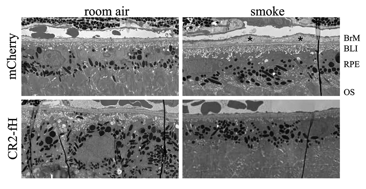
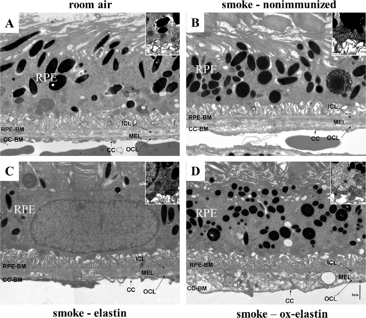
Purpose: Age-related macular degeneration is a slowly progressing disease. Studies have tied disease risk to an overactive complement system. We have previously demonstrated that pathology in two mouse models, the choroidal neovascularization (CNV) model and the smoke-induced ocular pathology (SIOP) model, can be reduced by specifically inhibiting the alternative complement pathway (AP). Here we report on the development of a novel injury-site targeted inhibitor of the alternative pathway, and its characterization in models of retinal degeneration.

Methods: Expression of the danger associated molecular pattern, a modified annexin IV, in injured ARPE-19 cells was confirmed by immunohistochemistry and complementation assays using B4 IgM mAb. Subsequently, a construct was prepared consisting of B4 single chain antibody (scFv) linked to a fragment of the alternative pathway inhibitor, fH (B4-scFv-fH). ARPE-19 cells stably expressing B4-scFv-fH were microencapsulated and administered intravitreally or subcutaneously into C57BL/6 J mice, followed by CNV induction or smoke exposure. Progression of CNV was analyzed using optical coherence tomography, and SIOP using structure-function analyses. B4-scFv-fH targeting and AP specificity was assessed by Western blot and binding experiments.

Results: B4-scFv-fH was secreted from encapsulated RPE and inhibited complement in RPE monolayers. B4-scFv-fH capsules reduced CNV and SIOP, and western blotting for C3a, C3d, IgM and IgG confirmed a reduction in complement activation and antibody binding in RPE/choroid.

Conclusions: Data supports a role for natural antibodies and neoepitope expression in ocular disease, and describes a novel strategy to target AP-specific complement inhibition to diseased tissue in the eye.

Precis: AMD risk is tied to an overactive complement system, and ocular injury is reduced by alternative pathway (AP) inhibition in experimental models. We developed a novel inhibitor of the AP that targets an injury-specific danger associated molecular pattern, and characterized it in disease models.

Keywords: Alternative pathway inhibitor; Choroidal neovascularization; Complement system; Encapsulated ARPE-19 cells; Natural antibody-mediated targeting; Smoke-induced ocular pathology.

Subretinal Rather Than Intravitreal Adeno-Associated Virus–Mediated Delivery of a Complement Alternative Pathway Inhibitor Is Effective in a Mouse Model of RPE Damage

We have a new manuscript out in iOVS, Subretinal Rather Than Intravitreal Adeno-Associated Virus–Mediated Delivery of a Complement Alternative Pathway Inhibitor Is Effective in a Mouse Model of RPE Damage. (pdf here)

Authors: Balasubramaniam Annamalai; Nathaniel Parsons; Crystal Nicholson; Elisabeth Obert; Bryan W. Jones @BWJones; and Bärbel Rohrer.

Abstract:

Purpose: The risk for age-related macular degeneration has been tied to an overactive complement system. Despite combined attempts by academia and industry to develop therapeutics that modulate the complement response, particularly in the late geographic atrophy form of advanced AMD, to date, there is no effective treatment. We have previously demonstrated that pathology in the smoke-induced ocular pathology (SIOP) model, a model with similarities to dry AMD, is dependent on activation of the alternative complement pathway and that a novel complement activation site targeted inhibitor of the alternative pathway can be delivered to ocular tissues via an adeno-associated virus (AAV).

Methods: Two different viral vectors for specific tissue targeting were compared: AAV5-VMD2-CR2-fH for delivery to the retinal pigment epithelium (RPE) and AAV2YF-smCBA-CR2-fH for delivery to retinal ganglion cells (RGCs). Efficacy was tested in SIOP (6 months of passive smoke inhalation), assessing visual function (optokinetic responses), retinal structure (optical coherence tomography), and integrity of the RPE and Bruch’s membrane (electron microscopy). Protein chemistry was used to assess complement activation, CR2-fH tissue distribution, and CR2-fH transport across the RPE.

Results: RPE- but not RGC-mediated secretion of CR2-fH was found to reduce SIOP and complement activation in RPE/choroid. Bioavailability of CR2-fH in RPE/choroid could be confirmed only after AAV5-VMD2-CR2-fH treatment, and inefficient, adenosine triphosphate–dependent transport of CR2-fH across the RPE was identified.

Conclusions: Our results suggest that complement inhibition for AMD-like pathology is required basal to the RPE and argues in favor of AAV vector delivery to the RPE or outside the blood-retina barrier.

Model-Based Comparison of Current Flow in Rod Bipolar Cells of Healthy and Early-Stage Degenerated Retina

We have a new manuscript out in Experimental Eye Research, Model-Based Comparison of Current Flow in Rod Bipolar Cells of Healthy and Early-Stage Degenerated Retina. (pdf here)

Authors: Pragya Kosta, Ege Iseri, Kyle Loizos, Javad Paknahad, Rebecca L. Pfeiffer @BeccaPfeiffer19, Crystal L. Sigulinsky @CLSigulinsky, James R. Anderson, Bryan W. Jones @BWJones, and Gianluca Lazzi.

Abstract: Retinal degenerative diseases, such as retinitis pigmentosa, are generally thought to initiate with the loss of photoreceptors, though recent work suggests that plasticity and remodeling occurs prior to photoreceptor cell loss. This degeneration subsequently leads to death of other retinal neurons, creating functional alterations and extensive remodeling of retinal networks. Retinal prosthetic devices stimulate the surviving retinal cells by applying external current using implanted electrodes. Although these devices restore partial vision, the quality of restored vision is limited. Further knowledge about the precise changes in degenerated retina as the disease progresses is essential to understand how current flows in retinas undergoing degenerative disease and to improve the performance of retinal prostheses. We developed computational models that describe current flow from rod photoreceptors to rod bipolar cells


Model-based Comparison of Current Flow in Rod Bipolar Cells of Healthy and Early-Stage Degenerated Retina

Chapter: Retinal Connectomics

We have a new chapter out in the Elsevier book series The Senses, 2021.

Authors are myself, Bryan W. Jones @BWJones and Robert E. Marc @robertmarc60.

Abstract: The retina is both a light sensor and a highly complex image-processing device – like supercomputers at the backs of eyes. The retina is also wonderfully compact with all circuitry (glia, neurons, synapses and gap junctions) required to compute sensory input, making it a convenient model for understanding the rest of the nervous system. This is also true for disease, with early evidence indicating retina may be a good model for studying progressive neural degenerative diseases. Modern ultrastructural approaches to the study of neural connections is a relatively new !eld has been termed “connectomics”. Connectomics approaches applied to the retina is termed retinal connectomics. These approaches are relatively new !elds that leverage modern technologies in light and ultrastructural imaging, computational storage, and data management to allow tracking of neuronal identity and connectivity, delivering a robust edge/node network map of circuit topologies. Understanding circuit topologies is critical to understanding how retinas process information, and how information processing is corrupted in disease. This chapter summarizes early history, discusses technical aspects of imaging connectomes, justi!es the importance of why connectomics approaches are important, particularly in retina, discusses what has been learned from early efforts in connectomics, and points the way to the next steps.

Please email me: bryan.jones@m.cc.utah.edu if you would like a pdf of the chapter.


A pathoconnectome of early neurodegeneration: Network changes in retinal degeneration

We have a new manuscript out in Experimental Eye Research, A pathoconnectome of early neurodegeneration: Network changes in retinal degeneration. (pdf here)

Authors: Rebecca L. Pfeiffer @BeccaPfeiffer19, James R. Anderson, Jeebika Dahal, Jessica C. Garcia, Jia-Hui Yang, Crystal L. Sigulinsky @CLSigulinsky, Kevin Rapp, Daniel P. Emrich, Carl B. Watt, Hope AB Johnstun, Alexis R. Houser, Robert E. Marc @robertmarc60, and Bryan W. Jones @BWJones.

Abstract: Connectomics has demonstrated that synaptic networks and their topologies are precise and directly correlate with physiology and behavior. The next extension of connectomics is pathoconnectomics: to map neural network synaptology and circuit topologies corrupted by neurological disease in order to identify robust targets for therapeutics. In this report, we characterize a pathoconnectome of early retinal degeneration. This pathoconnectome was generated using serial section transmission electron microscopy to achieve an ultrastructural connectome with 2.18nm/px resolution for accurate identification of all chemical and gap junctional synapses. We observe aberrant connectivity in the rod-network pathway and novel synaptic connections deriving from neurite sprouting. These observations reveal principles of neuron responses to the loss of network components and can be extended to other neurodegenerative diseases.


Network Architecture of Gap Junctional Coupling among Parallel Processing Channels in the Mammalian Retina

We have a new manuscript out in The Journal of Neuroscience, Network Architecture of Gap Junctional Coupling among Parallel Processing Channels in the Mammalian Retina.

Authors: Crystal L. Sigulinsky @CLSigulinsky, James R. Anderson, Ethan Kerzner @EthanKerzner, Christopher N. Rapp @ChrisNRapp, Rebecca L. Pfeiffer @BeccaPfeiffer19, Taryn M. Rodman, Daniel P. Emrich, Kevin D. Rapp, Noah T. Nelson @nooneelseinhere, J. Scott Lauritzen, Miriah Meyer@miriah_meyer, Robert E. Marc @robertmarc60, and Bryan W. Jones @BWJones.

Abstract: Gap junctions are ubiquitous throughout the nervous system, mediating critical signal transmission and integration, as well as emergent network properties. In mammalian retina, gap junctions within the Aii amacrine cell-ON cone bipolar cell (CBC) network are essential for night vision, modulation of day vision, and contribute to visual impairment in retinal degenerations, yet neither the extended network topology nor its conservation is well established. Here, we map the network contribution of gap junctions using a high-resolution connectomics dataset of an adult female rabbit retina. Gap junctions are prominent synaptic components of ON CBC classes, constituting 5%–25% of all axonal synaptic contacts. Many of these mediate canonical transfer of rod signals from Aii cells to ON CBCs for night vision, and we find that the uneven distribution of Aii signals to ON CBCs is conserved in rabbit, including one class entirely lacking direct Aii coupling. However, the majority of gap junctions formed by ON CBCs unexpectedly occur between ON CBCs, rather than with Aii cells. Such coupling is extensive, creating an interconnected network with numerous lateral paths both within, and particularly across, these parallel processing streams. Coupling patterns are precise with ON CBCs accepting and rejecting unique combinations of partnerships according to robust rulesets. Coupling specificity extends to both size and spatial topologies, thereby rivaling the synaptic specificity of chemical synapses. These ON CBC coupling motifs dramatically extend the coupled Aii-ON CBC network, with implications for signal flow in both scotopic and photopic retinal networks during visual processing and disease.

Immunization Against Oxidized Elastin Exacerbates Structural and Functional Damage in Mouse Model of Smoke-Induced Ocular Injury

We have a new manuscript out in Investigative Ophthalmology & Visual Science, Immunization Against Oxidized Elastin Exacerbates Structural and Functional Damage in Mouse Model of Smoke-Induced Ocular Injury

Authors: Balasubramaniam Annamalai; Crystal Nicholson; Nathaniel Parsons; Sarah Stephenson; Carl Atkinson; Bryan Jones; and Bärbel Rohrer.

Purpose: Age-related macular degeneration (AMD) is the leading cause of blindness in Western populations. While an overactive complement system has been linked to pathogenesis, mechanisms contributing to its activation are largely unknown. In aged and AMD eyes, loss of the elastin layer (EL) of Bruch’s membrane (BrM) has been reported. Elastin antibodies are elevated in patients with AMD, the pathogenic significance of which is unclear. Here we assess the role of elastin antibodies using a mouse model of smoke-induced ocular pathology (SIOP), which similarly demonstrates EL loss.

Methods: C57BL/6J mice were immunized with elastin or elastin peptide oxidatively modified by cigarette smoke (ox-elastin). Mice were then exposed to cigarette smoke or air for 6 months. Visual function was assessed by optokinetic response, retinal morphology by spectral-domain optical coherence tomography and electron microscopy, and complement activation and antibody deposition by Western blot.

Results: Ox-elastin IgG and IgM antibodies were elevated in ox-elastin immunized mice following 6 months of smoke, whereas elastin immunization had a smaller effect. Ox-elastin immunization exacerbated smoke-induced vision loss, with thicker BrM and more damaged retinal pigment epithelium (RPE) mitochondria compared with mice immunized with elastin or nonimmunized controls. These changes were correlated with increased levels of IgM, IgG2, IgG3, and complement activation products in RPE/choroid.

Conclusions: These data demonstrate that SIOP mice generate elastin-specific antibodies and that immunization with ox-elastin exacerbates ocular pathology. Elastin antibodies represented complement fixing isotypes that, together with the increased presence of complement activation seen in immunized mice, suggest that elastin antibodies exert pathogenic effects through mediating complement activation.

Müller Cell Metabolic Signatures: Evolutionary Conservation and Disruption in Disease

We have a new manuscript out in Trends in Endocrinology & Metabolism, Müller Cell Metabolic Signatures: Evolutionary Conservation and Disruption in Disease.

Authors: Rebecca L. Pfeiffer @BeccaPfeiffer19, Robert E. Marc @robertmarc60, and Bryan William Jones @BWJones.

This manuscript functions as both a review and presents some exciting new data demonstrating how the glutamate cycle is disrupted during retinal degenerative disease.

Abstract: Müller cells are glia that play important regulatory roles in retinal metabolism. These roles have been evolutionarily conserved across at least 300 million years. Müller cells have a tightly locked metabolic signature in the healthy retina, which rapidly degrades in response to insult and disease. This variation in metabolic signature occurs in a chaotic fashion, involving some central metabolic pathways. The cause of this divergence of Müller cells, from a single class with a unique metabolic signature to numerous separable metabolic classes, is currently unknown and illuminates potential alternative metabolic pathways that may be revealed in disease. Understanding the impacts of this heterogeneity on degenerate retinas and the implications for the metabolic support of surrounding neurons will be critical to long-term integration of retinal therapeutics for the restoration of visual perception following photoreceptor degeneration.

Optic Cup Morphogenesis Requires Neural Crest-Mediated Basement Membrane Assembly

We have a new manuscript out in Development, Optic cup morphogenesis requires neural crest-mediated basement membrane assembly.

Authors: Chase D. Bryan @CDBE30, Macaulie A. Casey, Rebecca L. Pfeiffer @BeccaPfeiffer19, Bryan W. Jones @BWJones, and Kristen M. Kwan @BlockInTheBack

This is a collaborative project out of the Kwan lab that we helped out with some of the ultrastructural work and analysis.

Abstract: Organogenesis requires precise interactions between a developing tissue and its environment. In vertebrates, the developing eye is surrounded by a complex extracellular matrix as well as multiple mesenchymal cell populations. Disruptions to either the matrix or periocular mesenchyme can cause defects in early eye development, yet in many cases, the underlying mechanism is unknown. Here, using multidimensional imaging and computational analyses in zebrafish, we establish that cell movements in the developing optic cup require neural crest. Ultrastructural analysis reveals that basement membrane formation around the developing eye is also dependent on neural crest, but only specifically around the retinal pigment epithelium. Neural crest cells produce the extracellular matrix protein nidogen: impairing nidogen function disrupts eye development, and strikingly, expression of nidogen in the absence of neural crest partially restores optic cup morphogenesis. These results demonstrate that eye formation is regulated in part by extrinsic control of extracellular matrix assembly.

Pathoconnectome Analysis of Müller Cells in Early Retinal Remodeling

We have a new manuscript out in Clinical Neurophysiology, An Update on Retinal Prostheses. PubMedDirect Link PDF here.

Authors: Rebecca L Pfeiffer, James R Anderson, Daniel P Emrich, Jeebika Dahal, Crystal L Sigulinsky, Hope AB Morrison, Jia-Hui Yang, Carl B Watt, Kevin D Rapp, Mineo Kondo, Hiroko Terasaki, Jessica C Garcia, Robert E Marc, and Bryan W Jones.

Abstract: Glia play important roles in neural function, including but not limited to amino acid recycling, ion homeostasis, glucose metabolism, and waste removal. During retinal degeneration and subsequent retinal remodeling, Müller cells (MCs) are the first cells to show metabolic and morphological alterations in response to stress. Metabolic alterations in MCs chaotically progress in retina undergoing photoreceptor degeneration; however, what relationship these alterations have with neuronal stress, synapse maintenance, or glia-glia interactions is currently unknown. The work described here reconstructs a MC from a pathoconnectome of early retinalremodeling retinalpathoconnectome 1 (RPC1) and explores relationships between MC structural and metabolic phenotypes in the context of neighboring neurons and glia. Here we find variations in intensity of osmication inter- and intracellularly, variation in small molecule metabolic content of MCs, as well as morphological alterations of glial endfeet. RPC1 provides a framework to analyze these relationships in early retinal remodeling through ultrastructural reconstructions of both neurons and glia. These reconstructions, informed by quantitative metabolite labeling via computational molecular phenotyping (CMP), allow us to evaluate neural-glial interactions in early retinal degeneration with unprecedented resolution and sensitivity.