Category Archives: Retinal Disease

Subretinal Rather Than Intravitreal Adeno-Associated Virus–Mediated Delivery of a Complement Alternative Pathway Inhibitor Is Effective in a Mouse Model of RPE Damage

We have a new manuscript out in iOVS, Subretinal Rather Than Intravitreal Adeno-Associated Virus–Mediated Delivery of a Complement Alternative Pathway Inhibitor Is Effective in a Mouse Model of RPE Damage. (pdf here)

Authors: Balasubramaniam Annamalai; Nathaniel Parsons; Crystal Nicholson; Elisabeth Obert; Bryan W. Jones @BWJones; and Bärbel Rohrer.

Abstract:

Purpose: The risk for age-related macular degeneration has been tied to an overactive complement system. Despite combined attempts by academia and industry to develop therapeutics that modulate the complement response, particularly in the late geographic atrophy form of advanced AMD, to date, there is no effective treatment. We have previously demonstrated that pathology in the smoke-induced ocular pathology (SIOP) model, a model with similarities to dry AMD, is dependent on activation of the alternative complement pathway and that a novel complement activation site targeted inhibitor of the alternative pathway can be delivered to ocular tissues via an adeno-associated virus (AAV).

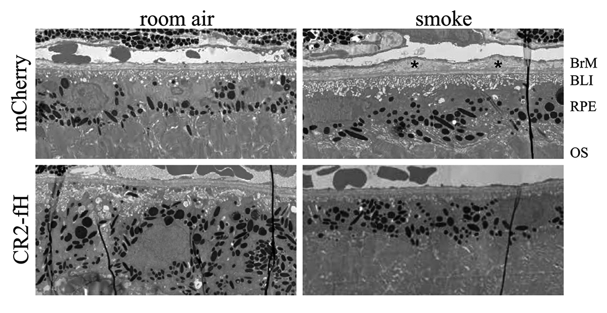
Methods: Two different viral vectors for specific tissue targeting were compared: AAV5-VMD2-CR2-fH for delivery to the retinal pigment epithelium (RPE) and AAV2YF-smCBA-CR2-fH for delivery to retinal ganglion cells (RGCs). Efficacy was tested in SIOP (6 months of passive smoke inhalation), assessing visual function (optokinetic responses), retinal structure (optical coherence tomography), and integrity of the RPE and Bruch’s membrane (electron microscopy). Protein chemistry was used to assess complement activation, CR2-fH tissue distribution, and CR2-fH transport across the RPE.

Results: RPE- but not RGC-mediated secretion of CR2-fH was found to reduce SIOP and complement activation in RPE/choroid. Bioavailability of CR2-fH in RPE/choroid could be confirmed only after AAV5-VMD2-CR2-fH treatment, and inefficient, adenosine triphosphate–dependent transport of CR2-fH across the RPE was identified.

Conclusions: Our results suggest that complement inhibition for AMD-like pathology is required basal to the RPE and argues in favor of AAV vector delivery to the RPE or outside the blood-retina barrier.

Model-Based Comparison of Current Flow in Rod Bipolar Cells of Healthy and Early-Stage Degenerated Retina

We have a new manuscript out in Experimental Eye Research, Model-Based Comparison of Current Flow in Rod Bipolar Cells of Healthy and Early-Stage Degenerated Retina. (pdf here)

Authors: Pragya Kosta, Ege Iseri, Kyle Loizos, Javad Paknahad, Rebecca L. Pfeiffer @BeccaPfeiffer19, Crystal L. Sigulinsky @CLSigulinsky, James R. Anderson, Bryan W. Jones @BWJones, and Gianluca Lazzi.

Abstract: Retinal degenerative diseases, such as retinitis pigmentosa, are generally thought to initiate with the loss of photoreceptors, though recent work suggests that plasticity and remodeling occurs prior to photoreceptor cell loss. This degeneration subsequently leads to death of other retinal neurons, creating functional alterations and extensive remodeling of retinal networks. Retinal prosthetic devices stimulate the surviving retinal cells by applying external current using implanted electrodes. Although these devices restore partial vision, the quality of restored vision is limited. Further knowledge about the precise changes in degenerated retina as the disease progresses is essential to understand how current flows in retinas undergoing degenerative disease and to improve the performance of retinal prostheses. We developed computational models that describe current flow from rod photoreceptors to rod bipolar cells


Model-based Comparison of Current Flow in Rod Bipolar Cells of Healthy and Early-Stage Degenerated Retina

A pathoconnectome of early neurodegeneration: Network changes in retinal degeneration

We have a new manuscript out in Experimental Eye Research, A pathoconnectome of early neurodegeneration: Network changes in retinal degeneration. (pdf here)

Authors: Rebecca L. Pfeiffer @BeccaPfeiffer19, James R. Anderson, Jeebika Dahal, Jessica C. Garcia, Jia-Hui Yang, Crystal L. Sigulinsky @CLSigulinsky, Kevin Rapp, Daniel P. Emrich, Carl B. Watt, Hope AB Johnstun, Alexis R. Houser, Robert E. Marc @robertmarc60, and Bryan W. Jones @BWJones.

Abstract: Connectomics has demonstrated that synaptic networks and their topologies are precise and directly correlate with physiology and behavior. The next extension of connectomics is pathoconnectomics: to map neural network synaptology and circuit topologies corrupted by neurological disease in order to identify robust targets for therapeutics. In this report, we characterize a pathoconnectome of early retinal degeneration. This pathoconnectome was generated using serial section transmission electron microscopy to achieve an ultrastructural connectome with 2.18nm/px resolution for accurate identification of all chemical and gap junctional synapses. We observe aberrant connectivity in the rod-network pathway and novel synaptic connections deriving from neurite sprouting. These observations reveal principles of neuron responses to the loss of network components and can be extended to other neurodegenerative diseases.


NeuroNex Grant

I am pleased to report that the The Marclab for Connectomics has been funded by the National Science Foundation with a 5 year grant as part of a large, international consortium to study synaptic weighting.  We are collaborating with the Erik Jorgensen laboratory here at the University of Utah, and will be exploring synapses in a model of retinal degeneration.  There is a nice writeup of the award on the Moran Eye Center website, here.

This is a wonderful opportunity to work with other colleagues that will be funded alongside us with this grant, including Uri Manor @manorlaboratory, Davi Bock @dddavi, Josh Vogelstein @neuro_data, Viren Jain @stardazed0, and others.  My thanks to Kristen Harris for heading up this initiative.

Müller Cell Metabolic Signatures: Evolutionary Conservation and Disruption in Disease

We have a new manuscript out in Trends in Endocrinology & Metabolism, Müller Cell Metabolic Signatures: Evolutionary Conservation and Disruption in Disease.

Authors: Rebecca L. Pfeiffer @BeccaPfeiffer19, Robert E. Marc @robertmarc60, and Bryan William Jones @BWJones.

This manuscript functions as both a review and presents some exciting new data demonstrating how the glutamate cycle is disrupted during retinal degenerative disease.

Abstract: Müller cells are glia that play important regulatory roles in retinal metabolism. These roles have been evolutionarily conserved across at least 300 million years. Müller cells have a tightly locked metabolic signature in the healthy retina, which rapidly degrades in response to insult and disease. This variation in metabolic signature occurs in a chaotic fashion, involving some central metabolic pathways. The cause of this divergence of Müller cells, from a single class with a unique metabolic signature to numerous separable metabolic classes, is currently unknown and illuminates potential alternative metabolic pathways that may be revealed in disease. Understanding the impacts of this heterogeneity on degenerate retinas and the implications for the metabolic support of surrounding neurons will be critical to long-term integration of retinal therapeutics for the restoration of visual perception following photoreceptor degeneration.

Pathoconnectome Analysis of Müller Cells in Early Retinal Remodeling

We have a new manuscript out in Clinical Neurophysiology, An Update on Retinal Prostheses. PubMedDirect Link PDF here.

Authors: Rebecca L Pfeiffer, James R Anderson, Daniel P Emrich, Jeebika Dahal, Crystal L Sigulinsky, Hope AB Morrison, Jia-Hui Yang, Carl B Watt, Kevin D Rapp, Mineo Kondo, Hiroko Terasaki, Jessica C Garcia, Robert E Marc, and Bryan W Jones.

Abstract: Glia play important roles in neural function, including but not limited to amino acid recycling, ion homeostasis, glucose metabolism, and waste removal. During retinal degeneration and subsequent retinal remodeling, Müller cells (MCs) are the first cells to show metabolic and morphological alterations in response to stress. Metabolic alterations in MCs chaotically progress in retina undergoing photoreceptor degeneration; however, what relationship these alterations have with neuronal stress, synapse maintenance, or glia-glia interactions is currently unknown. The work described here reconstructs a MC from a pathoconnectome of early retinalremodeling retinalpathoconnectome 1 (RPC1) and explores relationships between MC structural and metabolic phenotypes in the context of neighboring neurons and glia. Here we find variations in intensity of osmication inter- and intracellularly, variation in small molecule metabolic content of MCs, as well as morphological alterations of glial endfeet. RPC1 provides a framework to analyze these relationships in early retinal remodeling through ultrastructural reconstructions of both neurons and glia. These reconstructions, informed by quantitative metabolite labeling via computational molecular phenotyping (CMP), allow us to evaluate neural-glial interactions in early retinal degeneration with unprecedented resolution and sensitivity.


Persistent Remodeling And Neurodegeneration In Late-Stage Retinal Degeneration

We have a new manuscript out in Progress In Retinal And Eye Research, Persistent Remodeling And Neurodegeneration In Late-Stage Retinal Degeneration.

Authors: Rebecca L. Pfeiffer, Robert E. Marc, and Bryan William Jones.

I’m really proud of the work that Becca did on this manuscript.  It does a couple of important things including summarizing the field of retinal remodeling, but also introducing some new data that plants a flag to define retinal degeneration and retinal remodeling as the first steps in what becomes progressive neurodegeneration.  We view the retina as an excellent model to begin exploring diseases like Alzheimer’s, Parkinson’s, and other progressive neurodegenerative diseases.  In addition, Becca also demonstrates a new potential mechanism for misfolded proteins, that may point the way for how proteinopathies spread.

Abstract: Retinal remodeling is a progressive series of negative plasticity revisions that arise from retinal degeneration, and are seen in retinitis pigmentosa, age-related macular degeneration and other forms of retinal disease. These processes occur regardless of the precipitating event leading to degeneration. Retinal remodeling then culminates in a late-stage neurodegeneration that is indistinguishable from progressive central nervous system (CNS) proteinopathies. Following long-term deafferentation from photoreceptor cell death in humans, and long-lived animal models of retinal degeneration, most retinal neurons reprogram, then die. Glial cells reprogram into multiple anomalous metabolic phenotypes. At the same time, survivor neurons display degenerative inclusions that appear identical to progressive CNS neurodegenerative disease, and contain aberrant α-synuclein (α-syn) and phosphorylated α-syn. In addition, ultrastructural analysis indicates a novel potential mechanism for misfolded protein transfer that may explain how proteinopathies spread. While neurodegeneration poses a barrier to prospective retinal interventions that target primary photoreceptor loss, understanding the progression and time-course of retinal remodeling will be essential for the establishment of windows of therapeutic intervention and appropriate tuning and design of interventions. Finally, the development of protein aggregates and widespread neurodegeneration in numerous retinal degenerative diseases positions the retina as a ideal platform for the study of proteinopathies, and mechanisms of neurodegeneration that drive devastating CNS diseases.

Coupling Architecture Of The AII/ON Cone Bipolar Cell Network In Degenerate Retina

This abstract was presented today, April 8th at the 2019 Association for Research in Vision and Opthalmology (ARVO) meetings in Vancouver, Canada as a platform presentation by Crystal L. Sigulinsky, Rebecca L. PfeifferJames R. Anderson, Daniel P. Emrich, Christopher Rapp, Jeebika Dahal, Jessica Garcia, Hope Morrison, Kevin D. Rapp, Jia-Hui Yang, Carl B. Watt, Robert E. Marc and Bryan W. Jones.

Purpose
In mouse models of retinal degeneration, connexin36-containing gap junctions in the Aii amacrine cell network appear to mediate aberrant hyperactivity within the retina. However, it remains unclear whether this hyperactivity reflects changes in the underlying circuitry or dysfunction of the normative circuitry. Our connectomics-based mapping of retinal circuitry in rabbit Retinal Connectome 1 (RC1) has dramatically expanded the coupled Aii network. In addition to canonical Aii-to-Aii and Aii-to-ON cone bipolar cell (CBC) coupling, we describe pervasive in- and cross-class coupling motifs among ON CBCs. This study examines the changes in these coupling motifs in RPC1, an ultrastructural retinal pathoconnectome from a rabbit model of retinitis pigmentosa.

Methods
RC1 and RPC1 are connectomes built by automated transmission electron microscopy at ultrastructural (2 nm/pixel) resolution. RC1 is a 0.25 mm diameter volume of retina from a 13 month old, light-adapted female Dutch Belted rabbit and serves as the healthy reference connectome. RPC1 is a 0.09 mm diameter volume of pathological retina from a 10 month old, male transgenic P347L model of autosomal dominant retinitis pigmentosa showing early phase 1 retinal remodeling, when rod photoreceptors are still present, but stressed. ON CBCs, Aii amacrine cells, and their coupling partners were annotated using the Viking application. Coupling motifs and features were explored with 3D rendering and graph visualization of connectivity. Gap junctions were validated by 0.25 nm resolution recapture with goniometric tilt when necessary.

Results
All major coupling motifs were observed. Several ON CBC classes retained their class-specific coupling profiles, accepting and rejecting specific combinations of Aii and ON CBC class partnerships. However, aberrant partnerships exist in the coupled network, including both loss of prominent motifs and acquisition of novel ones.

Conclusions
Clearly aberrant morphological and synaptic changes exist in RPC1, including changes in the coupling specificity and gap junction distributions of both Aii amacrine cells and ON CBCs. This indicates that the Aii/ON CBC circuit topology is already altered during early phase 1 remodeling, with substantial implications for therapeutic interventions for blinding diseases that depend upon the surviving retinal network in human patients.

Aii Amacrine Cell Connectivity in Degenerating Retina

This abstract was presented today, April 8th at the 2019 Association for Research in Vision and Opthalmology (ARVO) meetings in Vancouver, Canada by Jeebika Dahal, Rebecca L. Pfeiffer, Crystal L. Sigulinsky, James R. Anderson, Daniel P. Emrich, Hope Morrison, Jessica C. Garcia, Kevin D. Rapp, Jia-Hui Yang, Carl B. Watt, Mineo Kondo, Hiroko Terasaki, Robert E. Marc and Bryan W. Jones.

Full resolution version here.

Purpose
Aii amacrine cells (Aii ACs) function in mediating scotopic vision via connection of rod bipolar cells (Rod BCs) to cone bipolar cell pathways. The purpose of this project is to determine the effect of retinal degeneration (RD) on Aii AC networks. We explore this in a pathoconnectome of early RD (RPC1), using a connectome of healthy retina (RC1) as control. Cells in each volume are evaluated by comparison of morphology, synaptic connectivity, and eventually network analysis.

Methods
Tissue for RPC1 was collected from a 10 month old transgenic p347L rabbit model of autosomal dominant retinitis pigmentosa. RC1 was collected from a 13 month old Dutch-Belted rabbit, with no indications of degeneration. Tissue was fixed in a mixed aldehyde solution, before subsequent dehydration, osmication, and resin embedding. Volumes were sectioned at 70nm (RPC1) and 90nm (RC1) and placed on formvar grids. 1 section was reserved from every 30 TEM sections for computational molecular phenotyping where it was placed on a slide and probed for small molecules or proteins. TEM sections were captured at 2.18nm/px using SerialEM software on a JEOL JEM-1400 TEM. The RC1 volume has a diameter of 250µm and RPC1 has a diameter of 90µm. Both volumes were analyzed using the Viking software suite.

Results
In this study, Aii ACs from RPC1 were compared to RC1. Initial results indicate no distinct difference in the morphology other than arbor size, which are likely due to eccentricity differences between volumes. However, in RPC1, we observe multiple instances of Aii AC coupling with Rod BCs in the ON region of the IPL. In contrast, Rod BCs never form gap junctions in healthy retina.

Conclusions
Coupling between Aii ACs and Rod BCs in RPC1 is a unique change in retinal network topology occurring in early RD. Further exploration of network changes as a response to RD is warranted, as many therapeutic interventions currently in development rely upon maintenance of inner retinal circuitry. Prior research demonstrates Rod BCs extend dendrites towards cones and change their receptor expression as rods degenerate. Therefore, knowing the network changes involving Aii ACs and their associations with bipolar cells is crucial to understanding how photoreceptor degeneration affects inner retinal visual processing.

OFF-layer Branches of ON Cone Bipolar Cells in Early Retinal Degeneration

This abstract was presented today, April 8th at the 2019 Association for Research in Vision and Opthalmology (ARVO) meetings in Vancouver, Canada by Jessica C. Garcia, Rebecca L. Pfeiffer, Crystal L. Sigulinsky, James R. Anderson, Daniel P. Emrich, Jeebika Dahal, Hope Morrison, Kevin D. Rapp, Jia-Hui Yang, Carl B. Watt, Mineo Kondo, Hiroko Terasaki, Robert E. Marc and Bryan W. Jones.

Full resolution version here.

Purpose: Cone bipolar cells are customarily classified into superclasses of ON (ON-BCs) and OFF (OFF-BCs). ON or OFF specialization is further segregated by stratification within the inner plexiform layer. Retinal degeneration induces negative plasticity termed remodeling, that includes aberrant neurite extension from multiple cell types (rewiring) and ON-BCs switching their glutamate receptors to match that of OFF-BCs (reprogramming). Previous analysis in healthy retina shows that ON-BCs can make small ribbon synapses in the descending axon or, less frequently, simple single branch projections in the OFF layer. What impact remodeling has on these OFF-layer branches is unknown. In this study, we compare OFF branches from ON-BCs in a connectome of early retinal degeneration (RPC1) to our healthy retinal connectome (RC1).

Methods: Retinal tissues selected for RC1 and RPC1 were collected post-mortem from a 13 month old Dutch-belted healthy female rabbit and a male 10 month old transgenic P347L rabbit model of autosomal dominant retinitis pigmentosa, respectively. RPC1 shows signs of remodeling including rod outer-segment degeneration and aberrant neurite extension. Tissues were fixed in mixed aldehydes and subsequently osmicated, dehydrated, resin embedded, and sectioned at 90 nm (RC1) or 70 nm (RPC1). Sections were placed on formvar grids, stained, and imaged at 2nm/px on a JEOL JEM-1400 TEM using SerialEM software. 1 section was reserved from every 30 for Computational Molecular Phenotyping, and probed for small molecules. Both volumes were evaluated using the Viking software suite.

Results: Ribbons of ON-BCs formed in the OFF layer branches have been previously described to contact glycinergic amacrine cells (ACs), GABAergic ACs, ON ganglion cells, and intrinsically photosensitive ganglion cells. Initial analysis of OFF branches of ON-BCs in RPC1 demonstrate more complex branching than in RC1 and increased number of synapses on these branches. In contrast to the inconsistent OFF layer branch stratification observed in RC1, the OFF branches in RPC1 appear to stratify at a similar level. Evaluation of synaptic partners is ongoing.

Conclusions: Increased complexity and number of synapses found in the OFF branches of some ON-BCs ultimately may represent ON network corruption. Exploring synaptic partners will reveal potential network alterations in retinal degenerative disease.