Seeing in color
Our visual systems extract hundreds of colors via the actions of only three kinds of cone photoreceptors (blue-, green- and red-sensitive cones) arranged as a sensor sheet in the back of the eye. Harry Sperling and Robert Marc were the first to image the chromatic cone array ∞Pubmed
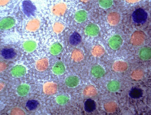
The retina
The array of cones is a sheet of sensors sandwiched between the supporting tissues of the eye and the neural retina that processes color information. The neural retina contains over 60 different kinds of cells and thousands to millions of copies of each, arranged in a glassy, transparent information processing system that feeds the brain. The light passes right through the neural retina.
Animal color vision: Can my dog see in color?
Animals don’t see in black and white. In fact most fishes, reptiles and birds have much richer color vision than you do. But we humans are at the pinnacle of mammalian vision. Most mammals see a beautifully tinted world, much like a dichromatic human. Though we refer to such people and animals as “color blind”, they aren’t blind at all. They just have a “thinner” but nevertheless elegant color space. Here is how the kittens Mango and Slate see the world.
A schematic of the human eye
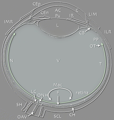
C cornea
CB ciliary body
CEp corneal epithelium
CEn corneal endothelium
CH choroid
F fovea
IMR insertion of the medial rectus muscle
ILR insertion of the lateral rectus muscle
IR iris
L Lens
LC Lamina cribrosa
LIM Limbus
Mac Macula
N Nasal half of the visual field
ONH optic nerve head
OT ora terminalis
OAV ophthalmic artery and vein
Pu pupil
PP Pars plana
SH Scleral Sheath
SCL Sclera
T Temporal half of the visual field
V vitreous
The world has no images …
… for it is all surface and light and suffusion. Patterns of light and hue mix, but there is no form in the mix. Images appear only through apertures.
Sensing of form is an exercise in the geometry of apertures. If I stand 5 meters from Ann and 10 meters from the tundra swan, she and I observe nearly identical images down to the yellow tear- drop of bill skin at the frontal canthus of its black eye, as it views us. Yet we see none of the same photons. The swan is encased in myriad potential images, each the same swan but all built of different photons; a bubble of ever-expanding swan light reflected perfectly from her surface. And the swan and I each see a singular Ann whose sphere of potential images radiates outward. These are well-worn photons, born deep in the sun and meandering to the surface, scattered atom-to-atom through the sky, off the water, the ice, yet still as potent as when created, only to be extinguished, absorbed in my eye. And betwixt these multihued, intersecting, propagating wave groups, there are six real, moving images … and six apertures … six geometry machines. Six eyes and six images.
Sensory perforations are everything. That is how sensory stimuli get past our barriers: through holes in the self. Eyes are perforated chambers like the camera obscura (the room of darkness) sealed from everything except a single cone of light traversing an aperture. In such magic chambers one could watch the Emperor’s horses at exercise on a wall … 25 centuries ago. The stream of never-ending photons weld a single point on a real object to its faint image on the wall within the room.
The key to images is casting off all but few beams …
… and squeezing them through apertures. Aperture size is critical as each contains a crossing of rays known as a focus. Large apertures blur: Each object point connects by straight rays to a spray of image points bounded by the upper and lower hole margins, forming a mess of misaligned foci. Decreasing aperture size reduces blurring, but small holes pass few photons and images are dimmed to uselessness. After all, one cannot “turn up” the Sun. And then there is spatial resolution to worry about. A sensor surface on which an image is cast … whether eye, film or digital camera chip … is composed of discrete sensing points, and image sharpness requires large sensors. A patch of 1000 sensing points will better define an image than one drawn by 100 points. In the camera obscura, images are enlarged by building bigger rooms. Doubling image area requires enlarging the room width from 10 feet to 14.14 feet from the aperture. But this new image is dimmer, with the same number of photons distributed across twice the area. To implement this scheme biologically would require eyes bigger than our bodies. While apertures are essential for image formation, but they are insufficient.
The solution is lensing
Lensing moves the focus of photon beams past the aperture, on the axis of view, allowing a wide aperture and superimposing many images more precisely. Lenses needn’t be very good to capture orders of magnitude more photons than a pinhole and to cast bright images. But there is only one way to move photons: slow them down. The speed of light (c) is constant within a medium, such as interstellar space or water. The velocity v of photons in water is about 3/4c in space … and on absorption the velocity drops to zero and the photon ceases to exist. Photons traversing different media successively slow and speed, and if the beam strikes the interface between two media at an angle, the beam must turn slightly in accord with its speed. This little law of physics easily allows inorganic and biological tissues to be lenses. Some of the best are curved structures, like the water-drop lenses with which Leeuwenhoek spied his animacules, themselves miniscule lensing cells chocked with watery proteins. The layering of transparent cells to form lensing elements is evolutionarily ancient and concisely summarized by Oyster in his spectacular opus, ∞The Human Eye