
Images and movies (unless otherwise noted) are free for educational use under: Creative Commons Attribution-Non Commercial-NoDerivs 3.0 Unported License.
Some images within presentations are licensed microstock images. Licensing information available on request.
Robert E. Marc :: 2016 Harvard AMD Conference Presentation :: marc-2016-harvard 141 Mb PDF
 Connectomics for K-12 Connectomics for K-12 |
 Brains in Cinema Brains in Cinema |
| Connectomics & Eternity |
 Marclab Journal Cover Gallery Marclab Journal Cover Gallery |
 Eye Candy Gallery |
 RC1 Connectome Navigation Movie (right-click or control-click and download)(self-playing moving) RC1 Connectome Navigation Movie (right-click or control-click and download)(self-playing moving)Play or Download 33 MB.mov |
 Connectomics Summary Movie (right-click or control-click and download)(self-playing moving) Connectomics Summary Movie (right-click or control-click and download)(self-playing moving)Play or Download 79 MB.mov |
Connectomics Summary Gallery |
 Marc 2012-07-Senses-vision flat lg.pdf (right-click or control-click and download) Marc 2012-07-Senses-vision flat lg.pdf (right-click or control-click and download)Download 11 MB PDF |
 IMAGE :: Journal of Microscopy Fig 11 300dpi 51 picas w X 46 picas h LZW compressed Download 9MB jpg. IMAGE :: Journal of Microscopy Fig 11 300dpi 51 picas w X 46 picas h LZW compressed Download 9MB jpg.
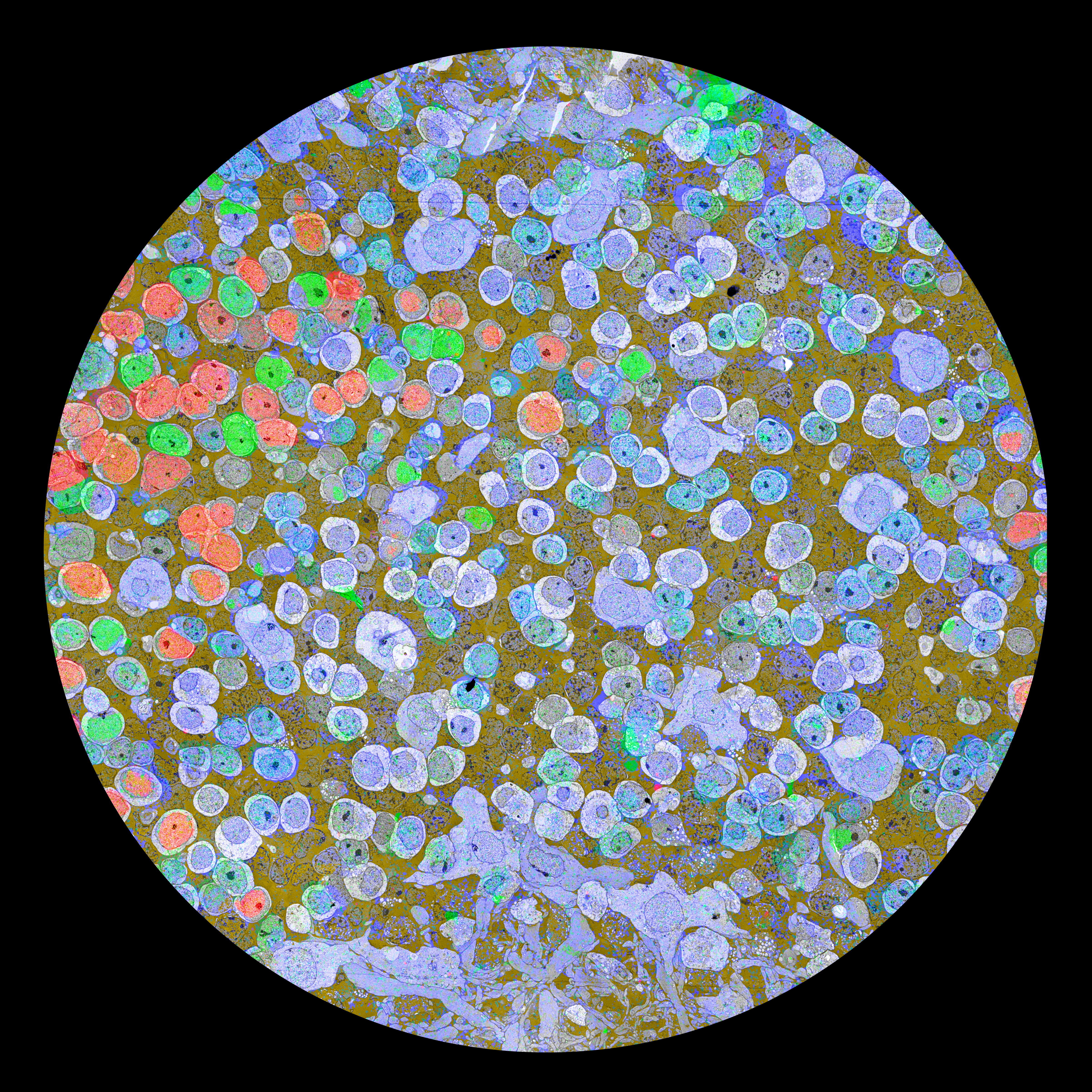
Caption: A keyframe image for a 90 section subvolume of retinal connectome RC1. RC1 is composed of over 340,000 transmission electron microscope (TEM) images from 341 sections spanning a 250 μm diameter tissue column, assembled into single analysis volume parsed by the Viking application. The subvolume, 17.8 μm wide with a native resolution of 2.18 nm/pixel, was designed as a public test volume for tracking algorithms. The six colored processes have different topologies that vary in tracking difficulty: yellow > green > red > lavender > blue > cyan. |
 REM Presentation at Photocamp Utah 2010 lecture MP4 (right-click or control-click and download) REM Presentation at Photocamp Utah 2010 lecture MP4 (right-click or control-click and download)Download MP4 (223 MB) © REMarc 2010 |
| IMAGE Connectome large (right-click or control-click and download) Download jpg as 23 MB © REMarc 2010 |
| PRESENTATION Marc 114th Japanese Society for Ophthalmology lecture 2010 lecture (right-click or control-click and download) Download pdf © REMarc 2010 |
© BWJones 2003 |
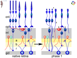
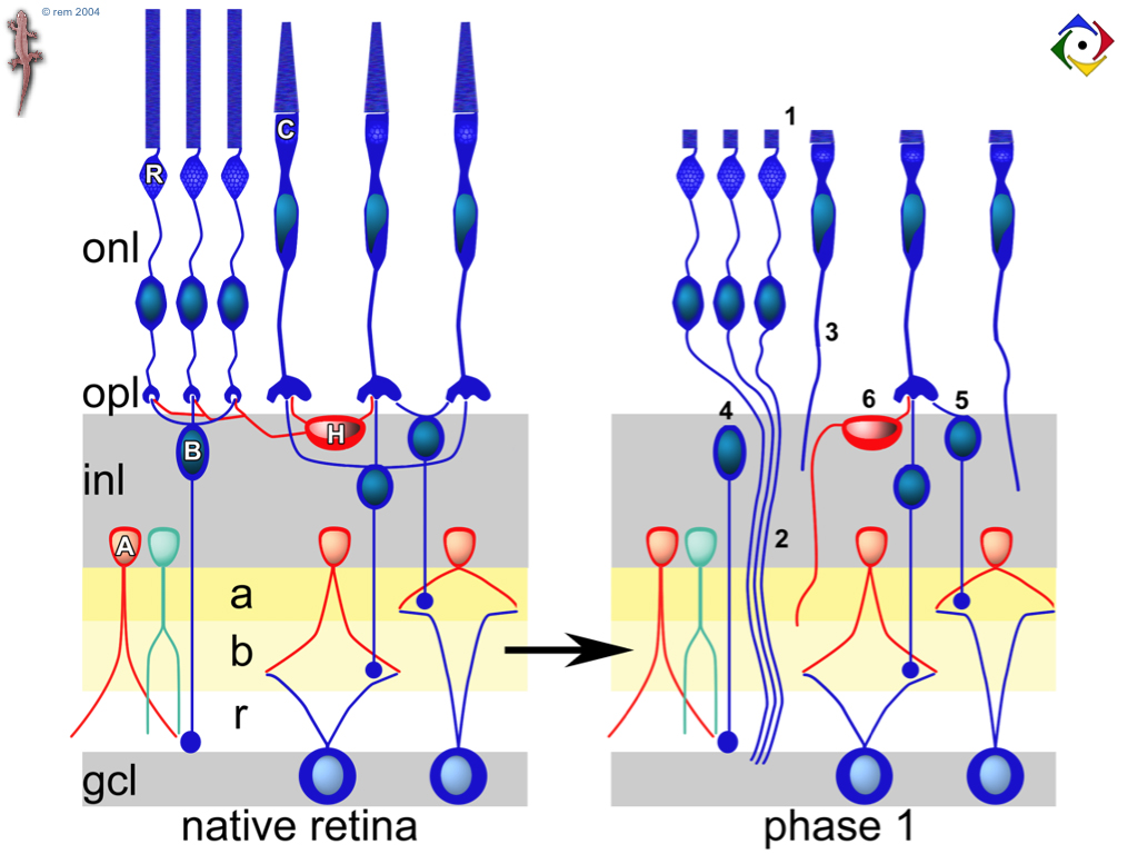
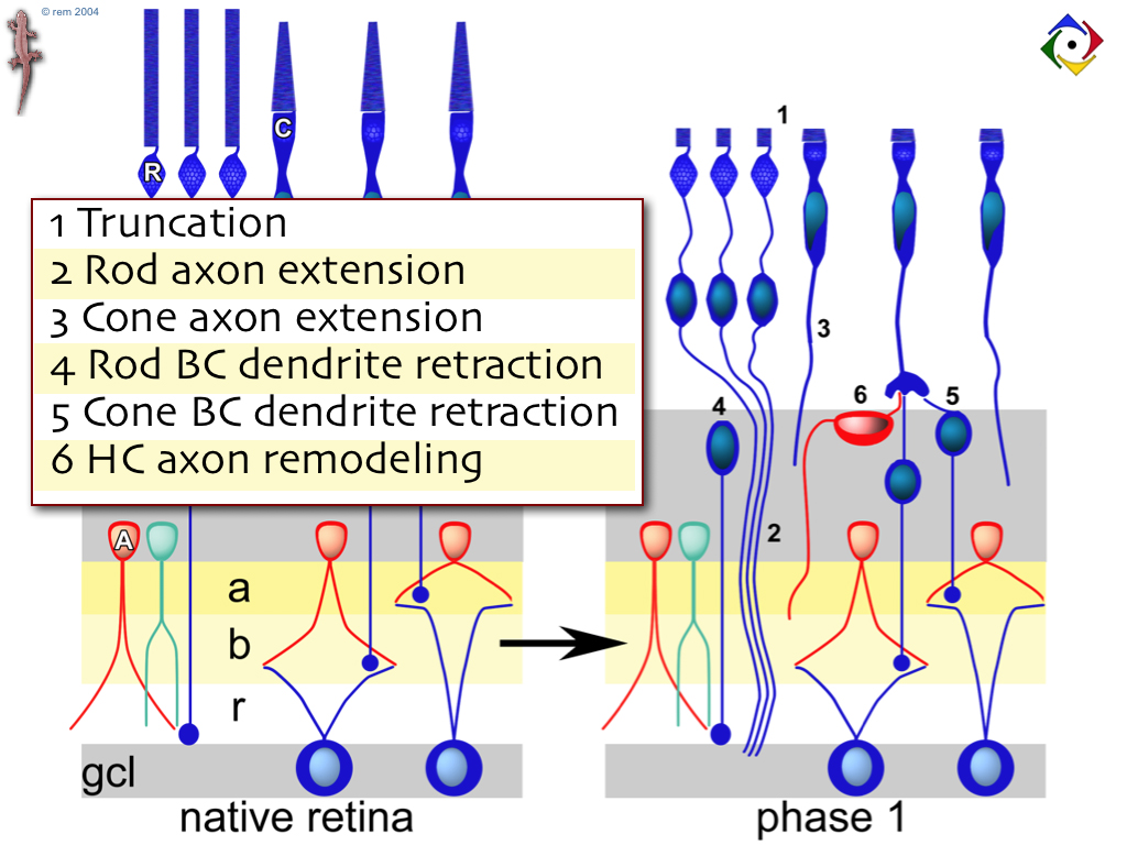
| IMAGE Remodeling diagrams: A set of static remodeling diagrams (*.tif) at full (1024 x 768) Keynote or Powerpoint resolution with LZW compression. All at 250-500 Kb and each has descriptive text.
|
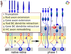 Normal → Phase 1 Remodeling Diagram text (right-click or control-click and download) Normal → Phase 1 Remodeling Diagram text (right-click or control-click and download) |
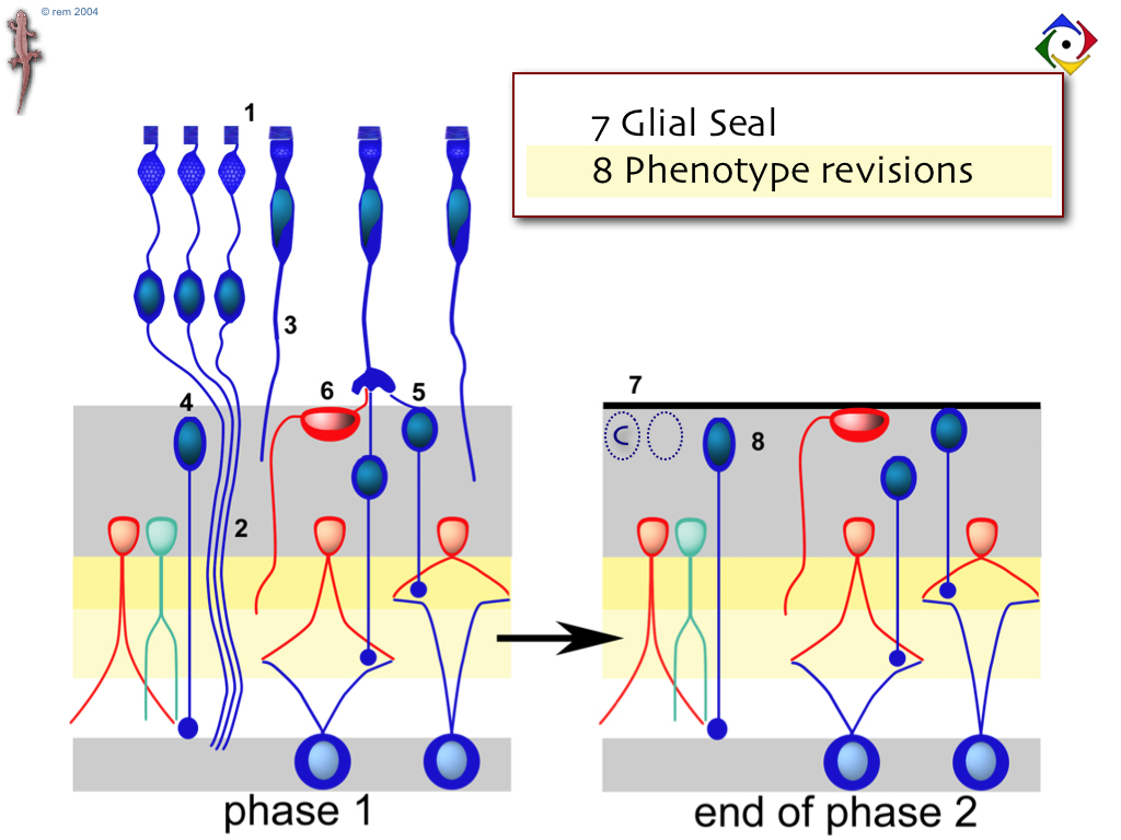
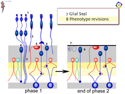 Phase 1 → Phase 2 Remodeling Diagram (right-click or control-click and download) Phase 1 → Phase 2 Remodeling Diagram (right-click or control-click and download) |
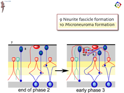 Phase 2 → Early Phase 3 Remodeling Diagram (right-click or control-click and download) Phase 2 → Early Phase 3 Remodeling Diagram (right-click or control-click and download) |
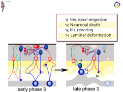 Early Phase 3 → Late Phase 3 Remodeling Diagram (right-click or control-click and download) Early Phase 3 → Late Phase 3 Remodeling Diagram (right-click or control-click and download)
© REM 2004 |
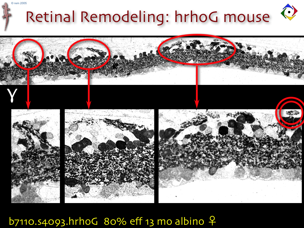
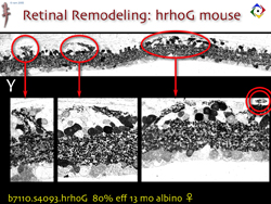 Microneuromas (right-click or control-click and download) Microneuromas (right-click or control-click and download)Microneuromas: An image of GABA signals showing extensive microneuroma formation in the hrhoG degeneration model (*.tif) at full (1024 x 768) Keynote or Powerpoint resolution with LZW compression. Microneuromas are rewiring tufts of varying size: as small as 10 µm to over 200 µm. They are not visible in normal histological preps but with hi res, hi def immunocytochem, they are clearly profuse. In some rodents they are present at 10,000-30,000 / retina. That only includes the larger, more obvious ones. The double circled tuft is actually in the RPE layer, outside the retina.© REM 2005 |
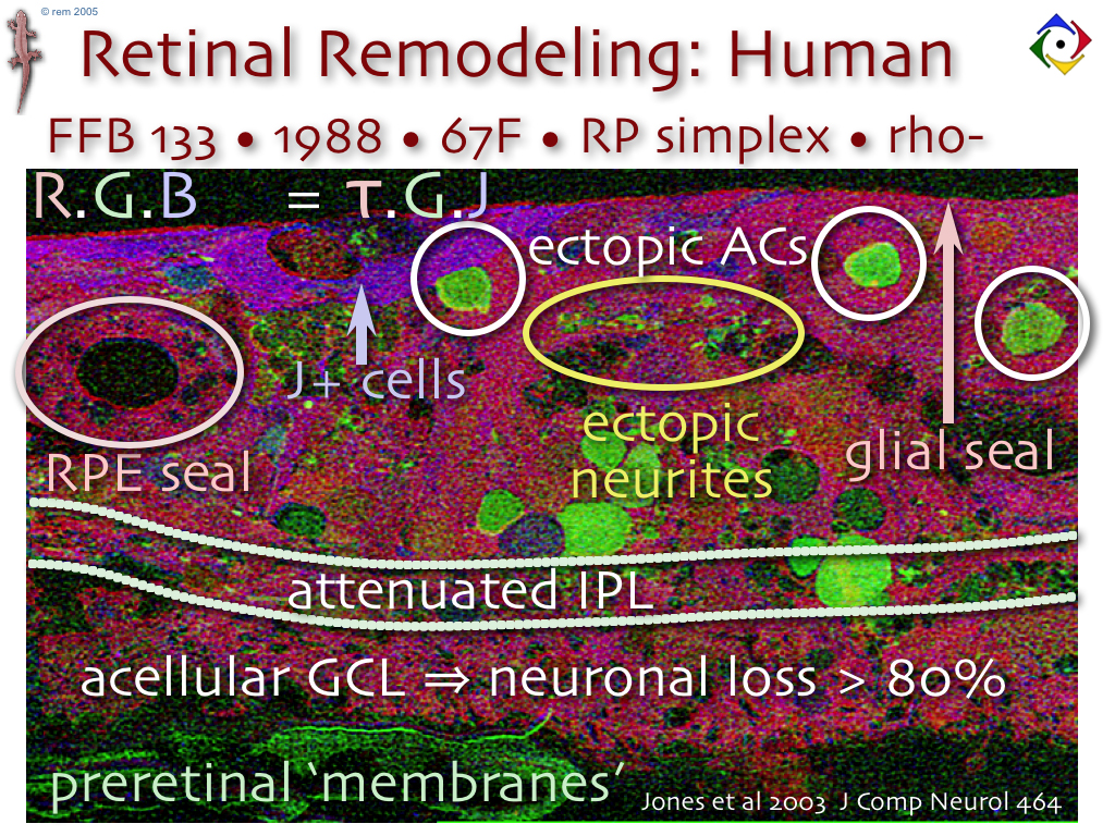
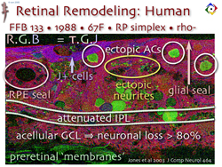 Human_remodeling (right-click or control-click and download) Human_remodeling (right-click or control-click and download)Advanced remodeling in the human retina: A fully labeled multispectral image (taurine=red, glycine=green, glutathione=blue) of human RP retina in advanced stages – the patient had been completely blind for decades. Full (1024 x 768) Keynote or Powerpoint resolution with LZW compression. This tissue bears no resemblance to retina nand was drawn from a dataset spanning 19 mm of retina. All was of similar disorganization.© REM 2005 |