We have a new collaborative manuscript out in iOVS, Primary Cilia in Amacrine Cells in Retinal Development. (pdf here)
Authors: Ke Ning; Brent E. Sendayen; Tia J. Kowal; Biao Wang; Bryan W. Jones @BWJones; Yang Hu; and Yang Sun.
Abstract:
Purpose: Primary cilia are conserved organelles found in polarized cells within the eye that regulate cell growth, migration, and differentiation. Although the role of cilia in photoreceptors is well-studied, the formation of cilia in other retinal cell types has received little attention. In this study, we examined the ciliary profile focused on the inner nuclear layer of retinas in mice and rhesus macaque primates.
Methods: Retinal sections or flatmounts from Arl13b-Cetn2 tg transgenic mice were immunostained for cell markers (Pax6, Sox9, Chx10, Calbindin, Calretinin, ChaT, GAD67, Prox1, TH, and vGluT3) and analyzed by confocal microscopy. Primate retinal sections were immunostained for ciliary and cell markers (Pax6 and Arl13b). Optical coherence tomography (OCT) and ERGs were used to assess visual function of Vift88 mice.
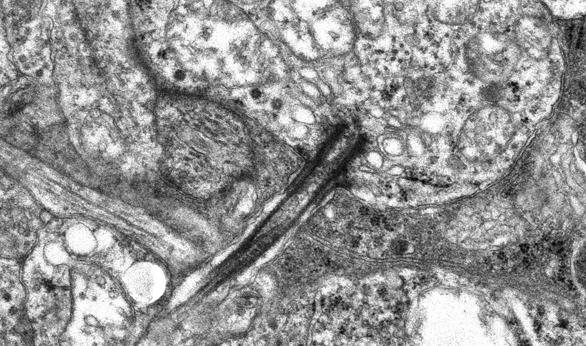
Results: During different stages of mouse postnatal eye development, we found that cilia are present in Pax6-positive amacrine cells, which were also observed in primate retinas. The cilia of subtypes of amacrine cells in mice were shown by immunostaining and electron microscopy. We also removed primary cilia from vGluT3 amacrine cells in mouse and found no significant vision defects. In addition, cilia were present in the outer limiting membrane, suggesting that a population of Müller glial cells forms cilia.
Conclusions: We report that several subpopulations of amacrine cells in inner nuclear layers of the retina form cilia during early retinal development in mice and primates.
