Category Archives: Ultrastructure

Distinctive Synaptic Structural Motifs Link Excitatory Retinal Interneurons To Diverse Postsynaptic Partner Types

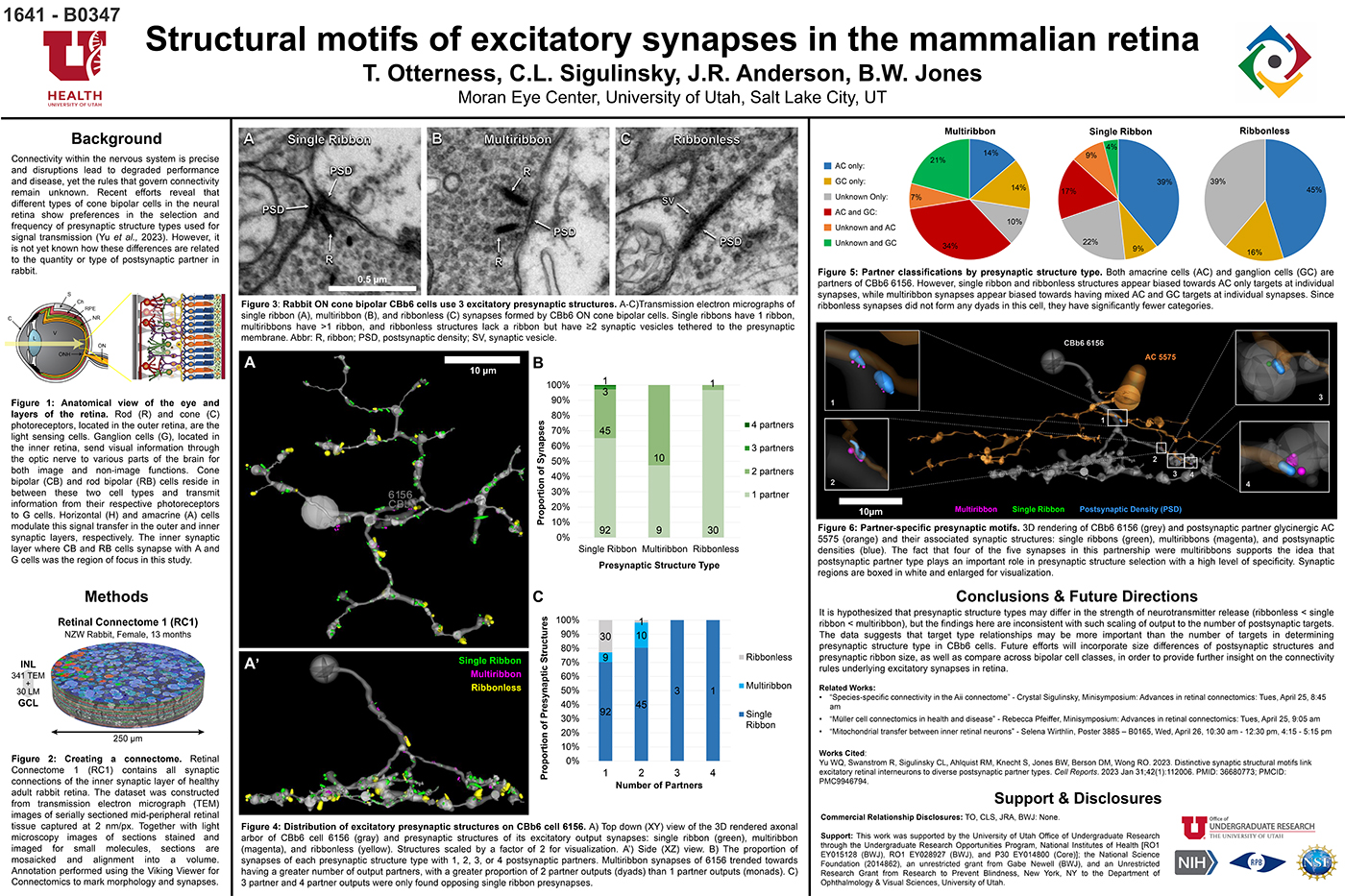
We have a new manuscript from the lab in Cell Reports, Distinctive Synaptic Structural Motifs Link Excitatory Retinal Interneurons To Diverse Postsynaptic Partner Types. This manuscript is in collaboration with the first author, Wan-Qing Yu @wanqing_yu, then co-authors Rachael Swanstrom, Crystal L. Sigulinsky @CSigulinsky, Richard M. Ahlquist, along with Sharm Knecht, myself Bryan W. Jones @BWJonesDavid M. Berson, and Rachel O. Wong. The PDF is here.

Abstract:
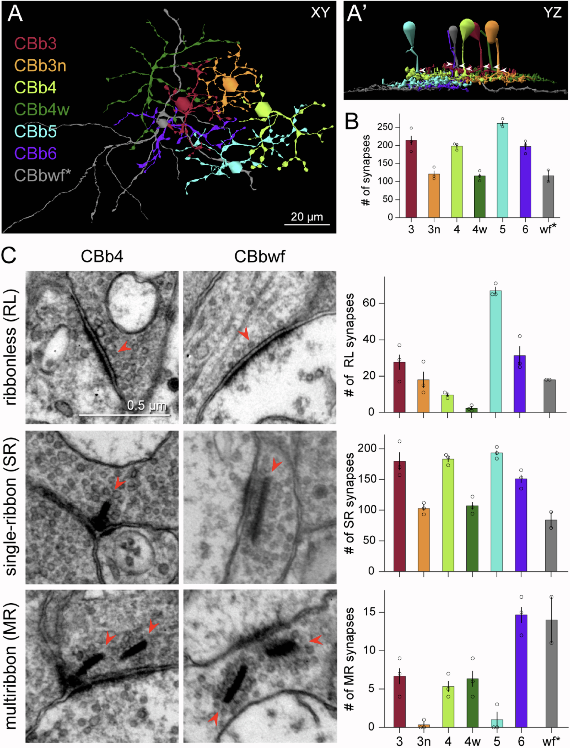
Neurons make converging and diverging synaptic connections with distinct partner types. Whether synapses involving separate partners demonstrate similar or distinct structural motifs is not yet well understood. We thus used serial electron microscopy in mouse retina to map output synapses of cone bipolar cells (CBCs) and compare their structural arrangements across bipolar types and postsynaptic partners. Three presynaptic configurations emerge—single-ribbon, ribbonless, and multiribbon synapses. Each CBC type exploits these arrangements in a unique combination, a feature also found among rabbit ON CBCs. Though most synapses are dyads, monads and triads are also seen. Altogether, mouse CBCs exhibit at least six motifs, and each CBC type uses these in a stereotypic pattern. Moreover, synapses between CBCs and particular partner types appear biased toward certain motifs. Our observations reveal synaptic strategies that diversify the output within and across CBC types, potentially shaping the distinct functions of retinal microcircuits.

Current Perspective on Retinal Remodeling: Implications for Therapeutics

We have a new paper out of the lab, a perspectives paper on Retinal Remodeling: Implications for Therapeutics. (pdf here).

Authors are Rebecca L. Pfeiffer @BeccaPfeiffer19, and Bryan W. Jones @BWJones.

Abstract: The retinal degenerative diseases retinitis pigmentosa and age-related macular degeneration are a leading cause of irreversible vision loss. Both present with progressive photoreceptor degeneration that is further complicated by processes of retinal remodeling. In this perspective, we discuss the current state of the field of retinal remodeling and its implications for vision-restoring therapeutics currently in development. Here, we discuss the challenges and pitfalls retinal remodeling poses for each therapeutic strategy under the premise that understanding the features of retinal remodeling in totality will provide a basic framework with which therapeutics can interface. Additionally, we discuss the potential for approaching therapeutics using a combined strategy of using diffusible molecules in tandem with other vision-restoring therapeutics. We end by discussing the potential of the retina and retinal remodeling as a model system for more broadly understanding the progression of neurodegeneration across the central nervous system.

Peptide-Based Immunotherapy Against Oxidized Elastin Ameliorates Pathology In Mouse Model Of Smoke-Induced Ocular Injury

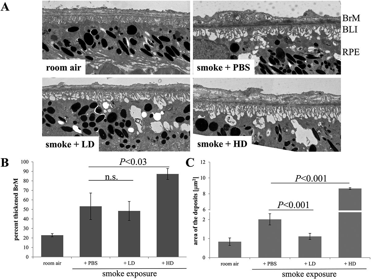
We have a new collaborative manuscript out in iOVS, Peptide-Based Immunotherapy Against Oxidized Elastin Ameliorates Pathology In Mouse Model Of Smoke-Induced Ocular Injury. (pdf here)

Authors: Bärbel Rohrer, Nathaniel Parsons , Balasubramaniam Annamalai , Crystal Nicholson , Elisabeth Obert , Bryan Jones @BWJones, and Andrew D Dick.

Abstract:

Purpose: Age-related macular degeneration (AMD), the leading cause of blindness in western populations, is associated with an overactive complement system, and an increase in circulating antibodies against certain epitopes, including elastin. As loss of the elastin layer of Bruch’s membrane (BrM) has been reported in aging and AMD, we previously showed that immunization with elastin peptide oxidatively modified by cigarette smoke (ox-elastin), exacerbated ocular pathology in the smoke-induced ocular pathology (SIOP) model. Here we asked whether ox-elastin peptide-based immunotherapy (PIT) ameliorates damage.

Methods: C57BL/6J mice were injected with ox-elastin peptide at two doses via weekly subcutaneous administration, while exposed to cigarette smoke for 6 months. FcγR-/- and uninjected C57BL/6J mice served as controls. Retinal morphology was assessed by by electron microscopy, and complement activation, antibody deposition and mechanisms of immunological tolerance were assessed by Western blotting and ELISA.

Results: Elimination of Fcγ receptors, preventing antigen/antibody-dependent cytotoxicity, protected against SIOP. Mice receiving PIT with low dose ox-elastin (LD-PIT) exhibited reduced humoral immunity, reduced complement activation and IgG/IgM deposition in the RPE/choroid, and largely a preserved BrM. While there is no direct evidence of ox-elastin pathogenicity, LD-PIT reduced IFNγ and increased IL-4 within RPE/choroid. High dose PIT was not protective.

Conclusions: These data further support ox-elastin role in ocular damage in in part via elastin-specific antibodies, and support the corollary that PIT with ox-elastin attenuates ocular pathology. Overall, damage is associated with complement activation, antibody-dependent cell-mediated cytotoxicity, and altered cytokine signature.

Keywords: Age-related macular degeneration; Complement; Elastin; Peptide-based immunotherapy; Smoking.

Primary Cilia in Amacrine Cells in Retinal Development

We have a new collaborative manuscript out in iOVS, Primary Cilia in Amacrine Cells in Retinal Development. (pdf here)

Authors: Ke Ning; Brent E. Sendayen; Tia J. Kowal; Biao WangBryan W. Jones @BWJones; Yang Hu; and Yang Sun.

Abstract:

Purpose: Primary cilia are conserved organelles found in polarized cells within the eye that regulate cell growth, migration, and differentiation. Although the role of cilia in photoreceptors is well-studied, the formation of cilia in other retinal cell types has received little attention. In this study, we examined the ciliary profile focused on the inner nuclear layer of retinas in mice and rhesus macaque primates.

Methods: Retinal sections or flatmounts from Arl13b-Cetn2 tg transgenic mice were immunostained for cell markers (Pax6, Sox9, Chx10, Calbindin, Calretinin, ChaT, GAD67, Prox1, TH, and vGluT3) and analyzed by confocal microscopy. Primate retinal sections were immunostained for ciliary and cell markers (Pax6 and Arl13b). Optical coherence tomography (OCT) and ERGs were used to assess visual function of Vift88 mice.

Results: During different stages of mouse postnatal eye development, we found that cilia are present in Pax6-positive amacrine cells, which were also observed in primate retinas. The cilia of subtypes of amacrine cells in mice were shown by immunostaining and electron microscopy. We also removed primary cilia from vGluT3 amacrine cells in mouse and found no significant vision defects. In addition, cilia were present in the outer limiting membrane, suggesting that a population of Müller glial cells forms cilia.

Conclusions: We report that several subpopulations of amacrine cells in inner nuclear layers of the retina form cilia during early retinal development in mice and primates.


Subretinal Rather Than Intravitreal Adeno-Associated Virus–Mediated Delivery of a Complement Alternative Pathway Inhibitor Is Effective in a Mouse Model of RPE Damage

We have a new manuscript out in iOVS, Subretinal Rather Than Intravitreal Adeno-Associated Virus–Mediated Delivery of a Complement Alternative Pathway Inhibitor Is Effective in a Mouse Model of RPE Damage. (pdf here)

Authors: Balasubramaniam Annamalai; Nathaniel Parsons; Crystal Nicholson; Elisabeth Obert; Bryan W. Jones @BWJones; and Bärbel Rohrer.

Abstract:

Purpose: The risk for age-related macular degeneration has been tied to an overactive complement system. Despite combined attempts by academia and industry to develop therapeutics that modulate the complement response, particularly in the late geographic atrophy form of advanced AMD, to date, there is no effective treatment. We have previously demonstrated that pathology in the smoke-induced ocular pathology (SIOP) model, a model with similarities to dry AMD, is dependent on activation of the alternative complement pathway and that a novel complement activation site targeted inhibitor of the alternative pathway can be delivered to ocular tissues via an adeno-associated virus (AAV).

Methods: Two different viral vectors for specific tissue targeting were compared: AAV5-VMD2-CR2-fH for delivery to the retinal pigment epithelium (RPE) and AAV2YF-smCBA-CR2-fH for delivery to retinal ganglion cells (RGCs). Efficacy was tested in SIOP (6 months of passive smoke inhalation), assessing visual function (optokinetic responses), retinal structure (optical coherence tomography), and integrity of the RPE and Bruch’s membrane (electron microscopy). Protein chemistry was used to assess complement activation, CR2-fH tissue distribution, and CR2-fH transport across the RPE.

Results: RPE- but not RGC-mediated secretion of CR2-fH was found to reduce SIOP and complement activation in RPE/choroid. Bioavailability of CR2-fH in RPE/choroid could be confirmed only after AAV5-VMD2-CR2-fH treatment, and inefficient, adenosine triphosphate–dependent transport of CR2-fH across the RPE was identified.

Conclusions: Our results suggest that complement inhibition for AMD-like pathology is required basal to the RPE and argues in favor of AAV vector delivery to the RPE or outside the blood-retina barrier.

Chapter: Retinal Connectomics

We have a new chapter out in the Elsevier book series The Senses, 2021.

Authors are myself, Bryan W. Jones @BWJones and Robert E. Marc @robertmarc60.

Abstract: The retina is both a light sensor and a highly complex image-processing device – like supercomputers at the backs of eyes. The retina is also wonderfully compact with all circuitry (glia, neurons, synapses and gap junctions) required to compute sensory input, making it a convenient model for understanding the rest of the nervous system. This is also true for disease, with early evidence indicating retina may be a good model for studying progressive neural degenerative diseases. Modern ultrastructural approaches to the study of neural connections is a relatively new !eld has been termed “connectomics”. Connectomics approaches applied to the retina is termed retinal connectomics. These approaches are relatively new !elds that leverage modern technologies in light and ultrastructural imaging, computational storage, and data management to allow tracking of neuronal identity and connectivity, delivering a robust edge/node network map of circuit topologies. Understanding circuit topologies is critical to understanding how retinas process information, and how information processing is corrupted in disease. This chapter summarizes early history, discusses technical aspects of imaging connectomes, justi!es the importance of why connectomics approaches are important, particularly in retina, discusses what has been learned from early efforts in connectomics, and points the way to the next steps.

Please email me: bryan.jones@m.cc.utah.edu if you would like a pdf of the chapter.


A pathoconnectome of early neurodegeneration: Network changes in retinal degeneration

We have a new manuscript out in Experimental Eye Research, A pathoconnectome of early neurodegeneration: Network changes in retinal degeneration. (pdf here)

Authors: Rebecca L. Pfeiffer @BeccaPfeiffer19, James R. Anderson, Jeebika Dahal, Jessica C. Garcia, Jia-Hui Yang, Crystal L. Sigulinsky @CLSigulinsky, Kevin Rapp, Daniel P. Emrich, Carl B. Watt, Hope AB Johnstun, Alexis R. Houser, Robert E. Marc @robertmarc60, and Bryan W. Jones @BWJones.

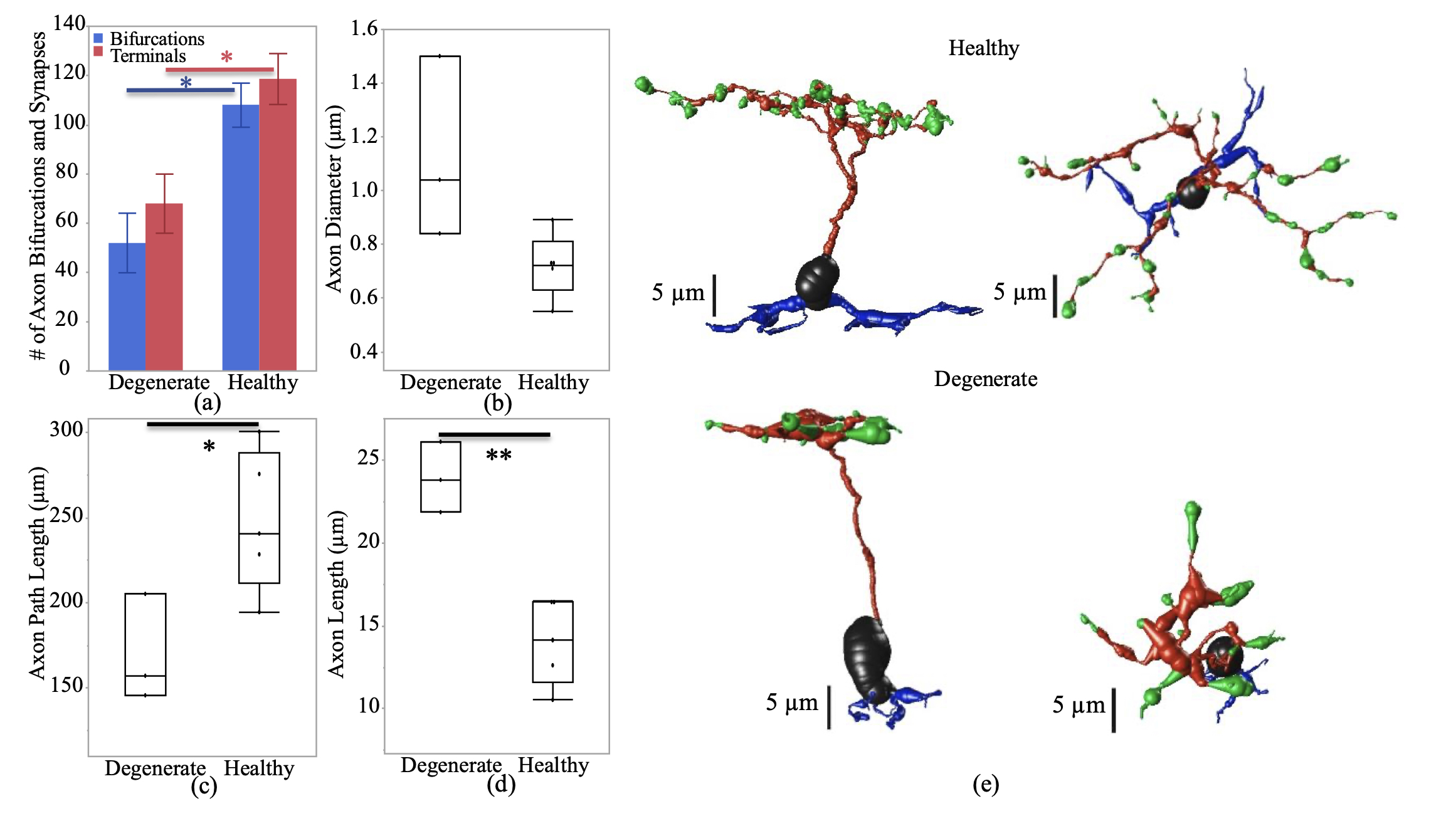
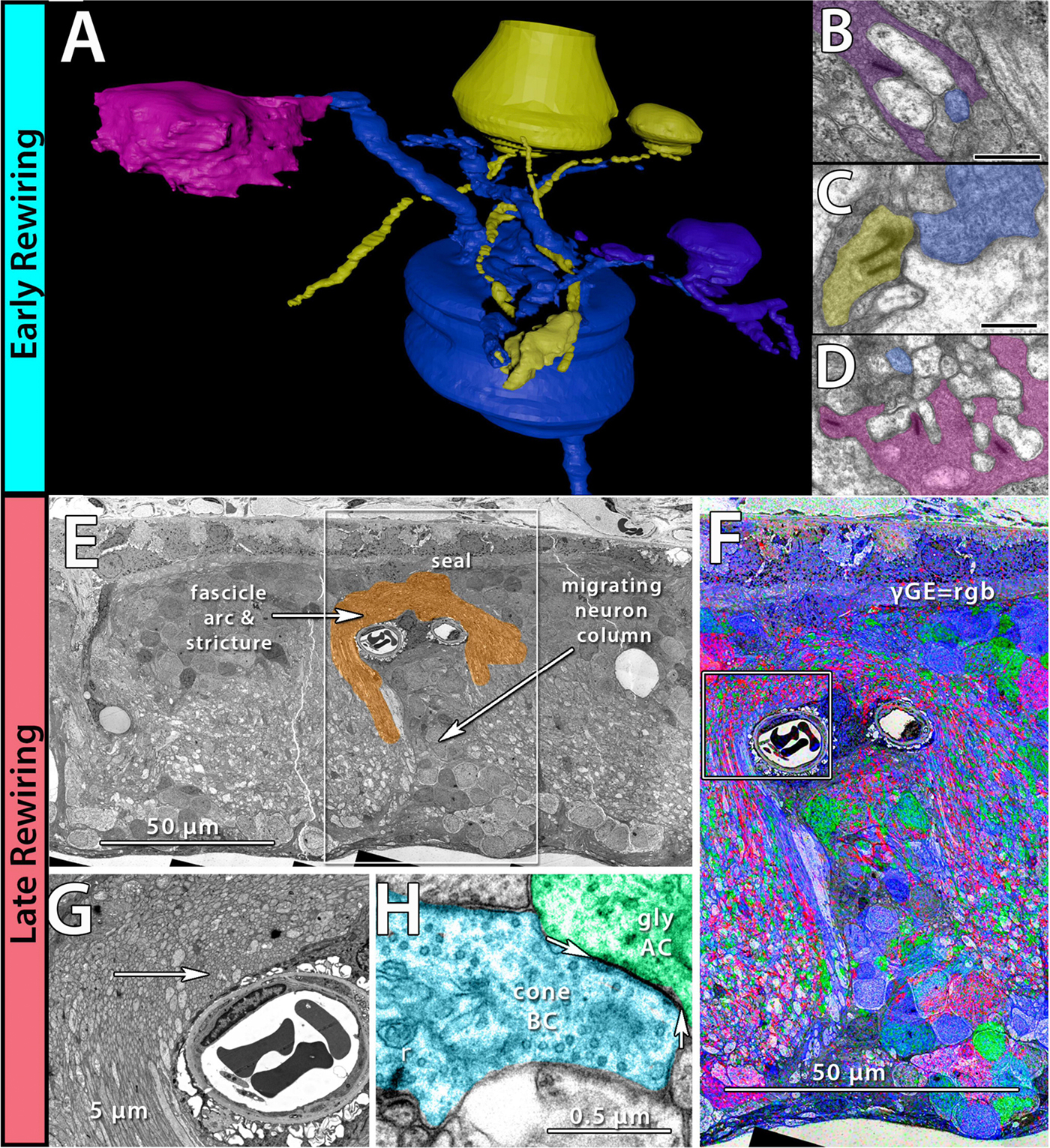
Abstract: Connectomics has demonstrated that synaptic networks and their topologies are precise and directly correlate with physiology and behavior. The next extension of connectomics is pathoconnectomics: to map neural network synaptology and circuit topologies corrupted by neurological disease in order to identify robust targets for therapeutics. In this report, we characterize a pathoconnectome of early retinal degeneration. This pathoconnectome was generated using serial section transmission electron microscopy to achieve an ultrastructural connectome with 2.18nm/px resolution for accurate identification of all chemical and gap junctional synapses. We observe aberrant connectivity in the rod-network pathway and novel synaptic connections deriving from neurite sprouting. These observations reveal principles of neuron responses to the loss of network components and can be extended to other neurodegenerative diseases.


NeuroNex Grant

I am pleased to report that the The Marclab for Connectomics has been funded by the National Science Foundation with a 5 year grant as part of a large, international consortium to study synaptic weighting.  We are collaborating with the Erik Jorgensen laboratory here at the University of Utah, and will be exploring synapses in a model of retinal degeneration.  There is a nice writeup of the award on the Moran Eye Center website, here.

This is a wonderful opportunity to work with other colleagues that will be funded alongside us with this grant, including Uri Manor @manorlaboratory, Davi Bock @dddavi, Josh Vogelstein @neuro_data, Viren Jain @stardazed0, and others.  My thanks to Kristen Harris for heading up this initiative.

Network Architecture of Gap Junctional Coupling among Parallel Processing Channels in the Mammalian Retina

We have a new manuscript out in The Journal of Neuroscience, Network Architecture of Gap Junctional Coupling among Parallel Processing Channels in the Mammalian Retina.

Authors: Crystal L. Sigulinsky @CLSigulinsky, James R. Anderson, Ethan Kerzner @EthanKerzner, Christopher N. Rapp @ChrisNRapp, Rebecca L. Pfeiffer @BeccaPfeiffer19, Taryn M. Rodman, Daniel P. Emrich, Kevin D. Rapp, Noah T. Nelson @nooneelseinhere, J. Scott Lauritzen, Miriah Meyer@miriah_meyer, Robert E. Marc @robertmarc60, and Bryan W. Jones @BWJones.

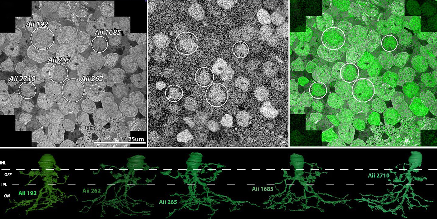
Abstract: Gap junctions are ubiquitous throughout the nervous system, mediating critical signal transmission and integration, as well as emergent network properties. In mammalian retina, gap junctions within the Aii amacrine cell-ON cone bipolar cell (CBC) network are essential for night vision, modulation of day vision, and contribute to visual impairment in retinal degenerations, yet neither the extended network topology nor its conservation is well established. Here, we map the network contribution of gap junctions using a high-resolution connectomics dataset of an adult female rabbit retina. Gap junctions are prominent synaptic components of ON CBC classes, constituting 5%–25% of all axonal synaptic contacts. Many of these mediate canonical transfer of rod signals from Aii cells to ON CBCs for night vision, and we find that the uneven distribution of Aii signals to ON CBCs is conserved in rabbit, including one class entirely lacking direct Aii coupling. However, the majority of gap junctions formed by ON CBCs unexpectedly occur between ON CBCs, rather than with Aii cells. Such coupling is extensive, creating an interconnected network with numerous lateral paths both within, and particularly across, these parallel processing streams. Coupling patterns are precise with ON CBCs accepting and rejecting unique combinations of partnerships according to robust rulesets. Coupling specificity extends to both size and spatial topologies, thereby rivaling the synaptic specificity of chemical synapses. These ON CBC coupling motifs dramatically extend the coupled Aii-ON CBC network, with implications for signal flow in both scotopic and photopic retinal networks during visual processing and disease.

Immunization Against Oxidized Elastin Exacerbates Structural and Functional Damage in Mouse Model of Smoke-Induced Ocular Injury

We have a new manuscript out in Investigative Ophthalmology & Visual Science, Immunization Against Oxidized Elastin Exacerbates Structural and Functional Damage in Mouse Model of Smoke-Induced Ocular Injury

Authors: Balasubramaniam Annamalai; Crystal Nicholson; Nathaniel Parsons; Sarah Stephenson; Carl Atkinson; Bryan Jones; and Bärbel Rohrer.

Purpose: Age-related macular degeneration (AMD) is the leading cause of blindness in Western populations. While an overactive complement system has been linked to pathogenesis, mechanisms contributing to its activation are largely unknown. In aged and AMD eyes, loss of the elastin layer (EL) of Bruch’s membrane (BrM) has been reported. Elastin antibodies are elevated in patients with AMD, the pathogenic significance of which is unclear. Here we assess the role of elastin antibodies using a mouse model of smoke-induced ocular pathology (SIOP), which similarly demonstrates EL loss.

Methods: C57BL/6J mice were immunized with elastin or elastin peptide oxidatively modified by cigarette smoke (ox-elastin). Mice were then exposed to cigarette smoke or air for 6 months. Visual function was assessed by optokinetic response, retinal morphology by spectral-domain optical coherence tomography and electron microscopy, and complement activation and antibody deposition by Western blot.

Results: Ox-elastin IgG and IgM antibodies were elevated in ox-elastin immunized mice following 6 months of smoke, whereas elastin immunization had a smaller effect. Ox-elastin immunization exacerbated smoke-induced vision loss, with thicker BrM and more damaged retinal pigment epithelium (RPE) mitochondria compared with mice immunized with elastin or nonimmunized controls. These changes were correlated with increased levels of IgM, IgG2, IgG3, and complement activation products in RPE/choroid.

Conclusions: These data demonstrate that SIOP mice generate elastin-specific antibodies and that immunization with ox-elastin exacerbates ocular pathology. Elastin antibodies represented complement fixing isotypes that, together with the increased presence of complement activation seen in immunized mice, suggest that elastin antibodies exert pathogenic effects through mediating complement activation.