Category Archives: Publications

NUDC Is Critical For Rod Photoreceptor Function, Maintenance, And Survival

We have a new manuscript in collaboration with the Gross Lab out of UAB in The FASEB Journal, (PubMed link here): NUDC is critical for rod photoreceptor function, maintenance, and survival. This manuscript is in collaboration with.  Authors are: Mary Anne Garner, Meredith G. Hubbard, Evan R. Boitet, Seth T. Hubbard, Anushree Gade, Guoxin Ying, Bryan W. Jones, Wolfgang Baehr, Alecia K. Gross. The PDF is here.

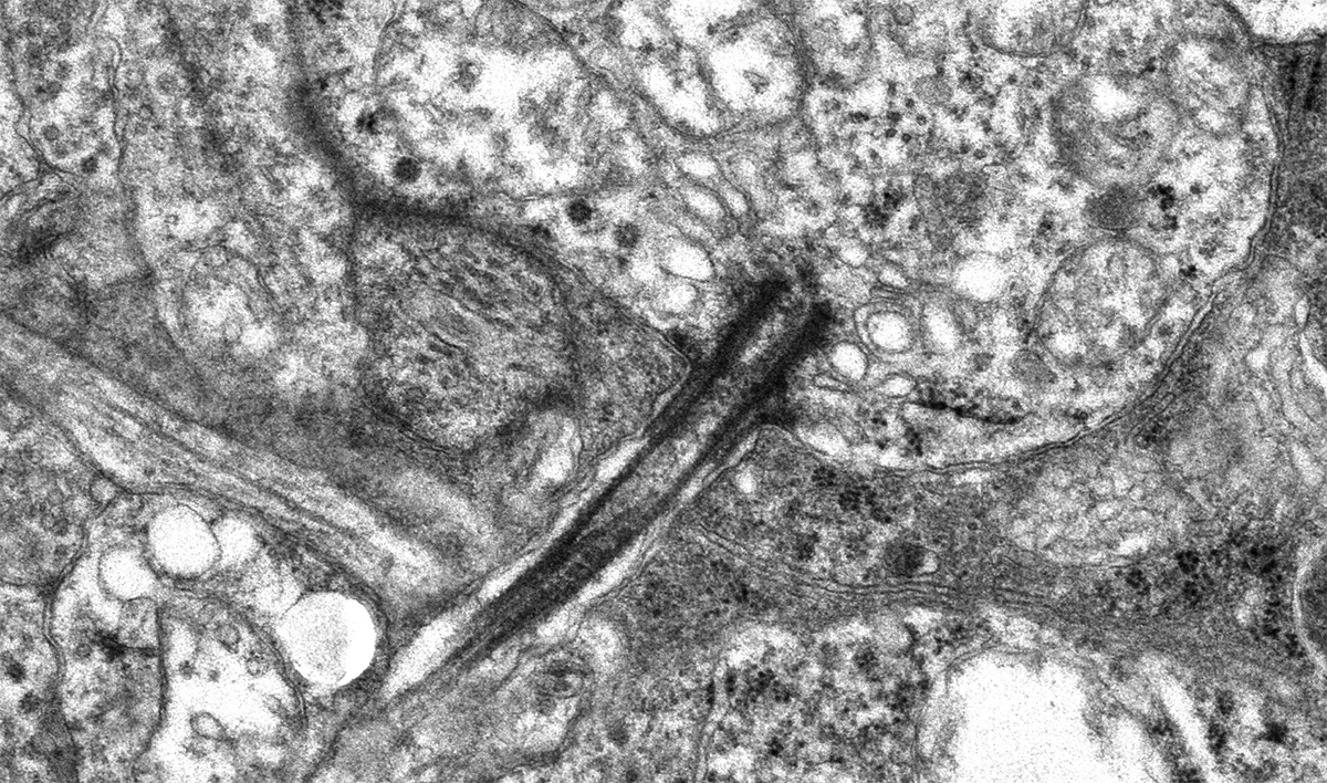
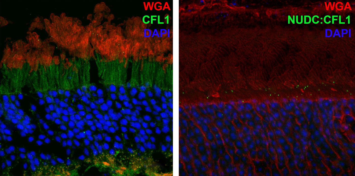
Abstract: NUDC (nuclear distribution protein C) is a mitotic protein involved in nuclear migration and cytokinesis across species. Considered a cytoplasmic dynein (henceforth dynein) cofactor, NUDC was shown to associate with the dynein motor complex during neuronal migration. NUDC is also expressed in postmitotic vertebrate rod photoreceptors where its function is unknown. Here, we examined the role of NUDC in postmitotic rod photoreceptors by studying the consequences of a conditional NUDC knockout in mouse rods (rNudC−/−). Loss of NUDC in rods led to complete photoreceptor cell death at 6 weeks of age. By 3 weeks of age, rNudC−/− function was diminished, and rhodopsin and mitochondria were mislocalized, consistent with dynein inhibition. Levels of outer segment proteins were reduced, but LIS1 (lissencephaly protein 1), a well-characterized dynein cofactor, was unaffected. Transmission electron microscopy revealed ultrastructural defects within the rods of rNudC−/− by 3 weeks of age. We investigated whether NUDC interacts with the actin modulator cofilin 1 (CFL1) and found that in rods, CFL1 is localized in close proximity to NUDC. In addition to its potential role in dynein trafficking within rods, loss of NUDC also resulted in increased levels of phosphorylated CFL1 (pCFL1), which would purportedly prevent depolymerization of actin. The absence of NUDC also induced an inflammatory response in Müller glia and microglia across the neural retina by 3 weeks of age. Taken together, our data illustrate the critical role of NUDC in actin cytoskeletal maintenance and dynein-mediated protein trafficking in a postmitotic rod photoreceptor.

Modeling Complex Age-Related Eye Disease

We have a new Progress in Retinal and Eye Research manuscript out in collaboration with my colleagues here at the Moran Eye Center.

Authors: Silke Becker, Zia L’Ecuyer, Bryan W Jones, Moussa A Zouache, Fiona S McDonnell, Frans Vinberg.

Abstract: Modeling complex eye diseases like age-related macular degeneration (AMD) and glaucoma poses significant challenges, since these conditions depend highly on age-related changes that occur over several decades, with many contributing factors remaining unknown. Although both diseases exhibit a relatively high heritability of >50%, a large proportion of individuals carrying AMD- or glaucoma-associated genetic risk variants will never develop these diseases. Furthermore, several environmental and lifestyle factors contribute to and modulate the pathogenesis and progression of AMD and glaucoma.

Several strategies replicate the impact of genetic risk variants, pathobiological pathways and environmental and lifestyle factors in AMD and glaucoma in mice and other species. In this review we will mostly discuss the most commonly available mouse models, which have and will likely continue to improve our understanding of the pathobiology of age-related eye diseases. Uncertainties persist whether small animal models can truly recapitulate disease progression and vision loss in patients, raising doubts regarding their usefulness when testing novel gene or drug therapies. We will elaborate on concerns that relate to shorter lifespan, body size and allometries, lack of macula and a true lamina cribrosa, as well as absence and sequence disparities of certain genes and differences in their chromosomal location in mice.

Since biological, rather than chronological, age likely predisposes an organism for both glaucoma and AMD, more rapidly aging organisms like small rodents may open up possibilities that will make research of these diseases more timely and financially feasible. On the other hand, due to the above-mentioned anatomical and physiological features, as well as pharmacokinetic and -dynamic differences small animal models are not ideal to study the natural progression of vision loss or the efficacy and safety of novel therapies. In this context, we will also discuss the advantages and pitfalls of alternative models that include larger species, such as non-human primates and rabbits, patient-derived retinal organoids, and human organ donor eyes.

Unbalanced Redox Status Network As An Early Pathological Event In Congenital Cataracts

We have a new manuscript from the lab in Experimental Eye Research, (PubMed link here). Metabolic changes and retinal remodeling in Heterozygous CRX mutant cats (CRXRDY/+). This manuscript is in collaboration with the Sheldon Rowan lab out of Tufts University.  Authors are: Eloy Bejarano, Elizabeth A Whitcomb, Rebecca L Pfeiffer (@BeccaPfeiffer19), Kristie L Rose, Maria José Asensio, José Antonio Rodríguez-Navarro, Alejandro Ponce-Mora, Antolín Canto, Inma Almansa, Kevin L Schey, Bryan W Jones (@BWJones), Allen Taylor, and Sheldon Rowan (@SheldonRowan). The PDF is here.

Abstract: The lens proteome undergoes dramatic composition changes during development and maturation. A defective developmental process leads to congenital cataracts that account for about 30% of cases of childhood blindness. Gene mutations are associated with approximately 50% of early-onset forms of lens opacity, with the remainder being of unknown etiology. To gain a better understanding of cataractogenesis, we utilized a transgenic mouse model expressing a mutant ubiquitin protein in the lens (K6W-Ub) that recapitulates most of the early pathological changes seen in human congenital cataracts. We performed mass spectrometry-based tandem-mass-tag quantitative proteomics in E15, P1, and P30 control or K6W-Ub lenses. Our analysis identified targets that are required for early normal differentiation steps and altered in cataractous lenses, particularly metabolic pathways involving glutathione and amino acids. Computational molecular phenotyping revealed that glutathione and taurine were spatially altered in the K6W-Ub cataractous lens. High-performance liquid chromatography revealed that both taurine and the ratio of reduced glutathione to oxidized glutathione, two indicators of redox status, were differentially compromised in lens biology. In sum, our research documents that dynamic proteome changes in a mouse model of congenital cataracts impact redox biology in lens. Our findings shed light on the molecular mechanisms associated with congenital cataracts and point out that unbalanced redox status due to reduced levels of taurine and glutathione, metabolites already linked to age-related cataract, could be a major underlying mechanism behind lens opacities that appear early in life.

Metabolic changes and retinal remodeling in Heterozygous CRX mutant cats (CRXRDY/+)

We have a new manuscript from the lab in Experimental Eye Research, (PubMed link here). Metabolic changes and retinal remodeling in Heterozygous CRX mutant cats (CRXRDY/+). This manuscript is in collaboration with the Simon Petersen-Jones lab out of Michigan State University.  Authors are: Laurence M. Occelli, Bryan W. Jones @BWJones, Taylor J. Cervantes, and Simon M. Petersen-Jones. The PDF is here.

Abstract: CRX is a transcription factor essential for normal photoreceptor development and survival. The CRXRdy cat has a naturally occurring truncating mutation in CRX and is a large animal model for dominant Leber congenital amaurosis. This study investigated retinal remodeling that occurs as photoreceptors degenerate. CRXRdy/+ cats from 6 weeks to 10 years of age were investigated. In vivo structural changes of retinas were analyzed by fundus examination, confocal scanning laser ophthalmoscopy and spectral domain optical coherence tomography. Histologic analyses including immunohistochemistry for computational molecular phenotyping with macromolecules and small molecules. Affected cats had a cone-led photoreceptor degeneration starting in the area centralis. Initially there was preservation of inner retinal cells such as bipolar, amacrine and horizontal cells but with time migration of the deafferented neurons occurred. Early in the process of degeneration glial activation occurs ultimately resulting in formation of a glial seal. With progression the macula-equivalent area centralis developed severe atrophy including loss of retinal pigmentary epithelium. Microneuroma formation occurs in advanced stages as more marked retinal remodeling occurred. This study indicates that retinal degeneration in the CrxRdy/+ cat retina follows the progressive, phased revision of retina that have been previously described for retinal remodeling. These findings suggest that therapy dependent on targeting inner retinal cells may be useful in young adults with preserved inner retinas prior to advanced stages of retinal remodeling and neuronal cell loss.

Impact of Retinal Degeneration on Response of ON and OFF Cone Bipolar Cells to Electrical Stimulation

We have a new manuscript from the lab in IEEE, Impact of Retinal Degeneration on Response of ON and OFF Cone Bipolar Cells to Electrical Stimulation. This manuscript is in collaboration with the Lazzi lab out of USC.  The first author, Shayan Farzad, Pragya Kosta, Ege Iseri, Steven T Walston, Jean-Marie C. Bouteiller,  Rebecca L. Pfeiffer @BeccaPfeiffer19, Crystal L. Sigulinsky @CSigulinsky, Jia-Hui Yang, Jessica C. Garcia, James R. Anderson, Bryan W. Jones @BWJones, and Gianluca Lazzi. The PDF is here.

Abstract: In retinal degenerative diseases, such as retinitis pigmentosa (RP) and age-related macular degeneration (AMD), the photoreceptors become stressed and start to degenerate in the early stages of the disease. Retinal prosthetic devices have been developed to restore vision in patients by applying electrical stimulation to the surviving retinal cells. However, these devices provide limited visual perception as the therapeutic interventions are generally considered in the later stages of the disease when only inner retinal layer cells are left. A potential treatment option for retinal degenerative diseases in the early stages can be stimulating bipolar cells, which receive presynaptic signals from photoreceptors. In this work, we constructed computational models of healthy and degenerated (both ON and OFF-type) cone bipolar cells (CBCs) with realistic morphologies extracted from connectomes of the healthy and early-stage degenerated rabbit retina. We examined these cells’ membrane potential and axon terminal calcium current differences when subjected to electrical stimulation. In addition, we investigated how differently healthy and degenerated cells behave with respect to various stimulation parameters, including pulse duration and cells’ distance from the stimulating electrode. The results suggested that regardless of the position of the OFF CBCs in the retina model, there is not a significant difference between the membrane potential of healthy and degenerate cells when electrically stimulated. However, the healthy ON CBC axon terminal membrane potential rising time-constant is shorter (0.29 ± 0.03 ms) than the degenerated cells (0.8 ± 0.07 ms). Moreover, the ionic calcium channels at the axon terminals of the cells have a higher concentration and higher current in degenerated cells (32.24 ± 6.12 pA) than the healthy cells (13.64 ± 2.88 pA) independently of the cell’s position.

Distinctive Synaptic Structural Motifs Link Excitatory Retinal Interneurons To Diverse Postsynaptic Partner Types

We have a new manuscript from the lab in Cell Reports, Distinctive Synaptic Structural Motifs Link Excitatory Retinal Interneurons To Diverse Postsynaptic Partner Types. This manuscript is in collaboration with the first author, Wan-Qing Yu @wanqing_yu, then co-authors Rachael Swanstrom, Crystal L. Sigulinsky @CSigulinsky, Richard M. Ahlquist, along with Sharm Knecht, myself Bryan W. Jones @BWJonesDavid M. Berson, and Rachel O. Wong. The PDF is here.

Abstract:
Neurons make converging and diverging synaptic connections with distinct partner types. Whether synapses involving separate partners demonstrate similar or distinct structural motifs is not yet well understood. We thus used serial electron microscopy in mouse retina to map output synapses of cone bipolar cells (CBCs) and compare their structural arrangements across bipolar types and postsynaptic partners. Three presynaptic configurations emerge—single-ribbon, ribbonless, and multiribbon synapses. Each CBC type exploits these arrangements in a unique combination, a feature also found among rabbit ON CBCs. Though most synapses are dyads, monads and triads are also seen. Altogether, mouse CBCs exhibit at least six motifs, and each CBC type uses these in a stereotypic pattern. Moreover, synapses between CBCs and particular partner types appear biased toward certain motifs. Our observations reveal synaptic strategies that diversify the output within and across CBC types, potentially shaping the distinct functions of retinal microcircuits.

Current Perspective on Retinal Remodeling: Implications for Therapeutics

We have a new paper out of the lab, a perspectives paper on Retinal Remodeling: Implications for Therapeutics. (pdf here).

Authors are Rebecca L. Pfeiffer @BeccaPfeiffer19, and Bryan W. Jones @BWJones.

Abstract: The retinal degenerative diseases retinitis pigmentosa and age-related macular degeneration are a leading cause of irreversible vision loss. Both present with progressive photoreceptor degeneration that is further complicated by processes of retinal remodeling. In this perspective, we discuss the current state of the field of retinal remodeling and its implications for vision-restoring therapeutics currently in development. Here, we discuss the challenges and pitfalls retinal remodeling poses for each therapeutic strategy under the premise that understanding the features of retinal remodeling in totality will provide a basic framework with which therapeutics can interface. Additionally, we discuss the potential for approaching therapeutics using a combined strategy of using diffusible molecules in tandem with other vision-restoring therapeutics. We end by discussing the potential of the retina and retinal remodeling as a model system for more broadly understanding the progression of neurodegeneration across the central nervous system.

Revival Of Light Signalling In The Postmortem Mouse And Human Retina

We have a new collaborative manuscript out in Nature, Revival of light signalling in the postmortem mouse and human retina. Full paper (here).

Authors: Fatima Abbas @neurofim, Silke Becker, Bryan W. Jones @BWJones, Ludovic S. Mure, Satchidananda Panda @SatchinPanda, Anne Hanneken & Frans Vinberg @fvinberg.

Abstract:
Death is defined as the irreversible cessation of circulatory, respiratory or brain activity. Many peripheral human organs can be transplanted from deceased donors using protocols to optimize viability. However, tissues from the central nervous system rapidly lose viability after circulation ceases, impeding their potential for transplantation. However, the time course and mechanisms causing neuronal death and the potential for revival remain poorly defined. Here, using the retina as a model of the central nervous system, we systemically examine the kinetics of death and neuronal revival. We demonstrate the swift decline of neuronal signalling and identify conditions for reviving synchronous in vivo-like trans-synaptic transmission in postmortem mouse and human retina. We measure light-evoked responses in human macular photoreceptors in eyes removed up to 5 h after death and identify modifiable factors that drive reversible and irreversible loss of light signalling after death. Finally, we quantify the rate-limiting deactivation reaction of phototransduction, a model G protein signalling cascade, in peripheral and macular human and macaque retina. Our approach will have broad applications and impact by enabling transformative studies in the human central nervous system, raising questions about the irreversibility of neuronal cell death, and providing new avenues for visual rehabilitation.

Peptide-Based Immunotherapy Against Oxidized Elastin Ameliorates Pathology In Mouse Model Of Smoke-Induced Ocular Injury

We have a new collaborative manuscript out in iOVS, Peptide-Based Immunotherapy Against Oxidized Elastin Ameliorates Pathology In Mouse Model Of Smoke-Induced Ocular Injury. (pdf here)

Authors: Bärbel Rohrer, Nathaniel Parsons , Balasubramaniam Annamalai , Crystal Nicholson , Elisabeth Obert , Bryan Jones @BWJones, and Andrew D Dick.

Abstract:

Purpose: Age-related macular degeneration (AMD), the leading cause of blindness in western populations, is associated with an overactive complement system, and an increase in circulating antibodies against certain epitopes, including elastin. As loss of the elastin layer of Bruch’s membrane (BrM) has been reported in aging and AMD, we previously showed that immunization with elastin peptide oxidatively modified by cigarette smoke (ox-elastin), exacerbated ocular pathology in the smoke-induced ocular pathology (SIOP) model. Here we asked whether ox-elastin peptide-based immunotherapy (PIT) ameliorates damage.

Methods: C57BL/6J mice were injected with ox-elastin peptide at two doses via weekly subcutaneous administration, while exposed to cigarette smoke for 6 months. FcγR-/- and uninjected C57BL/6J mice served as controls. Retinal morphology was assessed by by electron microscopy, and complement activation, antibody deposition and mechanisms of immunological tolerance were assessed by Western blotting and ELISA.

Results: Elimination of Fcγ receptors, preventing antigen/antibody-dependent cytotoxicity, protected against SIOP. Mice receiving PIT with low dose ox-elastin (LD-PIT) exhibited reduced humoral immunity, reduced complement activation and IgG/IgM deposition in the RPE/choroid, and largely a preserved BrM. While there is no direct evidence of ox-elastin pathogenicity, LD-PIT reduced IFNγ and increased IL-4 within RPE/choroid. High dose PIT was not protective.

Conclusions: These data further support ox-elastin role in ocular damage in in part via elastin-specific antibodies, and support the corollary that PIT with ox-elastin attenuates ocular pathology. Overall, damage is associated with complement activation, antibody-dependent cell-mediated cytotoxicity, and altered cytokine signature.

Keywords: Age-related macular degeneration; Complement; Elastin; Peptide-based immunotherapy; Smoking.

Primary Cilia in Amacrine Cells in Retinal Development

We have a new collaborative manuscript out in iOVS, Primary Cilia in Amacrine Cells in Retinal Development. (pdf here)

Authors: Ke Ning; Brent E. Sendayen; Tia J. Kowal; Biao WangBryan W. Jones @BWJones; Yang Hu; and Yang Sun.

Abstract:

Purpose: Primary cilia are conserved organelles found in polarized cells within the eye that regulate cell growth, migration, and differentiation. Although the role of cilia in photoreceptors is well-studied, the formation of cilia in other retinal cell types has received little attention. In this study, we examined the ciliary profile focused on the inner nuclear layer of retinas in mice and rhesus macaque primates.

Methods: Retinal sections or flatmounts from Arl13b-Cetn2 tg transgenic mice were immunostained for cell markers (Pax6, Sox9, Chx10, Calbindin, Calretinin, ChaT, GAD67, Prox1, TH, and vGluT3) and analyzed by confocal microscopy. Primate retinal sections were immunostained for ciliary and cell markers (Pax6 and Arl13b). Optical coherence tomography (OCT) and ERGs were used to assess visual function of Vift88 mice.

Results: During different stages of mouse postnatal eye development, we found that cilia are present in Pax6-positive amacrine cells, which were also observed in primate retinas. The cilia of subtypes of amacrine cells in mice were shown by immunostaining and electron microscopy. We also removed primary cilia from vGluT3 amacrine cells in mouse and found no significant vision defects. In addition, cilia were present in the outer limiting membrane, suggesting that a population of Müller glial cells forms cilia.

Conclusions: We report that several subpopulations of amacrine cells in inner nuclear layers of the retina form cilia during early retinal development in mice and primates.