Tag Archives: synapses

Structural Motifs Of Excitatory Synapses In The Mammalian Retina

This abstract was presented today, April 24th at the 2023 Association for Research in Vision and Opthalmology (ARVO) meetings in New Orleans, Louisiana by Taylor Otterness, Crystal Sigulinsky, James Anderson, and Bryan William Jones.

Full resolution version here.

Purpose
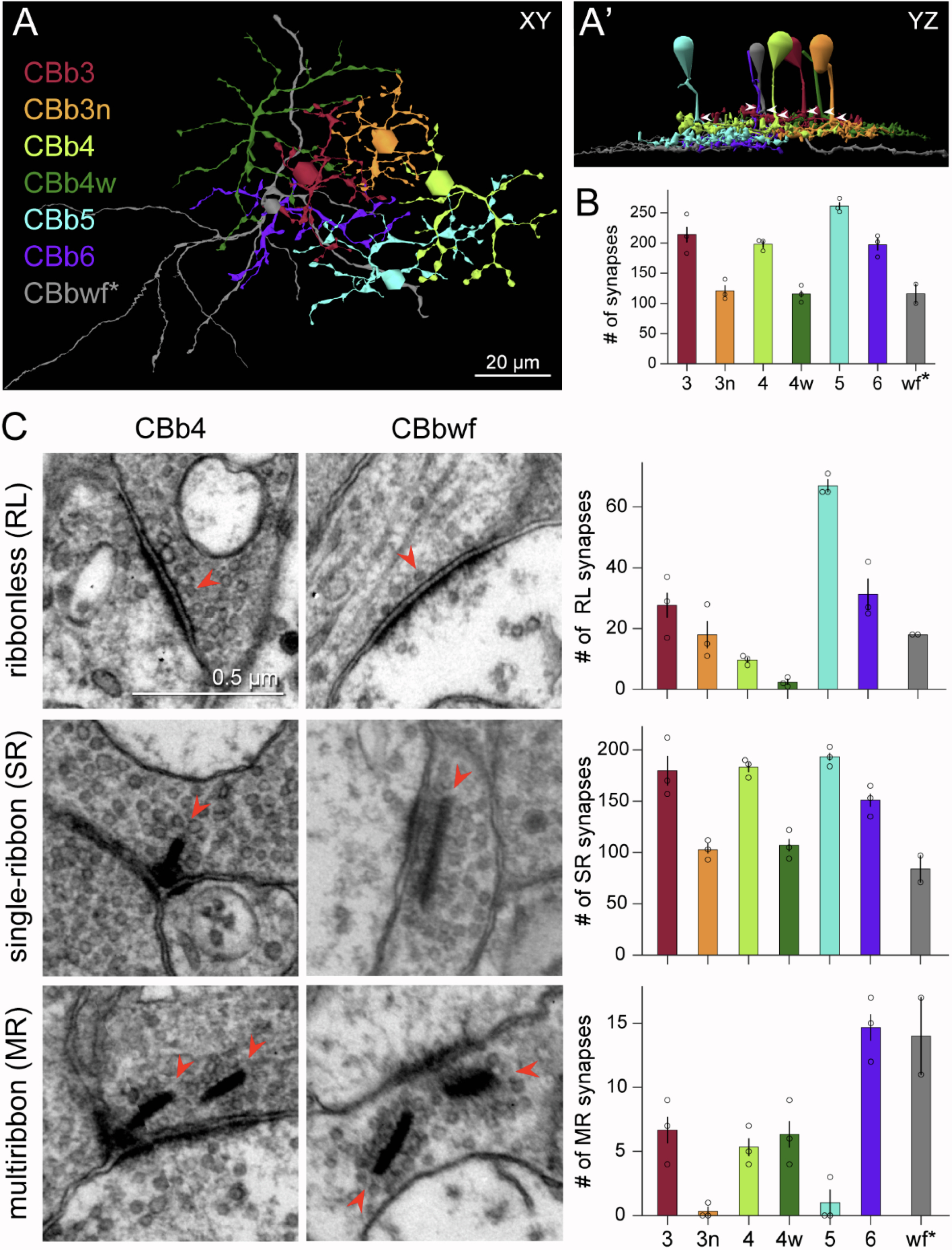
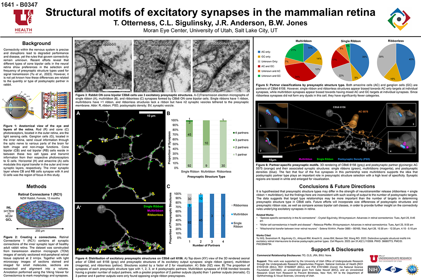
Connectivity within the nervous system is precise and disruptions lead to degraded performance and disease, yet the rules that govern connectivity remain unknown. Recent efforts reveal that different types of cone bipolar cells in the neural retina show preferences in the selection and frequency of presynaptic structure types used for signal transmission. However, it is not yet known how these differences are related to the quantity or type of postsynaptic partner. We used Retinal Connectome 1 (RC1) to analyze the synaptic output of rabbit CBb6 cells, a type of ON cone bipolar cell that forms excitatory synapses via diverse presynaptic structure types, to identify patterns in how these cells interact with their postsynaptic partners.

Methods
RC1 is a 0.25 mm diameter volume sampled from mid-peripheral retina of a 13 month old female Dutch-Belted rabbit, serially sectioned at 70 nm, and imaged at ultrastructural resolution (2nm/px) using transmission electron microscopy. Postsynaptic partners of CBb6 cell 6156’s presynaptic structures were annotated using the Viking Viewer for Connectomics. Statistical analyses were conducted in Microsoft Excel and investigated further with 3D rendering and graph visualization of connectivity.

Results
The factors tracked for comparison included presynaptic structure type, target number, and postsynaptic partner type. Multiribbon synapses of CBb6 cell 6156 trended towards having a greater number of output partners, with a greater proportion of dyads than monads. Despite this, triads and quadrads were only found opposing single ribbon synapses. As the different presynaptic structure types may differ in the strength of neurotransmitter release (ribbonless < single ribbon < multiribbon), these findings are inconsistent with scaling of output to the number of postsynaptic targets. Both amacrine cells (AC) and ganglion cells (GC) are postsynaptic partners of 6156. However, single ribbon and ribbonless structures appear biased towards AC only targets, while multiribbon synapses appear biased toward mixed AC and GC targets.

Conclusions
Target type relationships appear more important than the number of targets in determining presynaptic structure type in CBb6. Future efforts will examine size differences of postsynaptic structures and presynaptic ribbon size, and even compare across bipolar cell classes, in order to provide further insight on the connectivity rules underlying excitatory synapses.

Distinctive Synaptic Structural Motifs Link Excitatory Retinal Interneurons To Diverse Postsynaptic Partner Types

We have a new manuscript from the lab in Cell Reports, Distinctive Synaptic Structural Motifs Link Excitatory Retinal Interneurons To Diverse Postsynaptic Partner Types. This manuscript is in collaboration with the first author, Wan-Qing Yu @wanqing_yu, then co-authors Rachael Swanstrom, Crystal L. Sigulinsky @CSigulinsky, Richard M. Ahlquist, along with Sharm Knecht, myself Bryan W. Jones @BWJonesDavid M. Berson, and Rachel O. Wong. The PDF is here.

Abstract:
Neurons make converging and diverging synaptic connections with distinct partner types. Whether synapses involving separate partners demonstrate similar or distinct structural motifs is not yet well understood. We thus used serial electron microscopy in mouse retina to map output synapses of cone bipolar cells (CBCs) and compare their structural arrangements across bipolar types and postsynaptic partners. Three presynaptic configurations emerge—single-ribbon, ribbonless, and multiribbon synapses. Each CBC type exploits these arrangements in a unique combination, a feature also found among rabbit ON CBCs. Though most synapses are dyads, monads and triads are also seen. Altogether, mouse CBCs exhibit at least six motifs, and each CBC type uses these in a stereotypic pattern. Moreover, synapses between CBCs and particular partner types appear biased toward certain motifs. Our observations reveal synaptic strategies that diversify the output within and across CBC types, potentially shaping the distinct functions of retinal microcircuits.

Seasonal And Post-Trauma Remodeling Of The Ground Squirrel Retina

We have a new publication out, Seasonal and post-trauma remodeling in cone-dominant ground squirrel retina authored by Dana Merriman, Ben Sajdak, Wei Li and Bryan W. Jones.

Abstract:

With a photoreceptor mosaic containing ∼85% cones, the ground squirrel is one of the richest known mammalian sources of these important retinal cells. It also has a visual ecology much like the human’s. While the ground squirrel retina is understandably prominent in the cone biochemistry, physiology, and circuitry literature, far less is known about the remodeling potential of its retinal pigment epithelium, neurons, macroglia, or microglia. This review aims to summarize the data from ground squirrel retina to this point in time, and to relate them to data from other brain areas where appropriate. We begin with a survey of the ground squirrel visual system, making comparisons with traditional rodent models and with human. Because this animal’s status as a hibernator often goes unnoticed in the vision literature, we then present a brief primer on hibernation biology. Next we review what is known about ground squirrel retinal remodeling concurrent with deep torpor and with rapid recovery upon re-warming. Notable here is rapidly-reversible, temperature-dependent structural plasticity of cone ribbon synapses, as well as pre- and post-synaptic plasticity throughout diverse brain regions. It is not yet clear if retinal cell types other than cones engage in torpor-associated synaptic remodeling. We end with the small but intriguing literature on the ground squirrel retina’s remodeling responses to insult by retinal detachment. Notable for widespread loss of (cone) photoreceptors, there is surprisingly little remodeling of the RPE or Müller cells. Microglial activation appears minimal, and remodeling of surviving second- and third-order neurons seems absent, but both require further study. In contrast, traumatic brain injury in the ground squirrel elicits typical macroglial and microglial responses. Overall, the data to date strongly suggest a heretofore unrecognized, natural checkpoint between retinal deafferentiation and RPE and Müller cell remodeling events. As we continue to discover them, the unique ways by which ground squirrel retina responds to hibernation or injury may be adaptable to therapeutic use.