Author Archives: bwjones

Revival Of Light Signalling In The Postmortem Mouse And Human Retina

We have a new collaborative manuscript out in Nature, Revival of light signalling in the postmortem mouse and human retina. Full paper (here).

Authors: Fatima Abbas @neurofim, Silke Becker, Bryan W. Jones @BWJones, Ludovic S. Mure, Satchidananda Panda @SatchinPanda, Anne Hanneken & Frans Vinberg @fvinberg.

Abstract:
Death is defined as the irreversible cessation of circulatory, respiratory or brain activity. Many peripheral human organs can be transplanted from deceased donors using protocols to optimize viability. However, tissues from the central nervous system rapidly lose viability after circulation ceases, impeding their potential for transplantation. However, the time course and mechanisms causing neuronal death and the potential for revival remain poorly defined. Here, using the retina as a model of the central nervous system, we systemically examine the kinetics of death and neuronal revival. We demonstrate the swift decline of neuronal signalling and identify conditions for reviving synchronous in vivo-like trans-synaptic transmission in postmortem mouse and human retina. We measure light-evoked responses in human macular photoreceptors in eyes removed up to 5 h after death and identify modifiable factors that drive reversible and irreversible loss of light signalling after death. Finally, we quantify the rate-limiting deactivation reaction of phototransduction, a model G protein signalling cascade, in peripheral and macular human and macaque retina. Our approach will have broad applications and impact by enabling transformative studies in the human central nervous system, raising questions about the irreversibility of neuronal cell death, and providing new avenues for visual rehabilitation.

Dynein Dysregulation Due to the Absence of NUDC leads to Mitochondrial Mislocalization and Dysfunction in Rod Photoreceptors

This abstract was presented today, May 4th at the 2022  Association for Research in Vision and Opthalmology (ARVO) meetings in Denver, Colorado by Hailey Levi @drpepperis100, Meredith Hubbard, Mary Anne Garner, TJ Hollingsworth, Ke Jiang, Nat Nelson, Anushree Gade, Drue Benefield, Guoxin Ying, Wolfgang Baehr, Bryan Jones@BWJones, Anand Swaroop, Glenn Rowe, and Alecia Gross @alecia144g.

Proteomic changes in the lens of a congenital cataract mouse model lead to reduced levels of glutathione and taurine

This abstract was presented today, May 4th at the 2022  Association for Research in Vision and Opthalmology (ARVO) meetings in Denver, Colorado by Sheldon Rowan @SheldonRowan, Eloy Bejarano, Elizabeth Whitcomb, Rebecca Pfeiffer @BeccaPfeiffer19, Kristie Rose, Kevin Schey, Bryan Jones @BWJones, Allen Taylor.

Purpose: Congenital cataracts develop through multiple mechanisms, but often lead to common endpoints, including protein aggregation, impaired fiber cell differentiation, and absence of fiber cell denucleation. It is now apparent that other metabolic abnormalities associate with cataractogenesis, including reductions in levels of amino acids, glutathione, and taurine. Here, we analyze the proteome and metabolome of mice expressing a mutant ubiquitin protein (K6W-Ub) to determine the molecular mechanisms underlying formation of its congenital cataract.

Methods: C57BL/6J wild-type or cataractous K6W-Ub transgenic mouse lenses were dissected at E15.5, P1, or P30 and proteins were analyzed via MS-based tandem-mass-tag (TMT) quantitative proteomics. Small molecules were spatially quantified using computational molecular phenotyping (CMP), a tool that enables acquisition of free amino acid fingerprints for every cell in the lens. Validation of proteomics findings was also performed using Western blot analysis and immunohistochemistry.

Results: Proteomic analyses revealed pathways that were altered during lens differentiation, by expression of K6W-Ub, or both. Prominent pathways included glutathione metabolism; glycolysis/gluconeogenesis; and glycine, serine, and threonine metabolism. Within the glutathione metabolism pathway, GSTP1 and GGCT were most strongly downregulated by K6W-Ub. Other consistently downregulated proteins were PGAM2, GAMT, and HMOX1. Proteins that were upregulated by K6W-Ub expression belonged to pathways related to lysosome, autophagy, Alzheimer’s disease, and glycolysis/gluconeogenesis. Analysis of the metabolome via CMP revealed statistically significant decreases in taurine and glutathione and smaller decreases in glutamate, glutamine, aspartate, and valine in all ages of K6W-Ub lenses. Lens metabolites were spatially altered in the cataractous K6W-Ub lens.

Conclusions: K6W-Ub expressing lenses replicate many congenital cataract phenotypes and are useful disease models. The large reductions in levels of taurine and glutathione may be general signatures of cataract development, as human cataracts also have reduced glutathione and taurine. Key roles for amino acid metabolism and glycolysis/gluconeogenesis in cataractogenesis are emerging. Together our data point toward potential common metabolic/proteomic signatures of cataracts.

ARVO Mini-Symposium: Pathoconnectomics in Retinal Degeneration

Lab PI, Bryan Jones delivered a talk at the ARVO 2022 mini-symposium on Pathoconnectomics in Retinal Degeneration.

Abstract: Connectomics has demonstrated that synaptic networks and their topologies are precise and directly correlate with physiology and behavior. The next extension of connectomics is pathoconnectomics: to map neural network synaptology and circuit topologies corrupted by neurological disease in order to identify robust targets for therapeutics. The retina is ideal for pathoconnectomics approaches, and reveals common rules of how neural systems are wired, and how they break in neurodegenerative disease.

Happy New Year, 2022

“Those of us who survived, are now through the 2nd year of this pandemic.”

That sentence sounds like the introduction to a post apocalyptic story, and something that while not completely unexpected in my lifetime, still has a rawness to it, and an anger to it because of how the world has handled things.  Our distraction with political authoritarianism across the world, and an obsession with individual rights over that of society has made this far worse than it otherwise would in a healthy global society.  We are now looking at over 800,000 Americans are dead from COVID, with over 5.4 million dead worldwide, and we are not yet done.  If you had told me that these would be the numbers back in the first week of March 2020 when I shut the lab down to protect everyone in it, I might not have believed you.

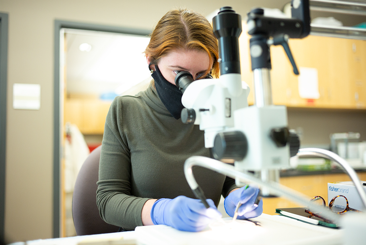
We started the year 2021 in the lab with a chiller for one of our electron microscopes that failed.  That took out half our electron microscopy infrastructure for a period of time, until we could get it back up and running, which of course was complicated by the pandemic. I’m grateful for Mark Kirkpatrick, and Kevin Mcilwrath from JEOL @JEOLUSA who helped us maintain the microscopes and keep them up and running. 2021 continued the hits with lots of new bureaucracy initiatives at the University of Utah with IT, and HR, and purchasing, and travel, that are an incredible tax on time and resources.  If I had enough resources to help balance out those demands, it would be acceptable, but as it is, I’m getting further and further away from the science, which is seriously bumming me out.  I got into this game to dig deep into the science. This skyrocketing administrative burden on top of all the other things that I’ve had to deal with as a result of the pandemic has really stressed things out.  Yet we’ve still been incredibly productive through the pandemic, mostly because of the work of personnel in the lab, Jia-Hui, Jamie, Nat @NatQuayleNelson, Crystal @CSigulinskyBecca @BeccaPfeiffer19, Jeebika, Selena, Olivia, Isabel and Gabe, all of whom I am immensely grateful for.

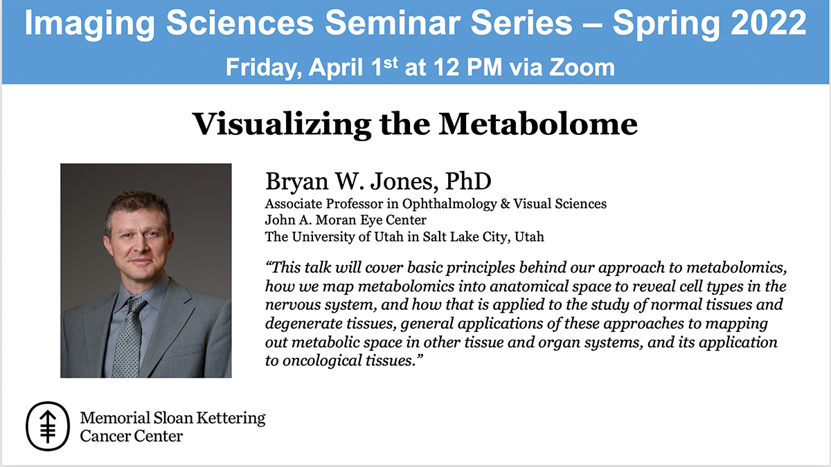
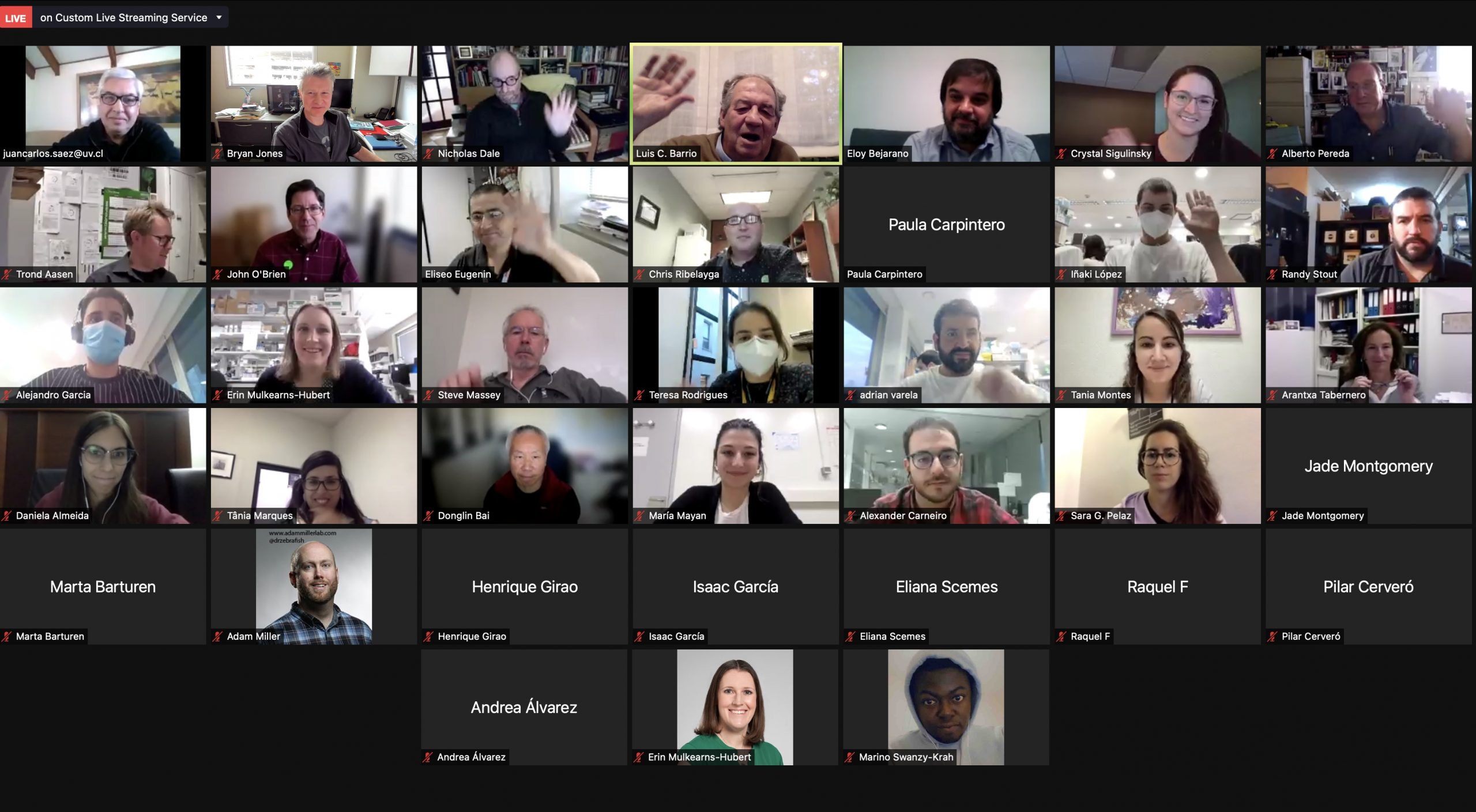
This year, despite the 2nd year of a global pandemic, we’ve still managed to publish a book chapter, 6 manuscripts, 2 abstracts, participated in teaching new faculty at the University of Utah School of Medicine, co-organized the Moran Eye Center Translational Research Day with Leah Owen, started faculty hiring initiatives, reviewed grants, started a new retinal connectomics initiative that will meet for the first time in January, submitted a grant renewal (triaged), submitted three other grants (not funded), taught classes for grad students, started serving on 3 PhD student committees, brought a couple of new undergraduates into the lab, had one undergraduate win Outstanding Undergraduate Researcher of The Year Award, had postdocs present at a couple of meetings, had a couple of PhD students rotate through the lab, gave 5 invited talks, initiated two more collaborations and more, all while struggling through unprecedented difficulties related to the pandemic.

The New Year 2022 looks promising, and we hope for a slow normalization of things. January will bring a new technician to the lab, and the first meeting of a select group of people for a new retinal connectomics initiative that we’ll be rolling out to a wider audience ASAP.  We’ll also have a new collaborative manuscript that has just been accepted in Nature.  Later in the first part of the year, we’ll have another three manuscripts submitted, a grant renewal that will go in, and we might have a new PhD student joining the lab as well as a new undergraduate. Hopefully later in the year, some of the irons that I have in the fire will return on those investments, and we’ll start to normalize some other things like meetings.  ARVO is planning on being a hybrid meeting, and I’ve agreed to a special interest group presentation there with Michael Redmond.  We’ll see what other meetings happen next year, pending outcomes from the pandemic.  I’d love to be able to send the postdocs to meetings this year.  ISER was pushed out to February of 2023 because of the pandemic, so that won’t be happening this year as was previously planned.  Further out in the year is harder to predict, but we should be finished with our third pathoconnectome, as well as our primate retinal connectome, paving the way for us to begin building our two human pathoconnectomes.

However this year ends up, we here at the Marclab for Connectomics wish you a Happy New Year for 2022, and much success and happiness.


Peptide-Based Immunotherapy Against Oxidized Elastin Ameliorates Pathology In Mouse Model Of Smoke-Induced Ocular Injury

We have a new collaborative manuscript out in iOVS, Peptide-Based Immunotherapy Against Oxidized Elastin Ameliorates Pathology In Mouse Model Of Smoke-Induced Ocular Injury. (pdf here)

Authors: Bärbel Rohrer, Nathaniel Parsons , Balasubramaniam Annamalai , Crystal Nicholson , Elisabeth Obert , Bryan Jones @BWJones, and Andrew D Dick.

Abstract:

Purpose: Age-related macular degeneration (AMD), the leading cause of blindness in western populations, is associated with an overactive complement system, and an increase in circulating antibodies against certain epitopes, including elastin. As loss of the elastin layer of Bruch’s membrane (BrM) has been reported in aging and AMD, we previously showed that immunization with elastin peptide oxidatively modified by cigarette smoke (ox-elastin), exacerbated ocular pathology in the smoke-induced ocular pathology (SIOP) model. Here we asked whether ox-elastin peptide-based immunotherapy (PIT) ameliorates damage.

Methods: C57BL/6J mice were injected with ox-elastin peptide at two doses via weekly subcutaneous administration, while exposed to cigarette smoke for 6 months. FcγR-/- and uninjected C57BL/6J mice served as controls. Retinal morphology was assessed by by electron microscopy, and complement activation, antibody deposition and mechanisms of immunological tolerance were assessed by Western blotting and ELISA.

Results: Elimination of Fcγ receptors, preventing antigen/antibody-dependent cytotoxicity, protected against SIOP. Mice receiving PIT with low dose ox-elastin (LD-PIT) exhibited reduced humoral immunity, reduced complement activation and IgG/IgM deposition in the RPE/choroid, and largely a preserved BrM. While there is no direct evidence of ox-elastin pathogenicity, LD-PIT reduced IFNγ and increased IL-4 within RPE/choroid. High dose PIT was not protective.

Conclusions: These data further support ox-elastin role in ocular damage in in part via elastin-specific antibodies, and support the corollary that PIT with ox-elastin attenuates ocular pathology. Overall, damage is associated with complement activation, antibody-dependent cell-mediated cytotoxicity, and altered cytokine signature.

Keywords: Age-related macular degeneration; Complement; Elastin; Peptide-based immunotherapy; Smoking.