Natural Immunoglobulin M-based Delivery of a Complement Alternative Pathway Inhibitor in Mouse Models of Retinal Degeneration

We have a new manuscript out in Experimental Eye Research, Natural Immunoglobulin M-based Delivery of a Complement Alternative Pathway Inhibitor in Mouse Models of Retinal Degeneration. (pdf here)

Authors: Balasubramaniam Annamalai, Nathaniel Parsons, Crystal Nicholson, Kusumam Joseph, Beth Coughlin, Xiaofeng Yang, Bryan W. Jones @BWJones, Stephen Tomlinson, and Bärbel Rohrer.

Abstract:

Purpose: Age-related macular degeneration is a slowly progressing disease. Studies have tied disease risk to an overactive complement system. We have previously demonstrated that pathology in two mouse models, the choroidal neovascularization (CNV) model and the smoke-induced ocular pathology (SIOP) model, can be reduced by specifically inhibiting the alternative complement pathway (AP). Here we report on the development of a novel injury-site targeted inhibitor of the alternative pathway, and its characterization in models of retinal degeneration.

Methods: Expression of the danger associated molecular pattern, a modified annexin IV, in injured ARPE-19 cells was confirmed by immunohistochemistry and complementation assays using B4 IgM mAb. Subsequently, a construct was prepared consisting of B4 single chain antibody (scFv) linked to a fragment of the alternative pathway inhibitor, fH (B4-scFv-fH). ARPE-19 cells stably expressing B4-scFv-fH were microencapsulated and administered intravitreally or subcutaneously into C57BL/6 J mice, followed by CNV induction or smoke exposure. Progression of CNV was analyzed using optical coherence tomography, and SIOP using structure-function analyses. B4-scFv-fH targeting and AP specificity was assessed by Western blot and binding experiments.

Results: B4-scFv-fH was secreted from encapsulated RPE and inhibited complement in RPE monolayers. B4-scFv-fH capsules reduced CNV and SIOP, and western blotting for C3a, C3d, IgM and IgG confirmed a reduction in complement activation and antibody binding in RPE/choroid.

Conclusions: Data supports a role for natural antibodies and neoepitope expression in ocular disease, and describes a novel strategy to target AP-specific complement inhibition to diseased tissue in the eye.

Precis: AMD risk is tied to an overactive complement system, and ocular injury is reduced by alternative pathway (AP) inhibition in experimental models. We developed a novel inhibitor of the AP that targets an injury-specific danger associated molecular pattern, and characterized it in disease models.

Keywords: Alternative pathway inhibitor; Choroidal neovascularization; Complement system; Encapsulated ARPE-19 cells; Natural antibody-mediated targeting; Smoke-induced ocular pathology.

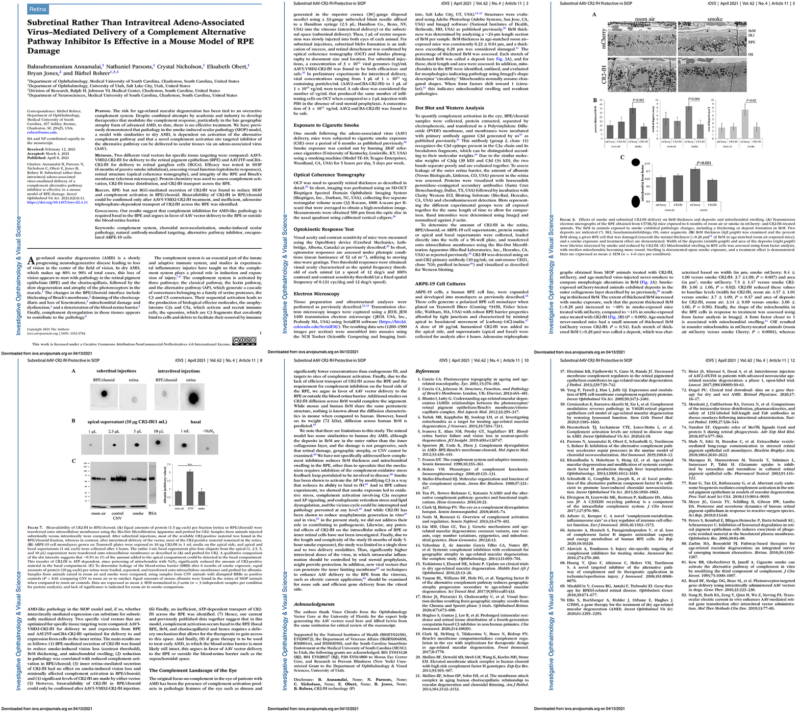
Subretinal Rather Than Intravitreal Adeno-Associated Virus–Mediated Delivery of a Complement Alternative Pathway Inhibitor Is Effective in a Mouse Model of RPE Damage

We have a new manuscript out in iOVS, Subretinal Rather Than Intravitreal Adeno-Associated Virus–Mediated Delivery of a Complement Alternative Pathway Inhibitor Is Effective in a Mouse Model of RPE Damage. (pdf here)

Authors: Balasubramaniam Annamalai; Nathaniel Parsons; Crystal Nicholson; Elisabeth Obert; Bryan W. Jones @BWJones; and Bärbel Rohrer.

Abstract:

Purpose: The risk for age-related macular degeneration has been tied to an overactive complement system. Despite combined attempts by academia and industry to develop therapeutics that modulate the complement response, particularly in the late geographic atrophy form of advanced AMD, to date, there is no effective treatment. We have previously demonstrated that pathology in the smoke-induced ocular pathology (SIOP) model, a model with similarities to dry AMD, is dependent on activation of the alternative complement pathway and that a novel complement activation site targeted inhibitor of the alternative pathway can be delivered to ocular tissues via an adeno-associated virus (AAV).

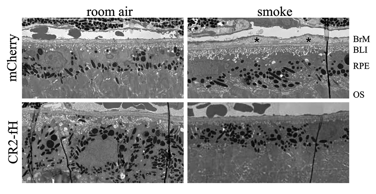
Methods: Two different viral vectors for specific tissue targeting were compared: AAV5-VMD2-CR2-fH for delivery to the retinal pigment epithelium (RPE) and AAV2YF-smCBA-CR2-fH for delivery to retinal ganglion cells (RGCs). Efficacy was tested in SIOP (6 months of passive smoke inhalation), assessing visual function (optokinetic responses), retinal structure (optical coherence tomography), and integrity of the RPE and Bruch’s membrane (electron microscopy). Protein chemistry was used to assess complement activation, CR2-fH tissue distribution, and CR2-fH transport across the RPE.

Results: RPE- but not RGC-mediated secretion of CR2-fH was found to reduce SIOP and complement activation in RPE/choroid. Bioavailability of CR2-fH in RPE/choroid could be confirmed only after AAV5-VMD2-CR2-fH treatment, and inefficient, adenosine triphosphate–dependent transport of CR2-fH across the RPE was identified.

Conclusions: Our results suggest that complement inhibition for AMD-like pathology is required basal to the RPE and argues in favor of AAV vector delivery to the RPE or outside the blood-retina barrier.

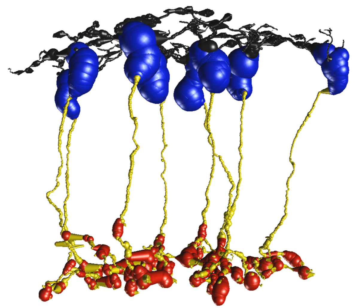
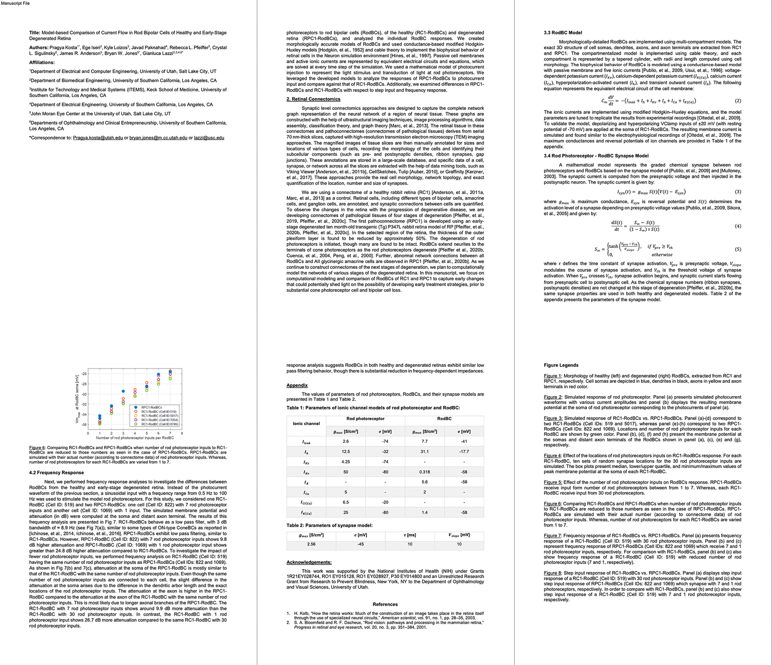
Model-Based Comparison of Current Flow in Rod Bipolar Cells of Healthy and Early-Stage Degenerated Retina

We have a new manuscript out in Experimental Eye Research, Model-Based Comparison of Current Flow in Rod Bipolar Cells of Healthy and Early-Stage Degenerated Retina. (pdf here)

Authors: Pragya Kosta, Ege Iseri, Kyle Loizos, Javad Paknahad, Rebecca L. Pfeiffer @BeccaPfeiffer19, Crystal L. Sigulinsky @CLSigulinsky, James R. Anderson, Bryan W. Jones @BWJones, and Gianluca Lazzi.

Abstract: Retinal degenerative diseases, such as retinitis pigmentosa, are generally thought to initiate with the loss of photoreceptors, though recent work suggests that plasticity and remodeling occurs prior to photoreceptor cell loss. This degeneration subsequently leads to death of other retinal neurons, creating functional alterations and extensive remodeling of retinal networks. Retinal prosthetic devices stimulate the surviving retinal cells by applying external current using implanted electrodes. Although these devices restore partial vision, the quality of restored vision is limited. Further knowledge about the precise changes in degenerated retina as the disease progresses is essential to understand how current flows in retinas undergoing degenerative disease and to improve the performance of retinal prostheses. We developed computational models that describe current flow from rod photoreceptors to rod bipolar cells


Model-based Comparison of Current Flow in Rod Bipolar Cells of Healthy and Early-Stage Degenerated Retina

Laboratory In A Pandemic

This past year has been remarkable in terms of the impact that COVID-19 has wrought, as well as its impact upon everyone in the team.  I am proud of the resiliency and huge efforts that people have gone through to keep science in the lab going, and cannot possibly relate how grateful I am to everyone.  They have struggled through working remotely, human resources problems, child-care issues, logistical issues related to access of research tools and data, equipment downtime due to maintenance issues, personal COVID infections, and outbreaks in their families, not being able to attend meetings, duties to the department and university to assist with the COVID-19 response, volunteering of time, money and resources to assist the COVI-19 response, having to be responsive to new IT demands, and so much more.

I had been tracking COVID-19 since January, but by March, I became particularly alarmed and shut the lab down, sending everyone home with computers in short order.  Having no idea how this would turn out, I did have visions of being able to get all caught up and do a ton of reading that I’ve not been able to do, and more.  2020 being 2020, that was just not possible, and the bureaucratic overhead of reporting and constant emails and videoconferencing has eaten up massive amounts of time.  This year, despite the global pandemic, we’ve still managed to publish 6 manuscripts, 3 pre-prints, 2 abstracts, and 1 chapter, and secured a new NSF grant.  We’ve continued to mentor graduate students, undergraduate students, and post-docs.  Again, I am incredibly proud of this team.

The danger with all this productivity in the face of a global pandemic is that we are burning our team out.  So, the plan was to take a couple weeks off, spend time with the closest people in our lives, recharge our batteries, and hit the ground running again in the New Year.  Reality intruded for me at least however, and I discovered a water leak from one of the electron microscope chillers that leaked into the walls.  I am investigating this now, but we are clearly down with half of our ultrastructural infrastructure unavailable until we can either get the chiller/microscope repaired or equipment replaced.  2020 just will not let go…  The lab is still working from home, but I’ve had to go into work and get some things done related to the water leak.

Outside of equipment failures and water leaks, working in the lab during 2020 has been a challenge.  After getting approval to have some people return to campus, we decided to bring technicians back for part time work in the lab and part time work at home, so long as they could keep distances while wearing masks while in the lab.  Not being able to be in the lab full time, and having to maintain physical distance from all others and wear masks and personal protective gear constantly has absolutely been an added burden.  However, we are fortunate in that we have space to spread out, but it has still been a massive challenge, getting work done.

Our lab has helped with the COVID-19 response by taking temperatures and performing health screenings of visitors and patients/visitors to the Moran Eye Center, a task all of our labs in the Moran Eye Center have been participating in.  It takes us away from our research duties, but also helps in some small way with the COVID-19 response.

Staying up on the work from students and post-docs has, like everything else, gone virtual.  This slows everything down of course and contributes to the sense of isolation.  Both Crystal @CSigulinsky and Becca @BeccaPfeiffer19 have been critical in this mentoring effort.  I can’t wait until we get this thing back under control and can meet in person again…

I’m also grateful for Mark Kirkpatrick, and Kevin Mcilwrath from JEOL @JEOLUSA who helped us maintain the microscopes and keep them up and running.  And when they went down, I am so grateful that Kevin could come in and help us get them back up and running.  We definitely had downtime as a result of the pandemic, but we would have had more, if it were not for Kevin Mcilwrath.  We will need him again, to get back up and running after the Christmas Eve chiller failure, and I am grateful for him and JEOL for making the efforts to keep us running.

We’ll see what the New Year brings.  The list of things to do starting in a few days is formidable.  Even though the vaccines are just now starting to roll out, we are still in the middle of a pandemic and will not be returning to normal functioning in the lab for months yet.  We have to perform repairs to our infrastructure, finish a manuscript I am working on, edits to a colleagues manuscript, getting a couple of manuscripts from post-docs going, a couple of new genetic models to create, a grant renewal to write, data to process for collaborations, getting our light microscopes back up and running, a grad student starting a rotation, undergrads presenting their research, and then completing work on all the other stuff that we normally have to do.

I am so grateful for Jia-Hui, Jamie, Hope, Nat, Crystal, Becca, Jeebika, Jessica, Selena and Olivia. You all made getting through this year possible.  Thank you for your teamwork, and your *hard* work.  Also, my undying gratitude to my chairman, and all the colleagues in my department.  What a brutal year, and my hopes are for more normalization, better leadership at the federal level, and mitigation of the COVID-19 pandemic as vaccines start to roll out.
Happy Weird Holidays, 2020 From The Marclab For Connectomics

Happy Weird Holidays, 2020 to you all from us here at the Marclab for Connectomics.

What a strange, and fundamentally challenging year this has been for so many.  It has been lonely, frustrating, painful, and discouraging for many of us, and I encourage all to be a bit more patient, kind, and compassionate to those around you, as you don’t know what they are dealing with.

This pandemic has exacerbated the challenges that many are dealing with financially, socially, and emotionally. It has led to incredible bureaucracy for many of us trying to run labs. Personnel issues as folks in labs struggle with the issues in their lives not only working from home, but working from home during a pandemic, including child care, or care for other family members. Struggles with personal illness (COVID and otherwise). Financial issues related to employment. Struggles with depression and isolation. Inability to collaborate, or attend meetings that are critical to ongoing projects. Loss of research resources. Inability to get equipment or service on equipment… For some, this pandemic has resulting in a complete cessation of research activities.

Lab productivity has certainly been hugely impacted for many labs and the implications of that for their NIH or NSF funding is still unclear. Our productivity has certainly suffered this year, and we are absolutely behind on promised data generation. I don’t know how we are going to deal with that yet. The goal is to try and catch up, and while our lab is nominally working right now, all undergraduate, and graduate students are working remotely. Postdocs are partially working in the lab, and technicians are splitting their time. Until all the vaccines roll out, it is unclear how much longer this will have to continue. Fortunately for us, we are heavily computational and we’ve been able to still be productive on existing data. We also have generous lab space that we can spread out in for those who are working on campus. So, for now, we are forging ahead, managing as best as we can and attempting to be helpful to others where we can by processing tissues for other labs where we can to help them with their productivity problems for instance, and as always, reviewing manuscripts and grants in-between trying to get our manuscripts and grants in.

This pandemic has exacerbated the challenges that many are dealing with financially, socially, emotionally, and scientifically. And while this life gives and it takes, it also gives us opportunities to make decisions that allow us to help improve the lives of others, and be a force for good.

Happy Holidays from the Marclab for Connectomics.

Jessica Garcia Is This Year’s Student Veteran Of The Year

Jessica Garcia is this year’s University of Utah Student Veteran of the Year. Jessica is an undergraduate student in the lab exploring the OFF-layer branches of ON cone bipolar cells in early retinal degeneration.

Jessica came to us by way of service with the US Navy, where she served as an aviation technician before attending the University of Utah.

Congratulations Jessica! We are so proud of you.

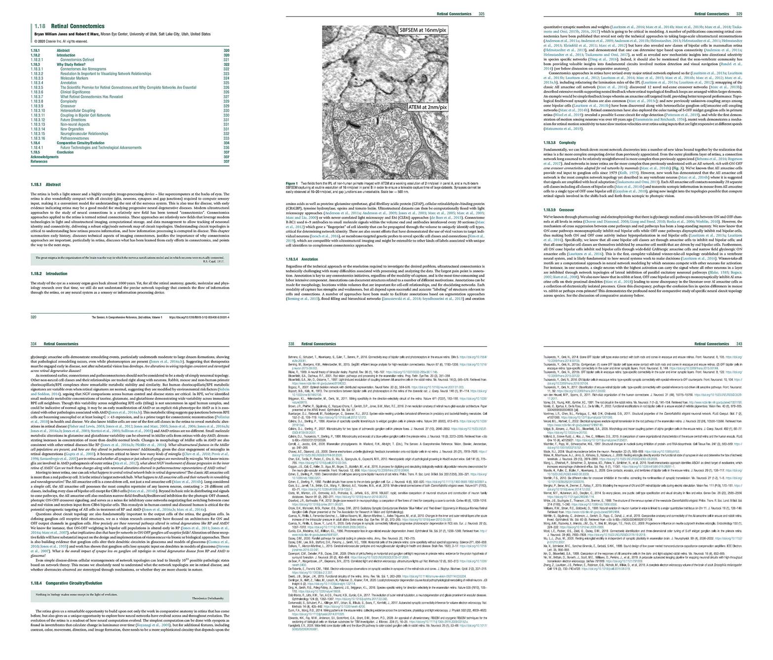
Chapter: Retinal Connectomics

We have a new chapter out in the Elsevier book series The Senses, 2021.

Authors are myself, Bryan W. Jones @BWJones and Robert E. Marc @robertmarc60.

Abstract: The retina is both a light sensor and a highly complex image-processing device – like supercomputers at the backs of eyes. The retina is also wonderfully compact with all circuitry (glia, neurons, synapses and gap junctions) required to compute sensory input, making it a convenient model for understanding the rest of the nervous system. This is also true for disease, with early evidence indicating retina may be a good model for studying progressive neural degenerative diseases. Modern ultrastructural approaches to the study of neural connections is a relatively new !eld has been termed “connectomics”. Connectomics approaches applied to the retina is termed retinal connectomics. These approaches are relatively new !elds that leverage modern technologies in light and ultrastructural imaging, computational storage, and data management to allow tracking of neuronal identity and connectivity, delivering a robust edge/node network map of circuit topologies. Understanding circuit topologies is critical to understanding how retinas process information, and how information processing is corrupted in disease. This chapter summarizes early history, discusses technical aspects of imaging connectomes, justi!es the importance of why connectomics approaches are important, particularly in retina, discusses what has been learned from early efforts in connectomics, and points the way to the next steps.

Please email me: bryan.jones@m.cc.utah.edu if you would like a pdf of the chapter.


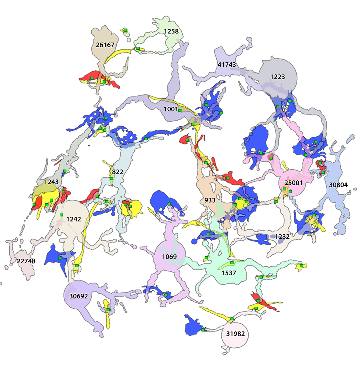
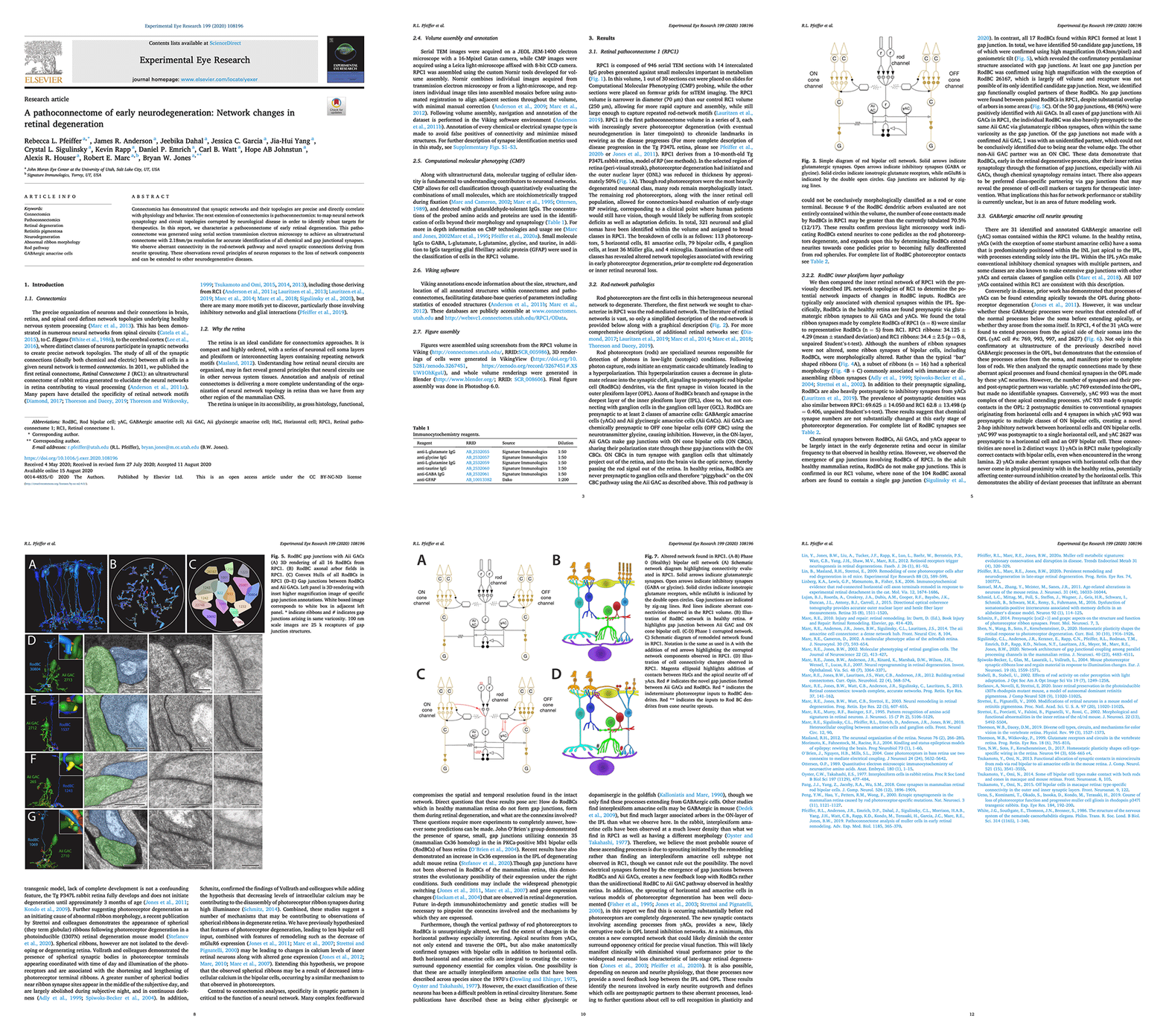
A pathoconnectome of early neurodegeneration: Network changes in retinal degeneration

We have a new manuscript out in Experimental Eye Research, A pathoconnectome of early neurodegeneration: Network changes in retinal degeneration. (pdf here)

Authors: Rebecca L. Pfeiffer @BeccaPfeiffer19, James R. Anderson, Jeebika Dahal, Jessica C. Garcia, Jia-Hui Yang, Crystal L. Sigulinsky @CLSigulinsky, Kevin Rapp, Daniel P. Emrich, Carl B. Watt, Hope AB Johnstun, Alexis R. Houser, Robert E. Marc @robertmarc60, and Bryan W. Jones @BWJones.

Abstract: Connectomics has demonstrated that synaptic networks and their topologies are precise and directly correlate with physiology and behavior. The next extension of connectomics is pathoconnectomics: to map neural network synaptology and circuit topologies corrupted by neurological disease in order to identify robust targets for therapeutics. In this report, we characterize a pathoconnectome of early retinal degeneration. This pathoconnectome was generated using serial section transmission electron microscopy to achieve an ultrastructural connectome with 2.18nm/px resolution for accurate identification of all chemical and gap junctional synapses. We observe aberrant connectivity in the rod-network pathway and novel synaptic connections deriving from neurite sprouting. These observations reveal principles of neuron responses to the loss of network components and can be extended to other neurodegenerative diseases.


NeuroNex Grant

I am pleased to report that the The Marclab for Connectomics has been funded by the National Science Foundation with a 5 year grant as part of a large, international consortium to study synaptic weighting.  We are collaborating with the Erik Jorgensen laboratory here at the University of Utah, and will be exploring synapses in a model of retinal degeneration.  There is a nice writeup of the award on the Moran Eye Center website, here.

This is a wonderful opportunity to work with other colleagues that will be funded alongside us with this grant, including Uri Manor @manorlaboratory, Davi Bock @dddavi, Josh Vogelstein @neuro_data, Viren Jain @stardazed0, and others.  My thanks to Kristen Harris for heading up this initiative.