Tag Archives: TEM

Simple Super-Resolution Microscopy

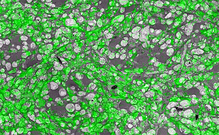
Super-resolution microscopy is a pretty big thing right now.  But there is more than one way to get super-resolution microscopy results.  There are a variety of approaches, most involving expensive new microscopes that preclude many scientists from participating in science that allows them to ask certain questions.  However, if they have access to a standard transmission election microscope and have antibodies that are glutaraldehyde tolerant, they can participate and ask questions that allow them to get around some of the inherent limitations imposed by physics.

In the image above for example, we have GABA labeling in green superimposed upon ultrastructural data showing us *which* processes in the inner plexiform layer of the retina are GABAergic.  Many of these processes are smaller than the wavelength of light.

There are multiple ways to get here of course with some very expensive microscopes offering dual light and electron microscopy approaches and yet other microscopes offering purely optical based solutions.  However, this is cheap and easy and accessible to many with the basic electron microscopy resources. Robert Marc first used this approach in back in 2000, and we subsequently used it for quite a bit of work for my Ph.D. dissertation in 2003, and notably in this paper.  It is also an integral technique associated with our connectomics efforts.

That said, I’ll need at some point soon to find the resources to get a traditional optical super-resolution microscopy solution to answer some questions we have in the lab on neural degenerative disease.
(Via Jonesblog)

SEM vs. TEM

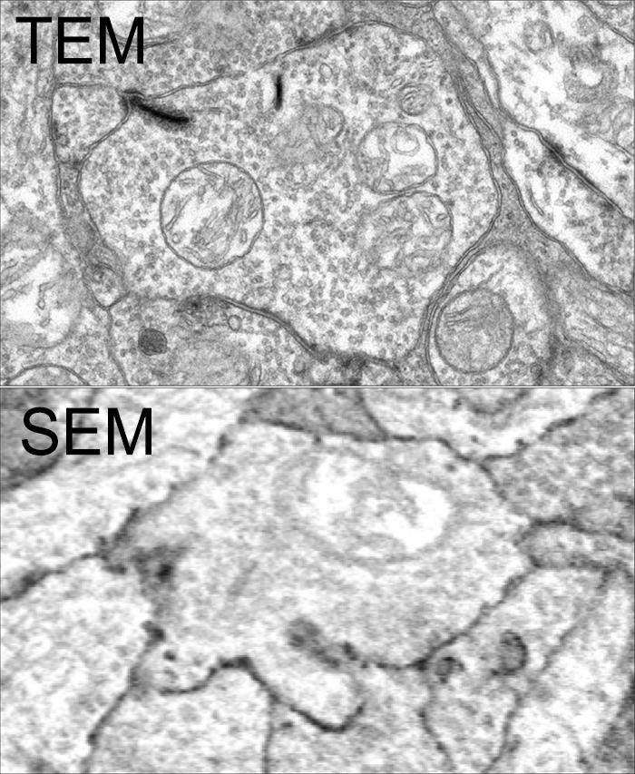
SEM vs. TEM is a tradeoff of convenience, resolution, cost and speed. The very physics of SEM signal integration means that the fundamental acquisition time for large canonical volume datasets are incompatible with 5 year grant cycles. SEM based approaches can potentially rival TEM, but dwell time/pixel increases logarithmically with resolution.

To give you some idea for the resolution differences at routine capture speeds, both of these above images capture a region within the inner plexiform layer of retina, looking at bipolar cell terminals. The TEM image was captured at a standard operating resolution of 2nm/pixel. The SEM image was captured at 16nm/pixel. You cannot see any gap junctions that might be present in the SEM image and you can only infer or guess at synaptic ribbons. And look at the texture!

You *can* get better resolution with SEM, but as I said before, the capture time increases logarithmically. To accomplish what we perform in 8-10 hours with a TEM, would take 108-115 hours on a current, cutting edge multi beam SEM. There are many other advantages of TEM including the ability to capture higher resolution images faster, be able to re-image in goniometric tilt series, be able to integrate molecular markers inside connectome volumes, and a TEM is about 1/3rd the cost of an SEM. Also, SEM images tend to be texturally poor as they are made from capturing electron backscatter of surfaces rather than made by projection of electrons through a small volume, and there is tremendous value in the texture of ultrastructural images. Ergo, this is why we use TEM.

This is not to say that SEM is not a great tool. It is just not the best tool for large scale connectomics where you have to have the resolution to capture all synapses and gap junctions, over large areas. For smaller volumes that do not require a canonical sampling of cell classes, SEM is absolutely an appropriate tool.

This content was originally published on Jonesblog.

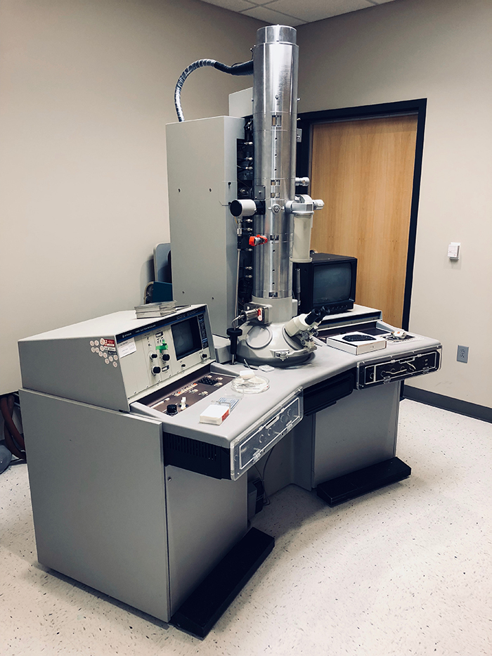
Hitachi H-600 Transmission Electron Microscope Retired

We are retiring our Hitachi H-600 Transmission Electron Microscope to make room for a new JEOL (@JEOLUSA) replacement to keep company with our other workhorse JEOL JEM-1400.  I have mixed feelings about retiring this microscope as this is the system we originally developed the first code to mosaic and register images and image slices for our connectomics work.

This fully functional and well cared for microscope will be made available through the University of Utah Surplus and Salvage as an auction if you are interested in bidding on it.  Contact me: bryan dot jones at m dot cc dot utah dot edu or @BWJones if you are interested in it.

Synapse Classification And Localization In Electron Micrographs

Synapse-classification_

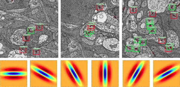
We have a new publication, Synapse Classification And Localization In Electron Micrographs in Pattern Recognition Letters.  Authors are: Vignesh JagadeeshJames Anderson, Bryan W. JonesRobert MarcSteven Fisher and B.S. Manjunath.

Abstract:  Classification and detection of biological structures in Electron Micrographs (EM) is a relatively new large scale image analysis problem. The primary challenges are in modeling diverse visual characteristics and development of scalable techniques. In this paper we propose novel methods for synapse detection and localization, an important problem in connectomics. We first propose an attribute based descriptor for characterizing synaptic junctions. These descriptors are task specific, low dimensional and can be scaled across large image sizes. Subsequently, techniques for fast localization of these junctions are proposed. Experimental results on images acquired from a mammalian retinal tissue compare favorably with state of the art descriptors used for object detection.