Tag Archives: electron microscopy

Kevin Rapp At TEM1

This is Kevin Rapp, a technician in the lab running some test captures on our original JEOL transmission electron microscope.  This microscope is likely the highest use electron microscope on planet Earth, having been run effectively non-stop for over a decade.  This microscope, purchased through a generous gift from Martha Ann Dumke Healey made possible our entire connectomics initiatives.

From Jonesblog.


Becca Pfeiffer At TEM2

This is Becca Pfeiffer setting up a test capture on our new JEOL electron microscope. We’ve customized this scope, like our previous scope, and it is taking us a little while to track down some variables with a piece of equipment this complex and hammer them down. My thanks to Becca, Jamie and Kevin for working through this together.

From Jonesblog.

Simple Super-Resolution Microscopy

Super-resolution microscopy is a pretty big thing right now.  But there is more than one way to get super-resolution microscopy results.  There are a variety of approaches, most involving expensive new microscopes that preclude many scientists from participating in science that allows them to ask certain questions.  However, if they have access to a standard transmission election microscope and have antibodies that are glutaraldehyde tolerant, they can participate and ask questions that allow them to get around some of the inherent limitations imposed by physics.

In the image above for example, we have GABA labeling in green superimposed upon ultrastructural data showing us *which* processes in the inner plexiform layer of the retina are GABAergic.  Many of these processes are smaller than the wavelength of light.

There are multiple ways to get here of course with some very expensive microscopes offering dual light and electron microscopy approaches and yet other microscopes offering purely optical based solutions.  However, this is cheap and easy and accessible to many with the basic electron microscopy resources. Robert Marc first used this approach in back in 2000, and we subsequently used it for quite a bit of work for my Ph.D. dissertation in 2003, and notably in this paper.  It is also an integral technique associated with our connectomics efforts.

That said, I’ll need at some point soon to find the resources to get a traditional optical super-resolution microscopy solution to answer some questions we have in the lab on neural degenerative disease.

(Via Jonesblog)

Retinal Remodeling And Metabolic Alterations in Human AMD

We have a new publication out (direct link, open access), Müller Cell Metabolic Chaos During Retinal Degeneration authored by Bryan W. JonesRebecca Pfeiffer, William Ferrell, Carl Watt, James Tucker, and Robert Marc.

Abstract:

Age-related macular degeneration (AMD) is a progressive retinal degeneration resulting in central visual field loss, ultimately causing debilitating blindness. AMD affects 18% of Americans from 65 to 74, 30% older than 74 years of age and is the leading cause of severe vision loss and blindness in Western populations. While many genetic and environmental risk factors are known for AMD, we currently know less about the mechanisms mediating disease progression. The pathways and mechanisms through which genetic and non-genetic risk factors modulate development of AMD pathogenesis remain largely unexplored. Moreover, current treatment for AMD is palliative and limited to wet/exudative forms. Retina is a complex, heterocellular tissue and most retinal cell classes are impacted or altered in AMD. Defining disease and stage-specific cytoarchitectural and metabolic responses in AMD is critical for highlighting targets for intervention. The goal of this article is to illustrate cell types impacted in AMD and demonstrate the implications of those changes, likely beginning in the retinal pigment epithelium (RPE), for remodeling of the the neural retina. Tracking heterocellular responses in disease progression is best achieved with computational molecular phenotyping (CMP), a tool that enables acquisition of a small molecule fingerprint for every cell in the retina. CMP uncovered critical cellular and molecular pathologies (remodeling and reprogramming) in progressive retinal degenerations such as retinitis pigmentosa (RP). We now applied these approaches to normal human and AMD tissues mapping progression of cellular and molecular changes in AMD retinas, including late-stage forms of the disease.

2-nm Resolution Anatomy of Retinal Neuro-Glial-Vascular Architecture

This abstract was presented today, May 2th at the 2016 Association for Research in Vision and Opthalmology (ARVO) meetings in Seattle, Washington by Jefferson R. Brown, Rebecca L. Pfeiffer, Crystal Sigulinsky, Felix Vazquez-Chona, Daniel Emrich, Bryan W. Jones, Robert E. Marc.

Abstract Number: 995

Author Block: Jefferson R. Brown, Rebecca L. Pfeiffer, Crystal Sigulinsky, Felix Vazquez-Chona, Daniel Emrich, Bryan W. Jones, Robert E. Marc
1 Dept of Ophthalmology, University of Utah, Salt Lake City, Utah, United States

Disclosure Block:Jefferson R. Brown, None; Rebecca L. Pfeiffer, None; Crystal Sigulinsky, None; Felix Vazquez-Chona, None; Daniel Emrich, None; Bryan W. Jones, None; Robert E. Marc, Signature Immunologics (Code I (Personal Financial Interest) )

Purpose:Retinal vasculature is strongly affected by degenerative pathologies and in turn, may also contribute to their progression. However, much of what we understand about the normal, healthy interaction between neurons, glia, and blood vessels at the ultrastructural level is limited to single section electron microscopy. The technology of serial section transmission electron microscopy (ssTEM) extends the high definition of TEM imaging into three dimensions to create volumes, allowing for more thorough visualization and analysis of the vascular-glial-neuronal complex.

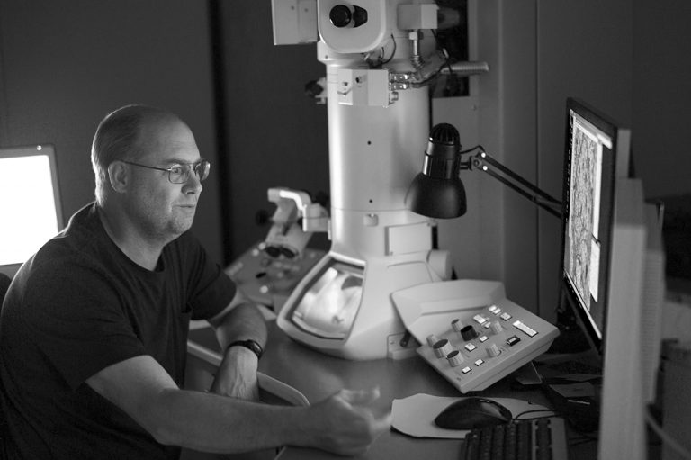
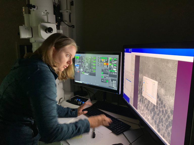
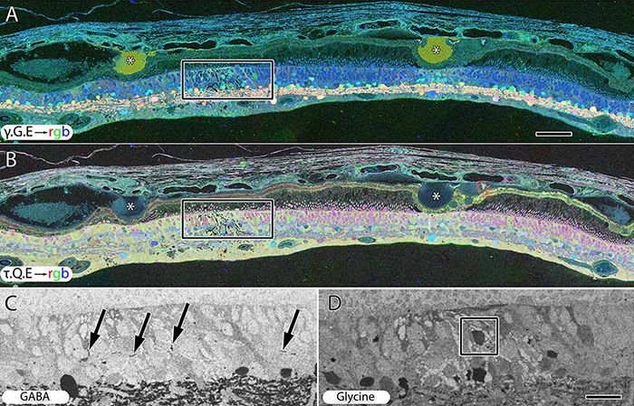
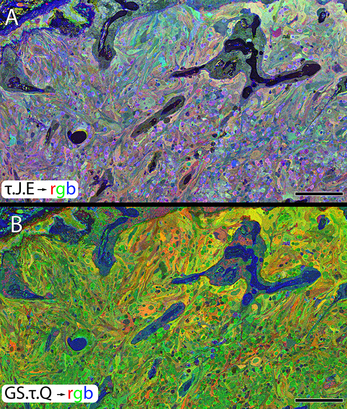
Methods:RC2 is a 40TB ssTEM volume of over 1,400 horizontal sections of retinal tissue derived from an adult female C57BL/6J mouse. The tissue sample is 250 um in diameter and spans the outer nuclear layer to the vitreal surface. Baseline resolution is 2.18nm per pixel. Visualization, navigation and metadata annotations of the database are made via the Viking software suite.

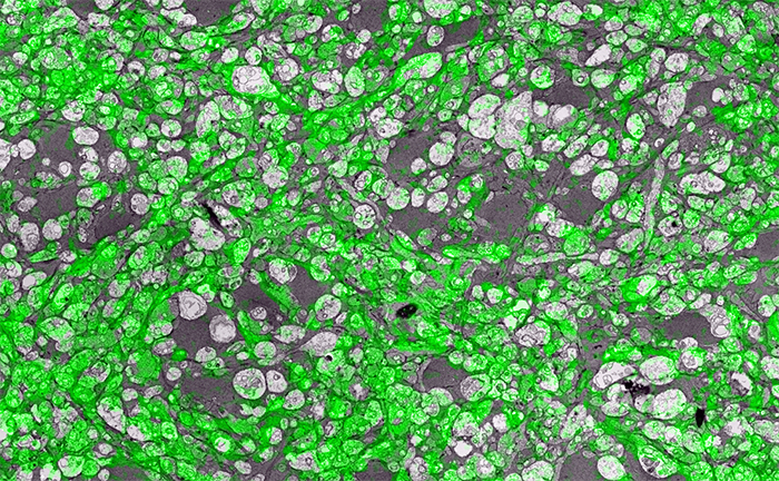
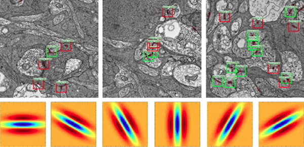
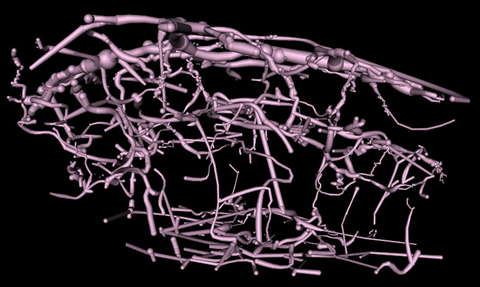
Results:Much of the retinal vascular basement membrane directly contacts Muller cells. In the ganglion cell layer, direct basement membrane contact with astrocytes is frequent. Microglia commonly contact the basement membrane, and occasionally direct contact of neurons onto basement membrane was observed. Full 3D reconstruction of all vascular pathways with associated endothelia and pericytes within the volume was completed, demonstrating that all the retinal capillary layers are continuous with one another [Figure].

Conclusions:The presence of occasional direct neuronal contact onto vascular basement membrane supports earlier work by Ochs and colleagues (2000) and suggests the blood-retina barrier does not universally involve retinal glia. However, since such contacts are extremely sparse, it remains to be seen whether this finding has biologic significance, though their existence suggests significance. The RC2 volume is a valuable resource to aid in discovery of defining characteristics of wild type neurovascular architecture.


The intro figure is a side view of reconstruction of all vasculature within the RC2 volume. Vessels at the top of the figure correspond to the outer plexiform layer, while those at the bottom correspond to the ganglion cell layer. This capillary plexus is one continuous structure. Visualization by VikingView software.

A Targeted Inhibitor Of The Alternative Complement Pathway Accelerates Recovery From Smoke-Induced Ocular Injury

We have a new publication out, A Targeted Inhibitor Of The Alternative Complement Pathway Accelerates Recovery From Smoke-Induced Ocular Injury authored by Alex Woodell, Bryan W. Jones, Tucker Williamson, Gloriane Schnabolk, Stephen Tomlinson, Carl Atkinson and Bärbel Rohrer.

Abstract:
PURPOSE. Morphological and genetic evidence exists that an overactive complement system driven by the complement alternative pathway (CAP) is involved in pathogenesis of age- related macular degeneration (AMD). Smoking is the only modifiable risk factor for AMD. As we have shown that smoke-related ocular pathology can be prevented in mice that lack an essential activator of CAP, we ask here whether this pathology can be reversed by increasing inhibition in CAP.

METHODS. Mice were exposed to either cigarette smoke (CS) or filtered air (6 hours/day, 5 days/week, 6 months). Smoke-exposed animals were then treated with the CAP inhibitor (CR2-fH) or vehicle control (PBS) for 3 months. Spatial frequency and contrast sensitivity were assessed by optokinetic response paradigms at 6 and 9 months; additional readouts included assessment of retinal morphology by electron microscopy (EM) and gene expression analysis by quantitative PCR.

RESULTS. The CS mice treated with CR2-fH showed significant improvement in contrast threshold compared to PBS-treated mice, whereas spatial frequency was unaffected by CS or pharmacological intervention. Treatment with CR2-fH in CS animals reversed thinning of the retina observed in PBS-treated mice as analyzed by spectral-domain optical coherence tomography, and reversed most morphological changes in RPE and Bruch’s membrane seen in CS animals by EM.

CONCLUSIONS. Taken together, these findings suggest that CAP inhibitors not only prevent, but have the potential to accelerate, the clearance of complement-mediated ocular injury. Improving our understanding of the regulation of the CAP pathway is paramount to developing novel treatment approaches for AMD.

Retinal Remodeling in Human Retinitis Pigmentosa

We have a new publication out (Direct Link, Free Open Access), Retinal Remodeling in Human Retinitis Pigmentosa authored by Bryan W. Jones, Rebecca Pfeiffer, Drew Ferrell, Carl Watt, Michael Marmor and Robert Marc.

Abstract: Retinitis Pigmentosa (RP) in the human is a progressive, currently irreversible neural degenerative disease usually caused by gene defects that disrupt the function or architecture of the photoreceptors. While RP can initially be a disease of photoreceptors, there is increasing evidence that the inner retina becomes progressively disorganized as the outer retina degenerates. These alterations have been extensively described in animal models, but remodeling in humans has not been as well characterized. This study, using computational molecular phenotyping (CMP) seeks to advance our understanding of the retinal remodeling process in humans. We describe cone mediated preservation of overall topology, retinal reprogramming in the earliest stages of the disease in retinal bipolar cells, and alterations in both small molecule and protein signatures of neurons and glia. Furthermore, while Müller glia appear to be some of the last cells left in the degenerate retina, they are also one of the first cell classes in the neural retina to respond to stress which may reveal mechanisms related to remodeling and cell death in other retinal cell classes. Also fundamentally important is the finding that retinal network topologies are altered. Our results suggest interventions that presume substantial preservation of the neural retina will likely fail in late stages of the disease. Even early intervention offers no guarantee that the interventions will be immune to progressive remodeling. Fundamental work in the biology and mechanisms of disease progression are needed to support vision rescue strategies.

Synapse Classification And Localization In Electron Micrographs

Synapse-classification_

We have a new publication, Synapse Classification And Localization In Electron Micrographs in Pattern Recognition Letters.  Authors are: Vignesh JagadeeshJames Anderson, Bryan W. JonesRobert MarcSteven Fisher and B.S. Manjunath.

Abstract:  Classification and detection of biological structures in Electron Micrographs (EM) is a relatively new large scale image analysis problem. The primary challenges are in modeling diverse visual characteristics and development of scalable techniques. In this paper we propose novel methods for synapse detection and localization, an important problem in connectomics. We first propose an attribute based descriptor for characterizing synaptic junctions. These descriptors are task specific, low dimensional and can be scaled across large image sizes. Subsequently, techniques for fast localization of these junctions are proposed. Experimental results on images acquired from a mammalian retinal tissue compare favorably with state of the art descriptors used for object detection.