Tag Archives: Müller cell

Müller Cell Connectomics In Health And Disease

This talk was presented today, April 25th at the 2023 Association for Research in Vision and Opthalmology (ARVO) meetings in New Orleans, Louisiana by Rebecca Pfeiffer as part of an ARVO Minisymposium Bryan William Jones organized.

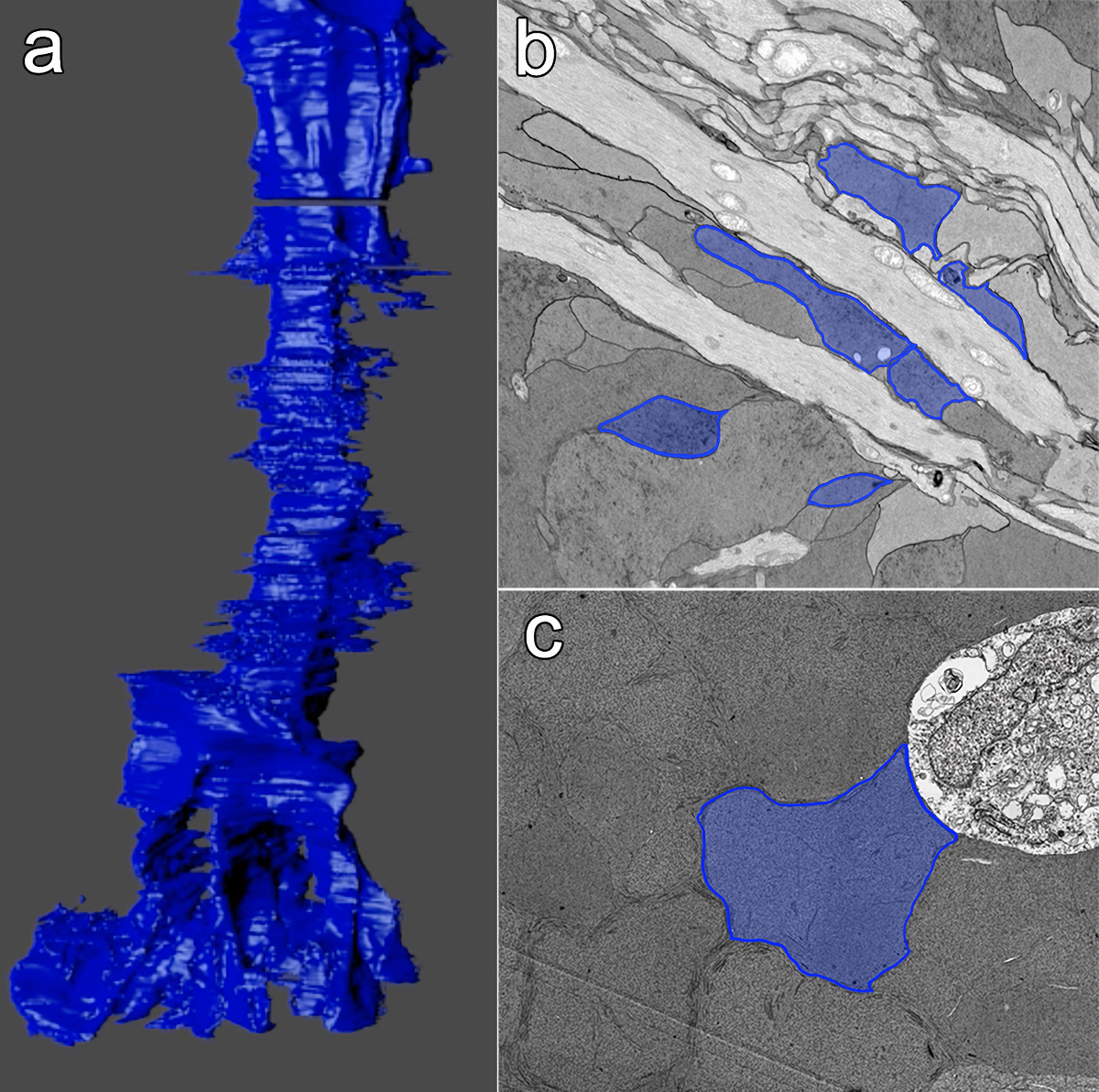
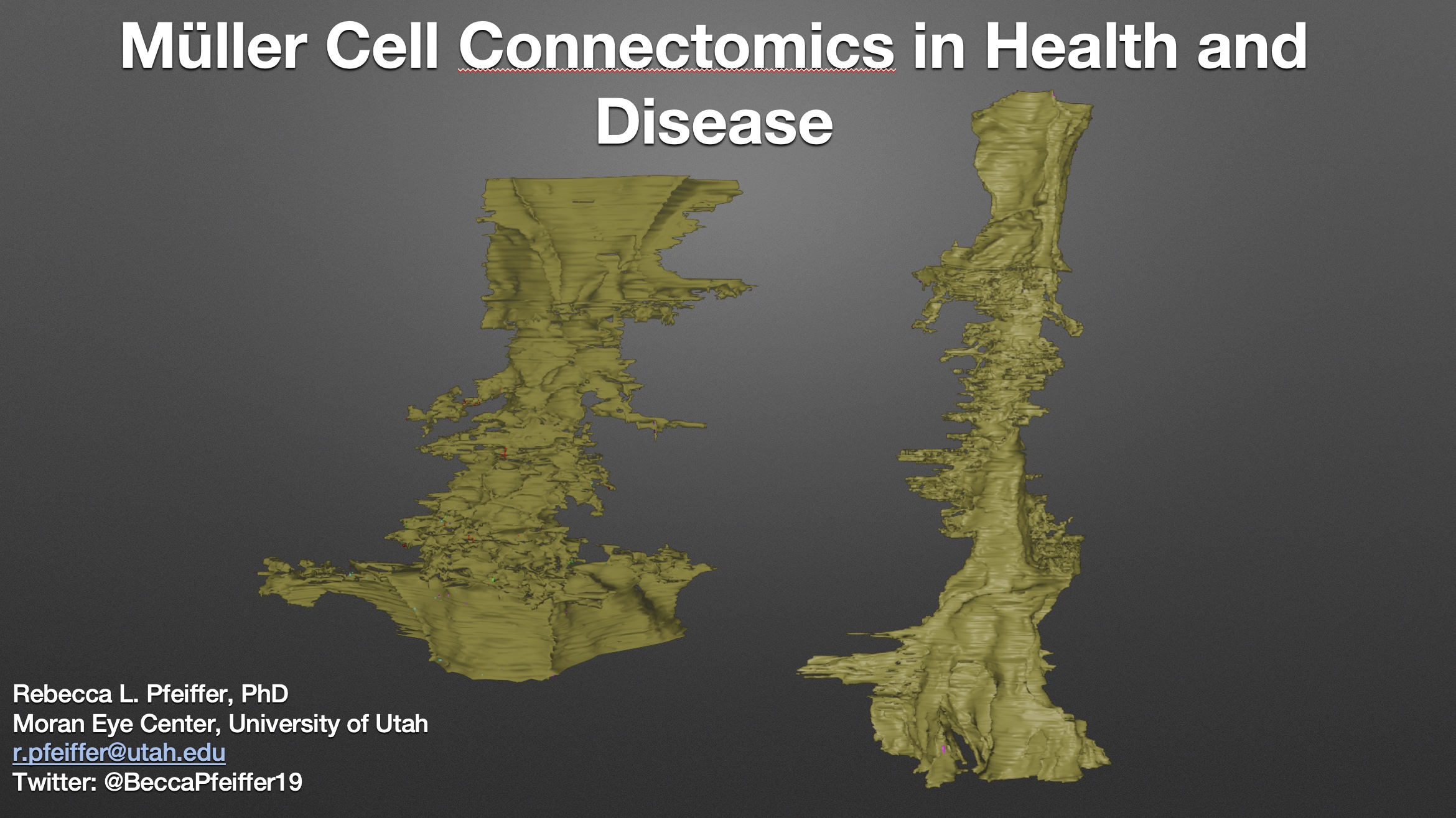
Abstract: Muller cells are a critical component of retinal function and rapidly change metabolically and morphologically in retinal disease. Of Muller cell functions, many require close physical relationships between the Muller cell and the synapses of the neurons they support. Despite this required neuro-glial relationship, little is known about the direct contacts between Muller cells and synapses in healthy or diseased retinas. In order to address this, I use a connectomics/pathoconnectomics approach to reconstruct Muller cells and their neighboring synapses. The retinas evaluated are from a healthy rabbit, retinal connectome 1 (RC1), and from the P347L rabbit model of retinitis pigmentosa, retinal pathoconnectome 1 (RPC1). Preliminary data demonstrate an increase in endfoot entanglement in RPC1 when compared with RC1, and direct synaptic contact analysis of both connectomes is ongoing.

Pathoconnectome Analysis of Müller Cells in Early Retinal Remodeling

We have a new manuscript out in Clinical Neurophysiology, An Update on Retinal Prostheses. PubMedDirect Link PDF here.

Authors: Rebecca L Pfeiffer, James R Anderson, Daniel P Emrich, Jeebika Dahal, Crystal L Sigulinsky, Hope AB Morrison, Jia-Hui Yang, Carl B Watt, Kevin D Rapp, Mineo Kondo, Hiroko Terasaki, Jessica C Garcia, Robert E Marc, and Bryan W Jones.

Abstract: Glia play important roles in neural function, including but not limited to amino acid recycling, ion homeostasis, glucose metabolism, and waste removal. During retinal degeneration and subsequent retinal remodeling, Müller cells (MCs) are the first cells to show metabolic and morphological alterations in response to stress. Metabolic alterations in MCs chaotically progress in retina undergoing photoreceptor degeneration; however, what relationship these alterations have with neuronal stress, synapse maintenance, or glia-glia interactions is currently unknown. The work described here reconstructs a MC from a pathoconnectome of early retinalremodeling retinalpathoconnectome 1 (RPC1) and explores relationships between MC structural and metabolic phenotypes in the context of neighboring neurons and glia. Here we find variations in intensity of osmication inter- and intracellularly, variation in small molecule metabolic content of MCs, as well as morphological alterations of glial endfeet. RPC1 provides a framework to analyze these relationships in early retinal remodeling through ultrastructural reconstructions of both neurons and glia. These reconstructions, informed by quantitative metabolite labeling via computational molecular phenotyping (CMP), allow us to evaluate neural-glial interactions in early retinal degeneration with unprecedented resolution and sensitivity.


Retinal Remodeling And Metabolic Alterations in Human AMD

We have a new publication out (direct link, open access), Müller Cell Metabolic Chaos During Retinal Degeneration authored by Bryan W. JonesRebecca Pfeiffer, William Ferrell, Carl Watt, James Tucker, and Robert Marc.

Abstract:

Age-related macular degeneration (AMD) is a progressive retinal degeneration resulting in central visual field loss, ultimately causing debilitating blindness. AMD affects 18% of Americans from 65 to 74, 30% older than 74 years of age and is the leading cause of severe vision loss and blindness in Western populations. While many genetic and environmental risk factors are known for AMD, we currently know less about the mechanisms mediating disease progression. The pathways and mechanisms through which genetic and non-genetic risk factors modulate development of AMD pathogenesis remain largely unexplored. Moreover, current treatment for AMD is palliative and limited to wet/exudative forms. Retina is a complex, heterocellular tissue and most retinal cell classes are impacted or altered in AMD. Defining disease and stage-specific cytoarchitectural and metabolic responses in AMD is critical for highlighting targets for intervention. The goal of this article is to illustrate cell types impacted in AMD and demonstrate the implications of those changes, likely beginning in the retinal pigment epithelium (RPE), for remodeling of the the neural retina. Tracking heterocellular responses in disease progression is best achieved with computational molecular phenotyping (CMP), a tool that enables acquisition of a small molecule fingerprint for every cell in the retina. CMP uncovered critical cellular and molecular pathologies (remodeling and reprogramming) in progressive retinal degenerations such as retinitis pigmentosa (RP). We now applied these approaches to normal human and AMD tissues mapping progression of cellular and molecular changes in AMD retinas, including late-stage forms of the disease.

Müller Cell Metabolic Chaos During Retinal Degeneration

We have a new publication out (direct link, open access), Müller Cell Metabolic Chaos During Retinal Degeneration authored by Rebecca PfeifferRobert Marc, Mineo Kondo, Hiroko Terasaki and Bryan W. Jones.

Abstract:

Müller cells play a critical role in retinal metabolism and are among the first cells to demonstrate metabolic changes in retinal stress or disease. The timing, extent, regulation, and impacts of these changes are not yet known. We evaluated metabolic phenotypes of Müller cells in the degenerating retina.

Retinas harvested from wild-type (WT) and rhodopsin Tg P347L rabbits were fixed in mixed aldehydes and resin embedded for computational molecular phenotyping (CMP). CMP facilitates small molecule fingerprinting of every cell in the retina, allowing evaluation of metabolite levels in single cells.

CMP revealed signature variations in metabolite levels across Müller cells from TgP347L retina. In brief, neighboring Müller cells demonstrated variability in taurine, glutamate, glutamine, glutathione, glutamine synthetase (GS), and CRALBP. This variability showed no correlation across metabolites, implying the changes are functionally chaotic rather than simply heterogeneous. The inability of any clustering algorithm to classify Müller cell as a single class in the TgP347L retina is a formal proof of metabolic variability in the present in degenerating retina.

Although retinal degeneration is certainly the trigger, Müller cell metabolic alterations are not a coherent response to the microenvironment. And while GS is believed to be the primary enzyme responsible for the conversion of glutamate to glutamine in the retina, alternative pathways appear to be unmasked in degenerating retina. Somehow, long term remodeling involves loss of Müller cell coordination and identity, which has negative implications for therapeutic interventions that target neurons alone.

Retinal Remodeling in Human Retinitis Pigmentosa

We have a new publication out (Direct Link, Free Open Access), Retinal Remodeling in Human Retinitis Pigmentosa authored by Bryan W. Jones, Rebecca Pfeiffer, Drew Ferrell, Carl Watt, Michael Marmor and Robert Marc.

Abstract: Retinitis Pigmentosa (RP) in the human is a progressive, currently irreversible neural degenerative disease usually caused by gene defects that disrupt the function or architecture of the photoreceptors. While RP can initially be a disease of photoreceptors, there is increasing evidence that the inner retina becomes progressively disorganized as the outer retina degenerates. These alterations have been extensively described in animal models, but remodeling in humans has not been as well characterized. This study, using computational molecular phenotyping (CMP) seeks to advance our understanding of the retinal remodeling process in humans. We describe cone mediated preservation of overall topology, retinal reprogramming in the earliest stages of the disease in retinal bipolar cells, and alterations in both small molecule and protein signatures of neurons and glia. Furthermore, while Müller glia appear to be some of the last cells left in the degenerate retina, they are also one of the first cell classes in the neural retina to respond to stress which may reveal mechanisms related to remodeling and cell death in other retinal cell classes. Also fundamentally important is the finding that retinal network topologies are altered. Our results suggest interventions that presume substantial preservation of the neural retina will likely fail in late stages of the disease. Even early intervention offers no guarantee that the interventions will be immune to progressive remodeling. Fundamental work in the biology and mechanisms of disease progression are needed to support vision rescue strategies.