Tag Archives: AII amacrine cell

Species-Specific Connectivity In The Aii Connectome

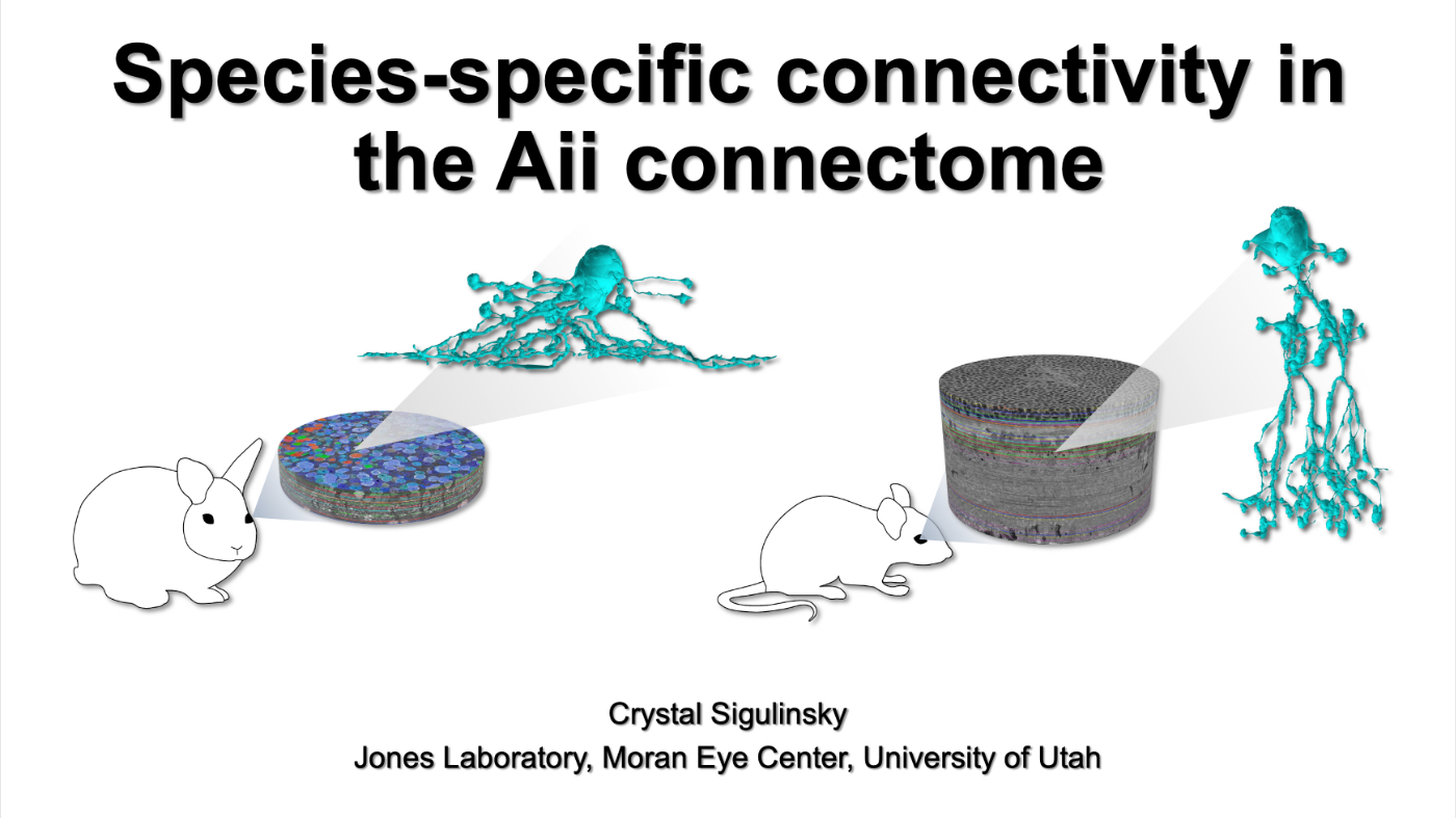
This talk was presented today, April 25th at the 2023 Association for Research in Vision and Opthalmology (ARVO) meetings in New Orleans, Louisiana by Crystal Sigulinsky as part of an ARVO Minisymposium organized by Bryan William Jones.

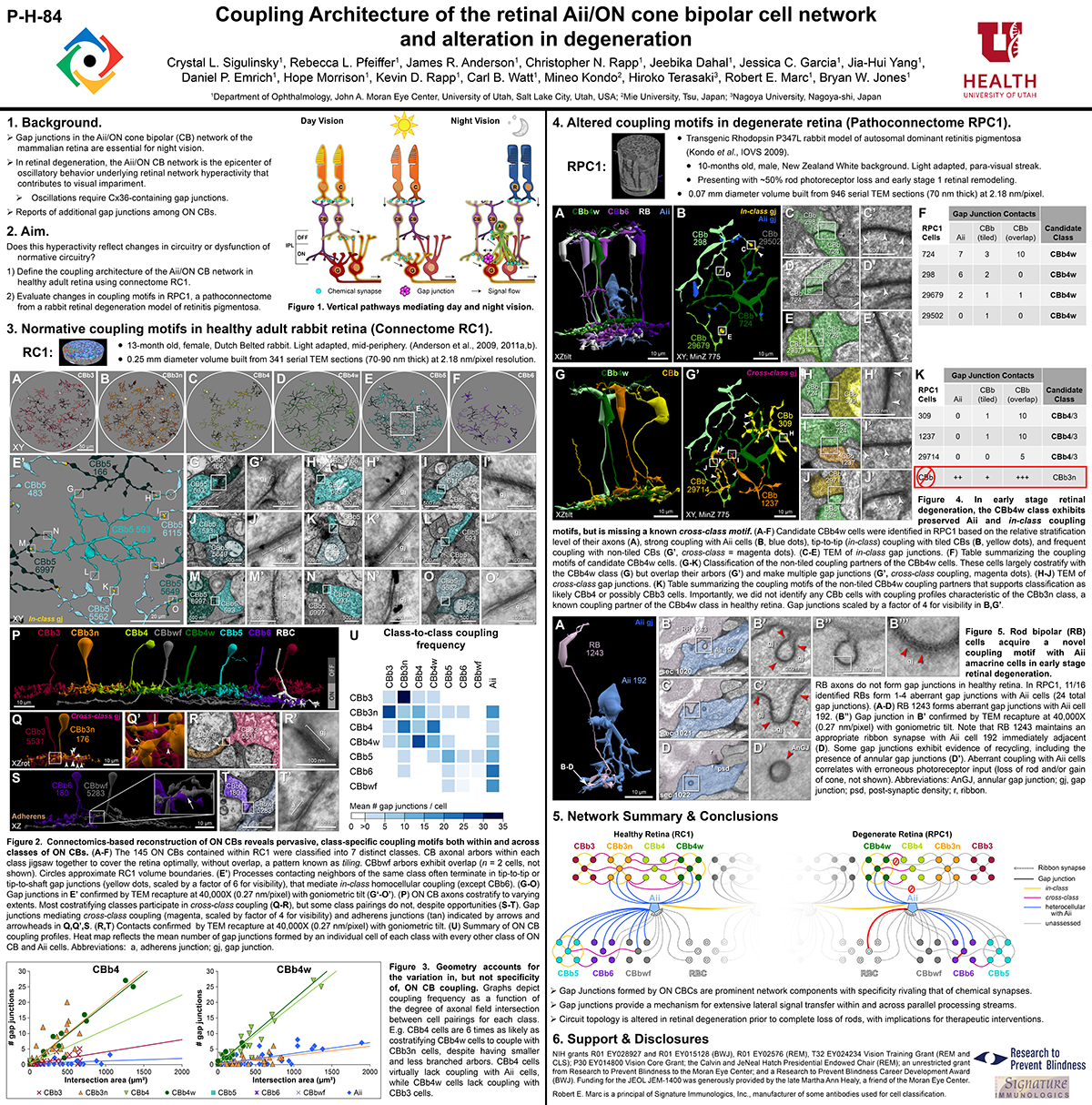
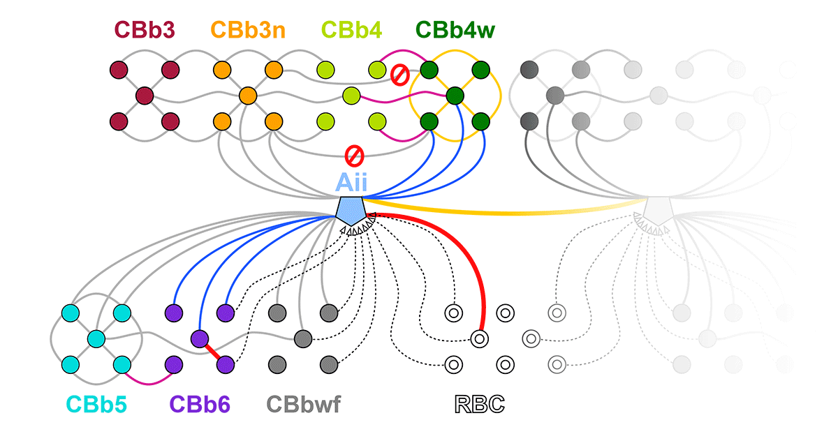
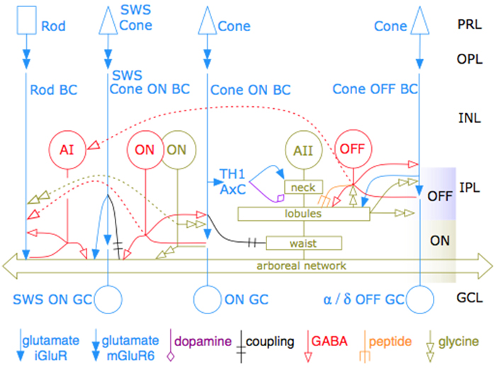
Abstract: Biomedical research relies heavily on animal models to study human disease and develop therapeutics. Understanding the architectural diversity in neural networks between humans and these model species is essential for choosing a relevant study model and interpreting conflicting results. Using comparative connectomics, we sought to map and compare the local neural network architecture of rabbit and mouse retinal Aii amacrine cells. This specialized narrow-field, multistratified, glycinergic interneuron has critical feedforward and feedback roles in both the photopic and scotopic retinal networks spanning the ON and OFF pathways, making it an ideal candidate for investigating species-specific differences in retinal networks. High-resolution, serial-section transmission electron microscopy (TEM) volumes of rabbit (RC1: female, 13- month, Dutch Belted) and mouse (RC2: female, 5-month, C57BL/6J) retinal tissue provided spatially-registered synaptic maps of Aii connectivity at directly comparable resolution and completeness. These reveal that despite species-specific morphologies, gross synaptology and compartmentalization appear conserved. Yet, rabbit and mouse Aii cells diverge in the weighting of their partnerships, most notably in their coupling profiles. Opposing biases in gap junction partnerships and their respective sizing rules indicate a greater relative output by mouse Aii cells to ON pathways than in rabbit. However, a unique topological conformation for a subset of conventional presynapses formed by Aii cell lobular dendrites with species-specific features and prevalence may influence signal output to specific partner classes within the OFF pathway and either nullify or exacerbate this difference in ON/OFF output. Additionally, rabbit Aii cells in RC1 showed greater Aii-Aii coupling than in mouse, which may suggest greater signal-to-noise compensation. Lastly, preliminary data suggest mouse Aii cells receive greater excitatory, but not inhibitory input/feedback from the OFF pathway than in rabbit. Together these data indicate that precise neural circuit architectures diverge between species and require detailed, comprehensive mapping to begin to dissect potential influence on signal flow.

Network Architecture of Gap Junctional Coupling among Parallel Processing Channels in the Mammalian Retina

We have a new manuscript out in The Journal of Neuroscience, Network Architecture of Gap Junctional Coupling among Parallel Processing Channels in the Mammalian Retina.

Authors: Crystal L. Sigulinsky @CLSigulinsky, James R. Anderson, Ethan Kerzner @EthanKerzner, Christopher N. Rapp @ChrisNRapp, Rebecca L. Pfeiffer @BeccaPfeiffer19, Taryn M. Rodman, Daniel P. Emrich, Kevin D. Rapp, Noah T. Nelson @nooneelseinhere, J. Scott Lauritzen, Miriah Meyer@miriah_meyer, Robert E. Marc @robertmarc60, and Bryan W. Jones @BWJones.

Abstract: Gap junctions are ubiquitous throughout the nervous system, mediating critical signal transmission and integration, as well as emergent network properties. In mammalian retina, gap junctions within the Aii amacrine cell-ON cone bipolar cell (CBC) network are essential for night vision, modulation of day vision, and contribute to visual impairment in retinal degenerations, yet neither the extended network topology nor its conservation is well established. Here, we map the network contribution of gap junctions using a high-resolution connectomics dataset of an adult female rabbit retina. Gap junctions are prominent synaptic components of ON CBC classes, constituting 5%–25% of all axonal synaptic contacts. Many of these mediate canonical transfer of rod signals from Aii cells to ON CBCs for night vision, and we find that the uneven distribution of Aii signals to ON CBCs is conserved in rabbit, including one class entirely lacking direct Aii coupling. However, the majority of gap junctions formed by ON CBCs unexpectedly occur between ON CBCs, rather than with Aii cells. Such coupling is extensive, creating an interconnected network with numerous lateral paths both within, and particularly across, these parallel processing streams. Coupling patterns are precise with ON CBCs accepting and rejecting unique combinations of partnerships according to robust rulesets. Coupling specificity extends to both size and spatial topologies, thereby rivaling the synaptic specificity of chemical synapses. These ON CBC coupling motifs dramatically extend the coupled Aii-ON CBC network, with implications for signal flow in both scotopic and photopic retinal networks during visual processing and disease.

Coupling architecture of the retinal Aii/ON cone bipolar cell network and alteration in degeneration

This poster was presented today, July 28th at the 2019 International Gap Junction Conference in Victoria, Canada by Crystal L. Sigulinsky, Rebecca L. PfeifferJames R. Anderson, Christopher Rapp, Jeebika Dahal, Jessica C Garcia, Jia-Hui Yang, Daniel P. Emrich, Hope Morrison, Kevin D. Rapp, Carl B. Watt, Mineo Kondo, Hiroko Terasaki, Robert E. Marc and Bryan W. Jones.

Almost full resolution version here.

Authors:

Crystal L Sigulinsky1, Rebecca L Pfeiffer1, James R. Anderson1, Christopher N. Rapp1, Jeebika Dahal1, Jessica C Garcia1, Jia-Hui Yang1, Daniel P. Emrich1, Hope Morrison1, Kevin D. Rapp1, Carl B. Watt1, Mineo Kondo2, Hiroko Terasaki3, Robert E. Marc1, Bryan W. Jones1
1Moran Eye Center/ Ophthalmology, University of Utah, Salt Lake City, Utah, United States; 2Mie University, Tsu, Japan; 3Nagoya University, Nagoya-shi, Japan;

ABSTRACT:

Background and aim:
Gap junctions are prevalent throughout the neural retina, with expression by every major neuronal class and at every level of signal processing. Yet, the functional roles and expressing cells/participating networks for many remain unknown. Spontaneous network spontaneous hyperactivity observed during retinal degeneration contributes to visual impairment and requires gap junctional coupling in the Aii amacrine cell/ON cone bipolar cell (CBC) network.  However, it remains unclear whether this hyperactivity reflects changes in the underlying circuitry or dysfunction of the normative circuitry. Here, we used connectomics-based mapping of retinal circuitry to 1) define the coupling architecture of the Aii/ON CBC network in healthy adult rabbit retina using connectome RC1 and 2) evaluate changes in coupling motifs in RPC1, a pathoconnectome from a rabbit retinal degeneration model.

Methods:

RC1 and RPC1 are connectomes built by automated transmission electron microscopy at ultrastructural (2 nm/pixel) resolution. RC1 is a 0.25 mm diameter volume of retina from a 13-month old, light adapted female Dutch Belted rabbit. RPC1 is a 0.07 mm diameter volume of degenerate retina from a transgenic P347L model of autosomal dominant retinitis pigmentosa (10-months old, male, New Zealand White background) presenting with ~50% rod loss. ON CBCs, Aii amacrine cells, and their coupling partners were annotated using the Viking application. Coupling motifs and features were explored with 3D rendering and network graph visualization. Gap junctions were validated by 0.25 nm resolution recapture with goniometric tilt when necessary.

Results:

Complete reconstruction of 37 ON CBCs in RC1 yielded 1339 gap junctions and revealed pervasive in- and cross-class coupling motifs among ON CBCs that produce complex network topologies within the coupled Aii network. Robust rulesets underlie class-specific coupling profiles with specificity defined beyond geometric opportunity. These coupling profiles enabled classification of all 145 ON CBCs contained within RC1 into 7 distinct classes. In RPC1, two ON CBC classes appear to retain their class-specific coupling profiles, accepting and rejecting specific combinations of Aii and ON CBC class partnerships. However, aberrant partnerships exist, including both loss of motifs and acquisition of novel ones.

Conclusions:

Gap junctions formed by ON CBCs are prominent network components, with specificity rivaling that of chemical synapses. These gap junctions not only subserve canonical signal transfer for night vision, but also extensive coupling within and across the parallel processing streams. Clearly aberrant morphological and synaptic changes exist in RPC1, including changes in the coupling specificity of both Aii and ON CBCs. Thus, circuit topology is altered prior to complete loss of rods, with substantial implications for therapeutic interventions for blinding diseases that depend upon the surviving retinal network.

Coupling Architecture Of The AII/ON Cone Bipolar Cell Network In Degenerate Retina

This abstract was presented today, April 8th at the 2019 Association for Research in Vision and Opthalmology (ARVO) meetings in Vancouver, Canada as a platform presentation by Crystal L. Sigulinsky, Rebecca L. PfeifferJames R. Anderson, Daniel P. Emrich, Christopher Rapp, Jeebika Dahal, Jessica Garcia, Hope Morrison, Kevin D. Rapp, Jia-Hui Yang, Carl B. Watt, Robert E. Marc and Bryan W. Jones.

Purpose
In mouse models of retinal degeneration, connexin36-containing gap junctions in the Aii amacrine cell network appear to mediate aberrant hyperactivity within the retina. However, it remains unclear whether this hyperactivity reflects changes in the underlying circuitry or dysfunction of the normative circuitry. Our connectomics-based mapping of retinal circuitry in rabbit Retinal Connectome 1 (RC1) has dramatically expanded the coupled Aii network. In addition to canonical Aii-to-Aii and Aii-to-ON cone bipolar cell (CBC) coupling, we describe pervasive in- and cross-class coupling motifs among ON CBCs. This study examines the changes in these coupling motifs in RPC1, an ultrastructural retinal pathoconnectome from a rabbit model of retinitis pigmentosa.

Methods
RC1 and RPC1 are connectomes built by automated transmission electron microscopy at ultrastructural (2 nm/pixel) resolution. RC1 is a 0.25 mm diameter volume of retina from a 13 month old, light-adapted female Dutch Belted rabbit and serves as the healthy reference connectome. RPC1 is a 0.09 mm diameter volume of pathological retina from a 10 month old, male transgenic P347L model of autosomal dominant retinitis pigmentosa showing early phase 1 retinal remodeling, when rod photoreceptors are still present, but stressed. ON CBCs, Aii amacrine cells, and their coupling partners were annotated using the Viking application. Coupling motifs and features were explored with 3D rendering and graph visualization of connectivity. Gap junctions were validated by 0.25 nm resolution recapture with goniometric tilt when necessary.

Results
All major coupling motifs were observed. Several ON CBC classes retained their class-specific coupling profiles, accepting and rejecting specific combinations of Aii and ON CBC class partnerships. However, aberrant partnerships exist in the coupled network, including both loss of prominent motifs and acquisition of novel ones.

Conclusions
Clearly aberrant morphological and synaptic changes exist in RPC1, including changes in the coupling specificity and gap junction distributions of both Aii amacrine cells and ON CBCs. This indicates that the Aii/ON CBC circuit topology is already altered during early phase 1 remodeling, with substantial implications for therapeutic interventions for blinding diseases that depend upon the surviving retinal network in human patients.

Comparative Anatomy And Connectivity Of The AII Amacrine Cell In Mouse And Rabbit Retina

This abstract was presented today, April 8th at the 2019 Association for Research in Vision and Opthalmology (ARVO) meetings in Vancouver, Canada by Selena Wirthlin, Crystal L. SigulinskyJames R. Anderson, Daniel P. Emrich, Christopher Rapp, Jeebika Dahal, Rebecca L. Pfeiffer, Kevin D. Rapp, Jia-Hui Yang, Carl B. Watt, Robert E. Marc and Bryan W. Jones.

Full resolution version here.

Purpose
Mouse retina structurally differs from rabbit retina, as it is thicker and vascularized, while the rabbit retina is thinner and avascular. The implications of these differences on neuronal morphology and connectivity is not known. This project compares the morphology and connectivity of the Aii amacrine cell (AC) with ultrastructural precision in connectomes of mouse (RC2) and rabbit (RC1) retina.

Methods
RC1 and RC2 are connectomes built by automated transmission electron microscopy at ultrastructural (2 nm/pixel) resolution. RC1 and RC2 are 0.25mm diameter volumes of retina. RC1 is from a 13 month old, female Dutch Belted rabbit. RC2 is from a 5 month old female C57BL/6J mouse. The Viking application was used to annotate Aii ACs in both connectomes.

Results
Mouse Aii ACs are noticeably elongated to span the thicker inner plexiform layer (IPL) and have a prominent neck region. Lobular appendages of Aii ACs in both species extend thin stalks from the soma, neck and proximal arboreal dendrites in the OFF sublamina, predominantly forming reciprocal synapses with OFF cone bipolar cells (BCs). In rabbits, multiple arboreal dendrites emerge from the base of the neck, branch and travel obliquely through the ON sublamina, and form gap junctions with ON cone BCs, neighbor Aii ACs, and itself. They extend laterally at the base of the IPL, collecting ribbon input from rod BCs. In contrast, mouse arboreal dendrites stem from a single primary dendrite that branches as it travels vertically through the IPL without self-branch interaction, terminating at variable depths that align with the more broadly ramified axon terminals of rod BCs. Conventional synapse to gap junction ratios reveal greater output in the OFF vs ON layer in mouse compared to rabbit. Notably, mouse Aii ACs form gap junctions with the descending axons of ON cone BCs as they pass its soma, in contrast to rabbit, where gap junctions do not form at contacts proximal to ON cone BC axon terminals.

Conclusions
Lateral expansion of rabbit Aii ACs may be attributable to eccentricity. However, morphological differences appear to mediate greater output to the OFF versus ON pathway in mouse. Synaptic partners are currently being analyzed. Comparative anatomy connectomics is essential for understanding possible implications of retinal structure on neuronal morphology and connectivity that may underlie network differences between the mouse and rabbit retina.

The AII Amacrine Cell Connectome: A Dense Network Hub

AII-connectome

We have a new publication in Frontiers in Neuroscience, The AII Amacrine Cell Connectome: A Dense Network Hub.  Authors are Robert E. MarcJames R. Anderson, Bryan W. Jones, Crystal Sigulinsky and J. Scott Lauritzen.

Abstract:  The mammalian AII retinal amacrine cell is a narrow-field, multistratified glycinergic neuron best known for its role in collecting scotopic signals from rod bipolar cells and distributing them to ON and OFF cone pathways in a crossover network via a combination of inhibitory synapses and heterocellular AII::ON cone bipolar cell gap junctions. Long considered a simple cell, a full connectomics analysis shows that AII cells possess the most complex interaction repertoire of any known vertebrate neuron, contacting at least 28 different cell classes, including every class of retinal bipolar cell. Beyond its basic role in distributing rod signals to cone pathways, the AII cell may also mediate narrow-field feedback and feedforward inhibition for the photopic OFF channel, photopic ON-OFF inhibitory crossover signaling, and serves as a nexus for a collection of inhibitory networks arising from cone pathways that likely negotiate fast switching between cone and rod vision. Further analysis of the complete synaptic counts for five AII cells shows that (1) synaptic sampling is normalized for anatomic target encounter rates; (2) qualitative targeting is specific and apparently errorless; and (3) that AII cells strongly differentiate partner cohorts by synaptic and/or coupling weights. The AII network is a dense hub connecting all primary retinal excitatory channels via precisely weighted drive and specific polarities. Homologs of AII amacrine cells have yet to be identified in non-mammalians, but we propose that such homologs should be narrow-field glycinergic amacrine cells driving photopic ON-OFF crossover via heterocellular coupling with ON cone bipolar cells and glycinergic synapses on OFF cone bipolar cells. The specific evolutionary event creating the mammalian AII scotopic-photopic hub would then simply be the emergence of large numbers of pure rod bipolar cells.