Tag Archives: Robert Marc

Store-Operated Calcium Entry In Müller Glia Is Controlled By Synergistic Activation Of TRPC And Orai Channels

We have a new publication out as collaborators with colleages, Store-Operated Calcium Entry In Müller Glia Is Controlled By Synergistic Activation Of TRPC And Orai Channels authored by Tünde Molnár, Oleg Yarishkin, Peter Barabas, Anthony Iuso, Bryan W. Jones, Robert Marc, Tam Phuong, and David Krizaj.

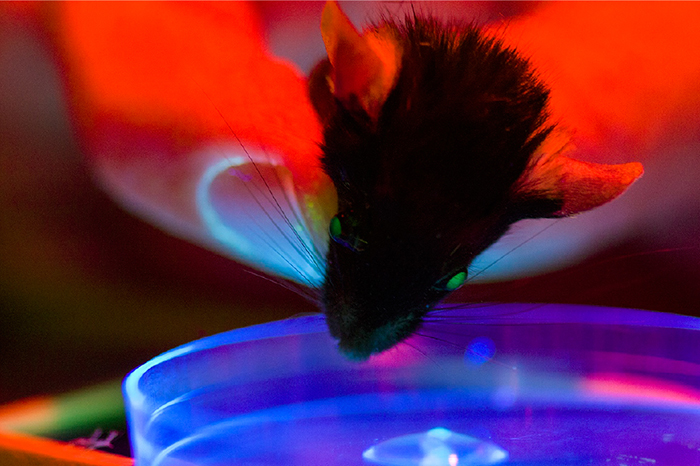
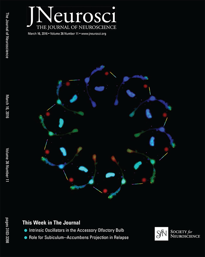
Bonus, we got the cover!  The image was created by Tam Phuong.


Significance: Store-operated Ca2+ signaling represents a major signaling pathway and source of cytosolic Ca2+ in astrocytes. Here, we show that the store-operated response in Müller cells, radial glia that perform key structural, signaling, osmoregulatory and mechanosensory functions within the retina, is mediated through synergistic activation of TRPC and Orai channels. The endfoot disproportionately expresses the depletion sensor STIM1, contains an extraordinarily high density of ER cisternae that shadow neuronal, astrocyte, vascular and axonal structures, interface with mitochondria but also originates SOCE-induced transcellular Ca2+ waves that propagate glial excitation into the proximal retina. These results identify a molecular mechanism that underlies complex interactions between the plasma membrane and calcium stores and contributes to radial glial function, regulation and response to mechanical stress.

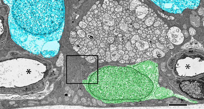
Abstract: The endoplasmic reticulum (ER) is at the epicenter of astrocyte Ca2+ signaling. We sought to identify the molecular mechanism underlying store-operated calcium entry (SOCE) that repletes ER Ca2+ stores in mouse Müller cells. Store depletion, induced through blockade of sequestration transporters in Ca2+-free saline, induced synergistic activation of canonical transient receptor potential (TRPC1) and Orai channels. Store-operated TRPC1 channels were identified by their electrophysiological properties, pharmacological blockers and ablation of the Trpc1 gene. ICRAC (Ca2+ release-activated) currents were identified by ion permeability, voltage-dependence and sensitivity to selective Orai antagonists Synta66 and GSK7975A. Depletion-evoked calcium influx was initiated at the Müller endfoot and apical process, triggering centrifugal propagation of Ca2+ waves into the cell body. EM analysis of the endfoot compartment showed high-density ER cisternae that shadow retinal ganglion cell (RGC) somata and axons, protoplasmic astrocytes, vascular endothelial cells and ER-mitochondrial contacts at the vitreal surface of the endfoot. The mouse retina expresses transcripts encoding both Stim and all known Orai genes; Müller glia predominantly express STIM1 whereas STIM2 is mainly confined to the outer plexiform and retinal ganglion cell layers. Elimination of TRPC1 facilitated Müller gliosis induced by the elevation of intraocular pressure (IOP), suggesting that TRPC channels might play a neuroprotective role during mechanical stress. These findings expand the current knowledge about store-operated signaling in astroglia, as well as calcium signaling pathways in Müller astroglia and functional roles these cells play in retinal physiology and pathology.
Webvision Chapter: Retinal Degeneration, Remodeling and Plasticity

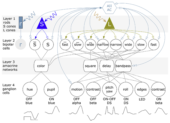
We have published a new chapter in Webvision, Retinal Degeneration, Remodeling and Plasticity that covers the history of the study of retinal degenerations and some of the implications for vision rescue.  Authors are Bryan W. Jones, Rebecca L. Pfeiffer and Robert E. Marc.  It will, like other Webvision chapters evolve over time, which is the whole point of Webvision, but we hope it will generate some discussion.

Retinal Prosthetics, Optogenetics and Photoswitches

Retinal-Prosthetics-Optogenetics-and-Photoswitches

We have a new publication, Retinal Prosthetics, Optogenetics and Photoswitches in ACS Chemical Neuroscience.  Authors are:  Robert E. MarcRebecca L. Pfeiffer, and Bryan W. Jones.

Abstract:

Three technologies have emerged as therapies to restore light sensing to profoundly blind patients suffering from late-stage retinal degenerations: (1) retinal prosthetics, (2) optogenetics, and (3) chemical photoswitches. Prosthetics are the most mature and the only approach in clinical practice. Prosthetic implants require complex surgical intervention and provide only limited visual resolution but can potentially restore navigational ability to many blind patients. Optogenetics uses viral delivery of type 1 opsin genes from prokaryotes or eukaryote algae to restore light responses in survivor neurons. Targeting and expression remain major problems, but are potentially soluble. Importantly, optogenetics could provide the ultimate in high-resolution vision due to the long persistence of gene expression achieved in animal models. Nevertheless, optogenetics remains challenging to implement in human eyes with large volumes, complex disease progression, and physical barriers to viral penetration. Now, a new generation of photochromic ligands or chemical photoswitches (azobenzene-quaternary ammonium derivatives) can be injected into a degenerated mouse eye and, in minutes to hours, activate light responses in neurons. These photoswitches offer the potential for rapidly and reversibly screening the vision restoration expected in an individual patient. Chemical photoswitch variants that persist in the cell membrane could make them a simple therapy of choice, with resolution and sensitivity equivalent to optogenetics approaches. A major complexity in treating retinal degenerations is retinal remodeling: pathologic network rewiring, molecular reprogramming, and cell death that compromise signaling in the surviving retina. Remodeling forces a choice between upstream and downstream targeting, each engaging different benefits and defects. Prosthetics and optogenetics can be implemented in either mode, but the use of chemical photoswitches is currently limited to downstream implementations. Even so, given the high density of human foveal ganglion cells, the ultimate chemical photoswitch treatment could deliver cost-effective, high-resolution vision for the blind.

Synapse Classification And Localization In Electron Micrographs

Synapse-classification_

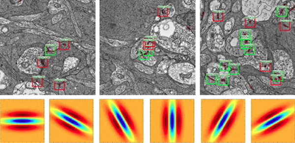
We have a new publication, Synapse Classification And Localization In Electron Micrographs in Pattern Recognition Letters.  Authors are: Vignesh JagadeeshJames Anderson, Bryan W. JonesRobert MarcSteven Fisher and B.S. Manjunath.

Abstract:  Classification and detection of biological structures in Electron Micrographs (EM) is a relatively new large scale image analysis problem. The primary challenges are in modeling diverse visual characteristics and development of scalable techniques. In this paper we propose novel methods for synapse detection and localization, an important problem in connectomics. We first propose an attribute based descriptor for characterizing synaptic junctions. These descriptors are task specific, low dimensional and can be scaled across large image sizes. Subsequently, techniques for fast localization of these junctions are proposed. Experimental results on images acquired from a mammalian retinal tissue compare favorably with state of the art descriptors used for object detection.

FASEB Bio-Art Competition Winner 2013

1254_40X_GBY_72dpi-1

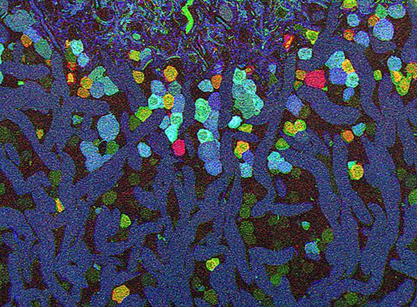
Bryan W. Jones and Robert E. Marc and were selected as 2013 FASEB BioArt Winners (Press release here).  This image shows a region of an amazingly complex retina from a goldfish (Carassius auratus auratus) analyzed using tools called Computational Molecular Phenotyping (CMP) that reveal the metabolic state of the all cell types in tissues.  These cells were labeled with antibodies for the presence of two fundamental amino acid metabolites (anti-glycine in red, anti-GABA in blue) and an amino acid tracer of physiologic activity (anti-AGB in green).   These labels allow us to visualize the metabolic state and therefore, classes of bipolaramacrine and horizontal cells.  More details on the image here.