Tag Archives: William D. Ferrell

Retinal Remodeling And Metabolic Alterations in Human AMD

We have a new publication out (direct link, open access), Müller Cell Metabolic Chaos During Retinal Degeneration authored by Bryan W. JonesRebecca Pfeiffer, William Ferrell, Carl Watt, James Tucker, and Robert Marc.

Abstract:

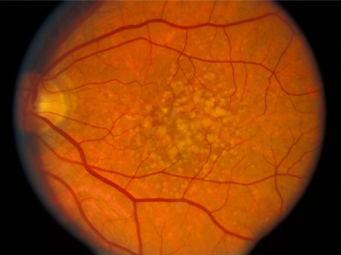
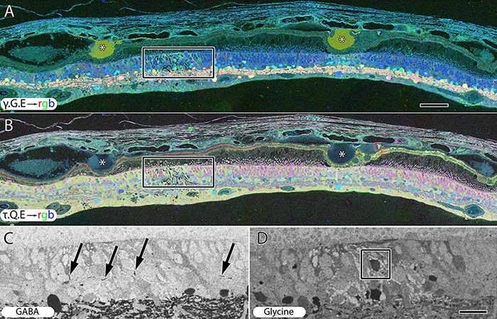
Age-related macular degeneration (AMD) is a progressive retinal degeneration resulting in central visual field loss, ultimately causing debilitating blindness. AMD affects 18% of Americans from 65 to 74, 30% older than 74 years of age and is the leading cause of severe vision loss and blindness in Western populations. While many genetic and environmental risk factors are known for AMD, we currently know less about the mechanisms mediating disease progression. The pathways and mechanisms through which genetic and non-genetic risk factors modulate development of AMD pathogenesis remain largely unexplored. Moreover, current treatment for AMD is palliative and limited to wet/exudative forms. Retina is a complex, heterocellular tissue and most retinal cell classes are impacted or altered in AMD. Defining disease and stage-specific cytoarchitectural and metabolic responses in AMD is critical for highlighting targets for intervention. The goal of this article is to illustrate cell types impacted in AMD and demonstrate the implications of those changes, likely beginning in the retinal pigment epithelium (RPE), for remodeling of the the neural retina. Tracking heterocellular responses in disease progression is best achieved with computational molecular phenotyping (CMP), a tool that enables acquisition of a small molecule fingerprint for every cell in the retina. CMP uncovered critical cellular and molecular pathologies (remodeling and reprogramming) in progressive retinal degenerations such as retinitis pigmentosa (RP). We now applied these approaches to normal human and AMD tissues mapping progression of cellular and molecular changes in AMD retinas, including late-stage forms of the disease.

Retinal Metabolic Response to Cigarette Smoke

This abstract was presented today at the Association for Research in Vision and Opthalmology (ARVO) meetings in Seattle, Washington by Alexandra D. Butler, William D. Ferrell, Alex Woodell, Carl Atkinson, Baerbel Rohrer, Robert E. Marc and Bryan W. Jones.

Purpose:  Smoking is the single largest risk factor for age-related macular degeneration, aside from age. Several of the main genetic risk factors for AMD are polymorphisms occurring in complement genes involved in the alternative, classical and common terminal pathways. To better understand the metabolic impact of smoking on the retina, we used computational molecular phenotyping (CMP) and examined the effects of cigarette smoke on wild type (wt) retinas and mice in which either the alternative pathway (complement factor B, CfB) or the common terminal pathway (complement component 3, C3) was removed.

Methods:  Mice were exposed to either cigarette smoke or filtered air. Cigarette smoke (CS) was generated using an automated cigarette-smoking machine (Model TE-10, Teague Enterprises, Davis, CA) by burning 3R4F reference cigarettes (2.45 mg nicotine per cigarette; purchased from the Tobacco Health Research Institute, University of Kentucky, Lexington, KY). Mice were exposed to CS for 6 hours/day, 5 days/week for 6 months. Age matched room filtered air exposed mice were used as controls. Eyes were enucleated immediately post-mortem, fixed in 1% paraformaldehyde, 2.5% glutaraldehyde, dehydrated in graded methanols, embedded in eponates and histologically analyzed with CMP.

Results:  Alterations in retinal small molecule signatures from mice exposed to cigarette smoke were observed compared to retinas from non-smoked mice in wt, CfB and C3 knockout mice. Signal changes with arginine, glutamine and glutathione progressively increased in the retinas of smoked exposed wt, CfB and C3 knockout mice, indicating increased response profiles to cell stress. Both Müller cells and photoreceptors of wt smoked retinas demonstrated changes relative to non- smoked retinas.

Conclusions:  Arginine, glutamine and glutathione, amino acids known to be involved in cellular stress responses, were increased in retinal neurons and glial cells upon smoke exposure. Eliminating essential components of the complement system, a cascade required for the maintenance of the immune privilege of the eye, appears to exacerbate responses to cigarette smoke in oxidative damage response related pathways. Understanding complement-dependent alterations in the eye will aid in our understanding of AMD pathology and may open new avenues for novel treatment strategies.

Support:  RPB CDA (BWJ), Thome AMD Grant (BWJ), NIH EY02576 (RM), NIH EY015128 (RM), NSF 0941717 (RM), NIH EY014800 Vision Core (RM), NIH EY019320 (BR), VA merit award RX000444 (BR), grant to MUSC from RPB