Category Archives: Daily

Simple Super-Resolution Microscopy

Super-resolution microscopy is a pretty big thing right now.  But there is more than one way to get super-resolution microscopy results.  There are a variety of approaches, most involving expensive new microscopes that preclude many scientists from participating in science that allows them to ask certain questions.  However, if they have access to a standard transmission election microscope and have antibodies that are glutaraldehyde tolerant, they can participate and ask questions that allow them to get around some of the inherent limitations imposed by physics.

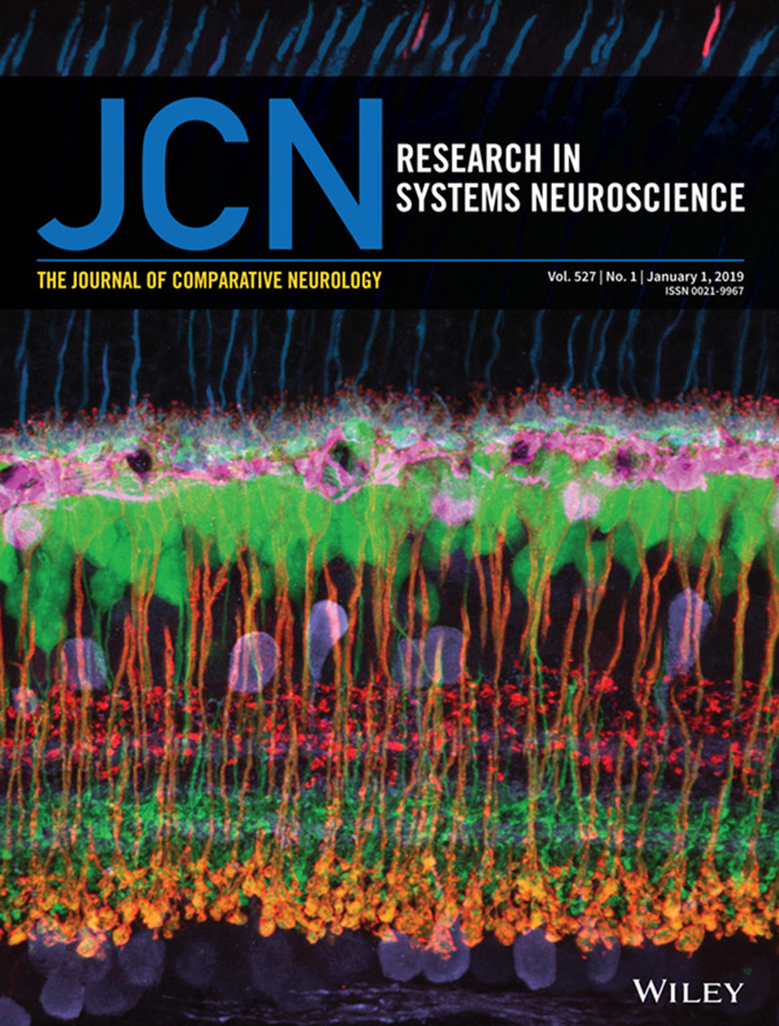
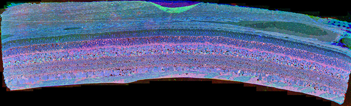
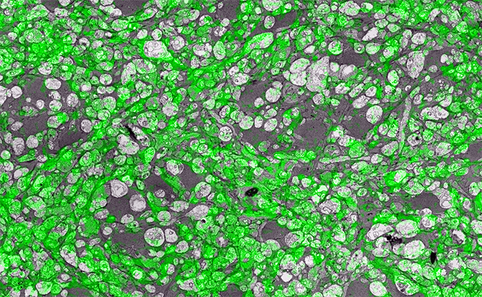
In the image above for example, we have GABA labeling in green superimposed upon ultrastructural data showing us *which* processes in the inner plexiform layer of the retina are GABAergic.  Many of these processes are smaller than the wavelength of light.

There are multiple ways to get here of course with some very expensive microscopes offering dual light and electron microscopy approaches and yet other microscopes offering purely optical based solutions.  However, this is cheap and easy and accessible to many with the basic electron microscopy resources. Robert Marc first used this approach in back in 2000, and we subsequently used it for quite a bit of work for my Ph.D. dissertation in 2003, and notably in this paper.  It is also an integral technique associated with our connectomics efforts.

That said, I’ll need at some point soon to find the resources to get a traditional optical super-resolution microscopy solution to answer some questions we have in the lab on neural degenerative disease.

(Via Jonesblog)

Off to RD2018 and ISER 2018

The Marclab for Connectomics is off to RD2018 and ISER 2018 in Killarney, Ireland and Belfast, Northern Ireland.  I’ll be organizing sessions on retinal degeneration, and I’m tremendously proud of the work Dr. Crystal Sigulinsky will be presenting from her work on gap junctional connectivity in retinal degenerations and the work Dr. Rebecca Pfeiffer (@BeccaPfeiffer19) will be presenting on her work on the retinal pathoconnectome in two talks on bipolar cells and Müller cells.

SEM vs. TEM

SEM vs. TEM is a tradeoff of convenience, resolution, cost and speed. The very physics of SEM signal integration means that the fundamental acquisition time for large canonical volume datasets are incompatible with 5 year grant cycles. SEM based approaches can potentially rival TEM, but dwell time/pixel increases logarithmically with resolution.

To give you some idea for the resolution differences at routine capture speeds, both of these above images capture a region within the inner plexiform layer of retina, looking at bipolar cell terminals. The TEM image was captured at a standard operating resolution of 2nm/pixel. The SEM image was captured at 16nm/pixel. You cannot see any gap junctions that might be present in the SEM image and you can only infer or guess at synaptic ribbons. And look at the texture!

You *can* get better resolution with SEM, but as I said before, the capture time increases logarithmically. To accomplish what we perform in 8-10 hours with a TEM, would take 108-115 hours on a current, cutting edge multi beam SEM. There are many other advantages of TEM including the ability to capture higher resolution images faster, be able to re-image in goniometric tilt series, be able to integrate molecular markers inside connectome volumes, and a TEM is about 1/3rd the cost of an SEM. Also, SEM images tend to be texturally poor as they are made from capturing electron backscatter of surfaces rather than made by projection of electrons through a small volume, and there is tremendous value in the texture of ultrastructural images. Ergo, this is why we use TEM.

This is not to say that SEM is not a great tool. It is just not the best tool for large scale connectomics where you have to have the resolution to capture all synapses and gap junctions, over large areas. For smaller volumes that do not require a canonical sampling of cell classes, SEM is absolutely an appropriate tool.

This content was originally published on Jonesblog.

Nornir listed on NITRC

Nornir is now registered with NeuroImaging Tools & Resources Collaboratory (NITRC).

Nornir’s takes large sets of overlapping images in 2D and produces registered (a.k.a. aligned) 2D mosaics and 3D volumes of any size and scale. Registered slices may be exported as a single large images or viewed/annoted with our Viking viewer.

Nornir has been used successfully on transmission electron microscopy, scanning electron microscopy images, and light microscopy images. Nornir supports interleaving different imaging methods into the same volume. Support for SerialEM, Objective Imaging, and Digital Micrograph (DM4) raw data is available. Adding formats is not complicated and the author will consider requests.

Nornir runs on fairly humble hardware for the task. A 32-core 64GB Xeon system built a ~60 TB 250um diameter 2.12nm/pixel volume from roughly 1400 slices. Nornir works incrementally, only updating data that has changed.

Installation is fairly simple and primarily uses Python’s PIP installer.

For further information: http://nornir.github.io/

Hitachi H-600 Transmission Electron Microscope Retired

We are retiring our Hitachi H-600 Transmission Electron Microscope to make room for a new JEOL (@JEOLUSA) replacement to keep company with our other workhorse JEOL JEM-1400.  I have mixed feelings about retiring this microscope as this is the system we originally developed the first code to mosaic and register images and image slices for our connectomics work.

This fully functional and well cared for microscope will be made available through the University of Utah Surplus and Salvage as an auction if you are interested in bidding on it.  Contact me: bryan dot jones at m dot cc dot utah dot edu or @BWJones if you are interested in it.

Congratulations Dr. Kerzner

Congratulations to Dr. Ethan Kerzner who successfully defended his PhD dissertation in the Viz Design Lab at the University of Utah’s Scientific Computing Institute.

Ethan’s work has been instrumental in helping us to understanding complex gap junctional networks in our retinal connectomics initiatives.  His Graffinity software package allowed us to explore multivariate graphs, and pull out complex relationships of neurons and gap junctions that would not have been easily possible with other approaches.

Ethan is now off to Google X, and we wish him the very best and look forward to many more interactions in the future.