Jessica Garcia Is This Year’s Student Veteran Of The Year

Jessica Garcia is this year’s University of Utah Student Veteran of the Year. Jessica is an undergraduate student in the lab exploring the OFF-layer branches of ON cone bipolar cells in early retinal degeneration.

Jessica came to us by way of service with the US Navy, where she served as an aviation technician before attending the University of Utah.

Congratulations Jessica! We are so proud of you.

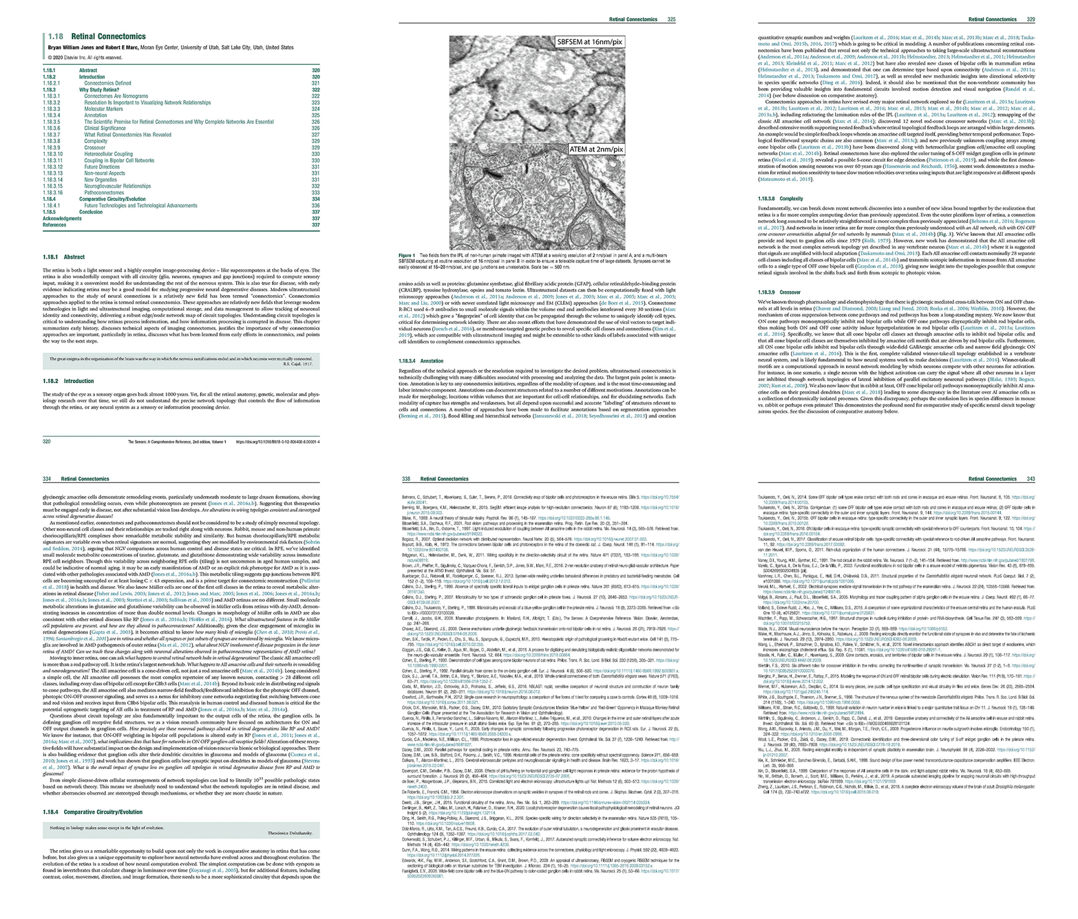
Chapter: Retinal Connectomics

We have a new chapter out in the Elsevier book series The Senses, 2021.

Authors are myself, Bryan W. Jones @BWJones and Robert E. Marc @robertmarc60.

Abstract: The retina is both a light sensor and a highly complex image-processing device – like supercomputers at the backs of eyes. The retina is also wonderfully compact with all circuitry (glia, neurons, synapses and gap junctions) required to compute sensory input, making it a convenient model for understanding the rest of the nervous system. This is also true for disease, with early evidence indicating retina may be a good model for studying progressive neural degenerative diseases. Modern ultrastructural approaches to the study of neural connections is a relatively new !eld has been termed “connectomics”. Connectomics approaches applied to the retina is termed retinal connectomics. These approaches are relatively new !elds that leverage modern technologies in light and ultrastructural imaging, computational storage, and data management to allow tracking of neuronal identity and connectivity, delivering a robust edge/node network map of circuit topologies. Understanding circuit topologies is critical to understanding how retinas process information, and how information processing is corrupted in disease. This chapter summarizes early history, discusses technical aspects of imaging connectomes, justi!es the importance of why connectomics approaches are important, particularly in retina, discusses what has been learned from early efforts in connectomics, and points the way to the next steps.

Please email me: bryan.jones@m.cc.utah.edu if you would like a pdf of the chapter.


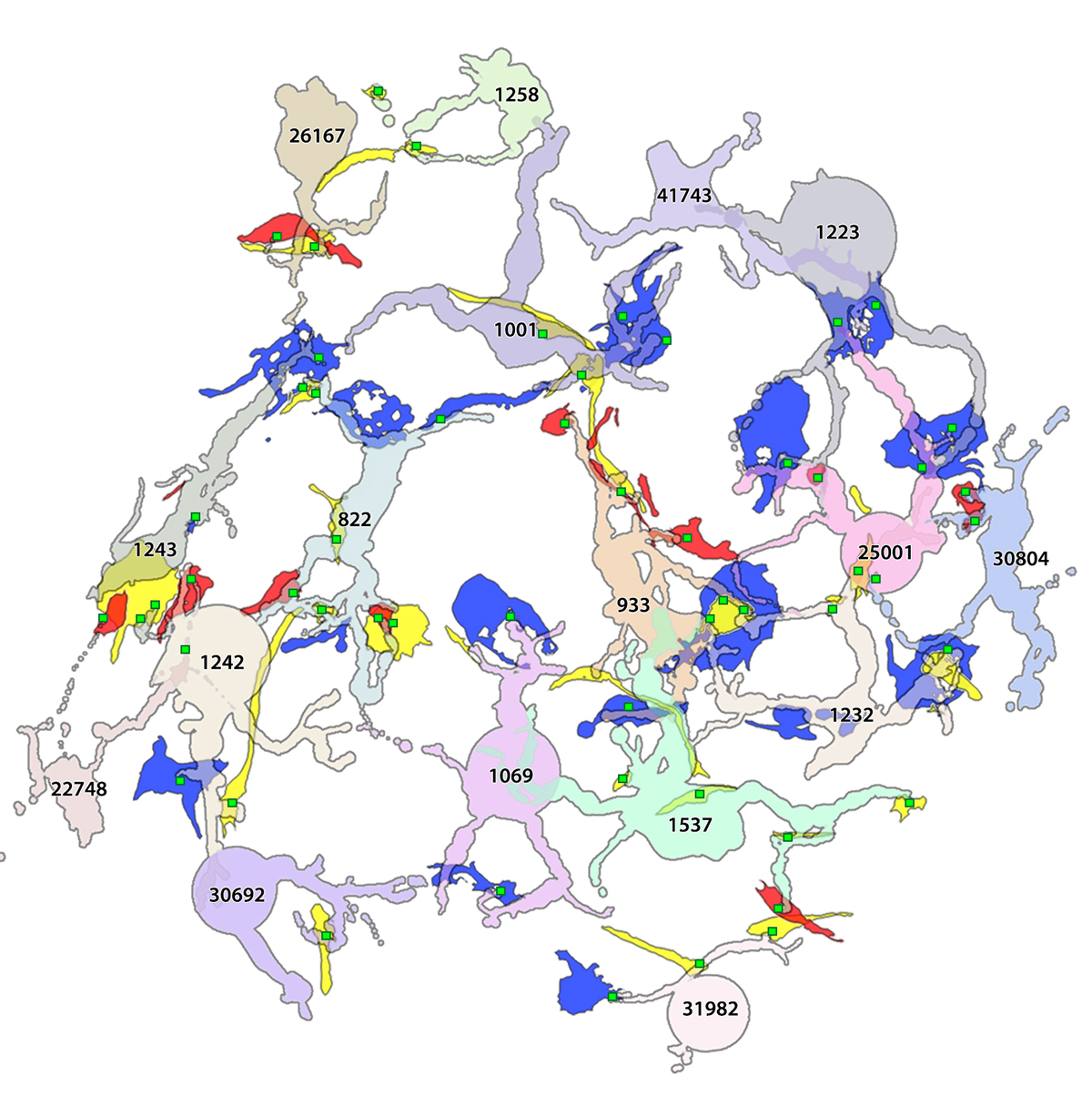
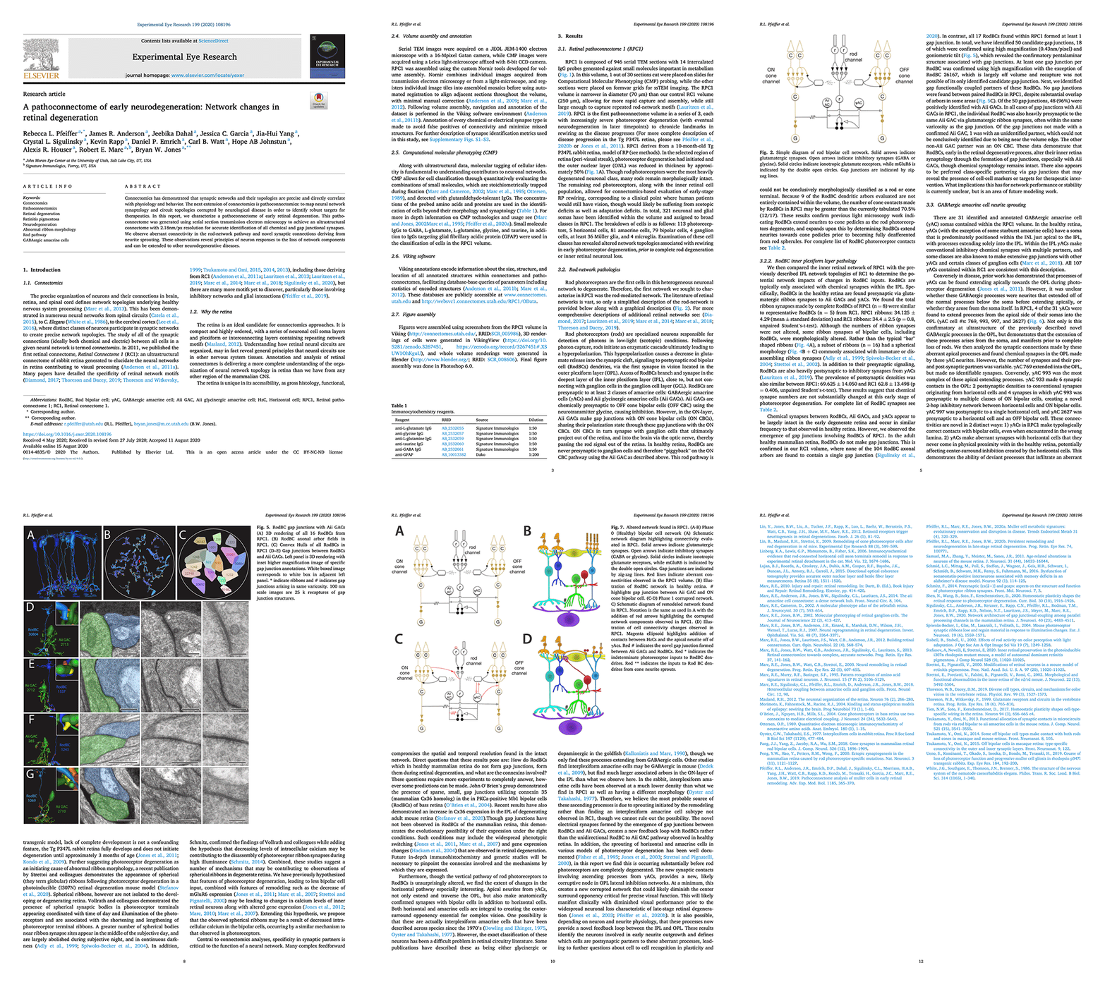
A pathoconnectome of early neurodegeneration: Network changes in retinal degeneration

We have a new manuscript out in Experimental Eye Research, A pathoconnectome of early neurodegeneration: Network changes in retinal degeneration. (pdf here)

Authors: Rebecca L. Pfeiffer @BeccaPfeiffer19, James R. Anderson, Jeebika Dahal, Jessica C. Garcia, Jia-Hui Yang, Crystal L. Sigulinsky @CLSigulinsky, Kevin Rapp, Daniel P. Emrich, Carl B. Watt, Hope AB Johnstun, Alexis R. Houser, Robert E. Marc @robertmarc60, and Bryan W. Jones @BWJones.

Abstract: Connectomics has demonstrated that synaptic networks and their topologies are precise and directly correlate with physiology and behavior. The next extension of connectomics is pathoconnectomics: to map neural network synaptology and circuit topologies corrupted by neurological disease in order to identify robust targets for therapeutics. In this report, we characterize a pathoconnectome of early retinal degeneration. This pathoconnectome was generated using serial section transmission electron microscopy to achieve an ultrastructural connectome with 2.18nm/px resolution for accurate identification of all chemical and gap junctional synapses. We observe aberrant connectivity in the rod-network pathway and novel synaptic connections deriving from neurite sprouting. These observations reveal principles of neuron responses to the loss of network components and can be extended to other neurodegenerative diseases.


NeuroNex Grant

I am pleased to report that the The Marclab for Connectomics has been funded by the National Science Foundation with a 5 year grant as part of a large, international consortium to study synaptic weighting.  We are collaborating with the Erik Jorgensen laboratory here at the University of Utah, and will be exploring synapses in a model of retinal degeneration.  There is a nice writeup of the award on the Moran Eye Center website, here.

This is a wonderful opportunity to work with other colleagues that will be funded alongside us with this grant, including Uri Manor @manorlaboratory, Davi Bock @dddavi, Josh Vogelstein @neuro_data, Viren Jain @stardazed0, and others.  My thanks to Kristen Harris for heading up this initiative.

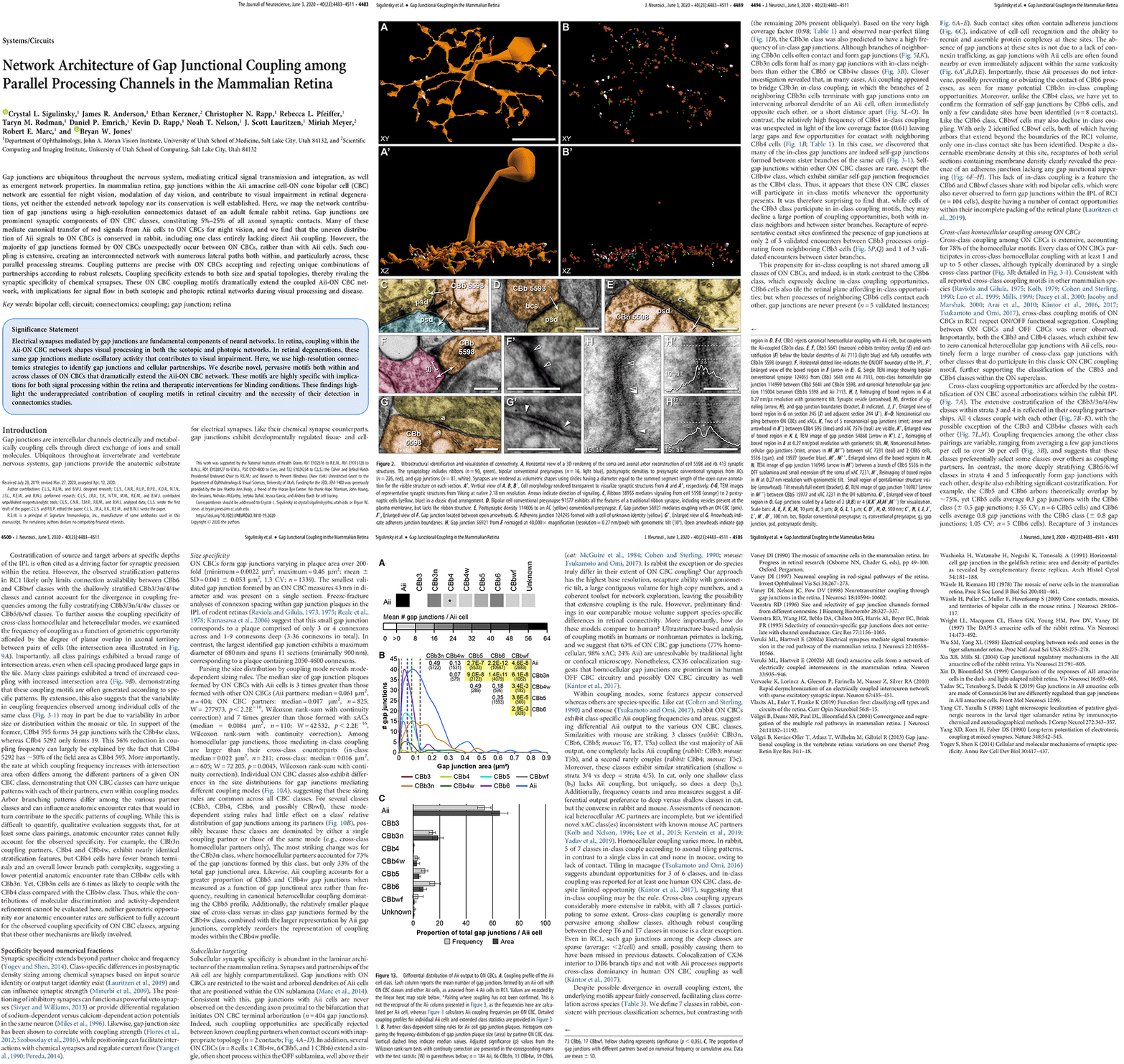
Network Architecture of Gap Junctional Coupling among Parallel Processing Channels in the Mammalian Retina

We have a new manuscript out in The Journal of Neuroscience, Network Architecture of Gap Junctional Coupling among Parallel Processing Channels in the Mammalian Retina.

Authors: Crystal L. Sigulinsky @CLSigulinsky, James R. Anderson, Ethan Kerzner @EthanKerzner, Christopher N. Rapp @ChrisNRapp, Rebecca L. Pfeiffer @BeccaPfeiffer19, Taryn M. Rodman, Daniel P. Emrich, Kevin D. Rapp, Noah T. Nelson @nooneelseinhere, J. Scott Lauritzen, Miriah Meyer@miriah_meyer, Robert E. Marc @robertmarc60, and Bryan W. Jones @BWJones.

Abstract: Gap junctions are ubiquitous throughout the nervous system, mediating critical signal transmission and integration, as well as emergent network properties. In mammalian retina, gap junctions within the Aii amacrine cell-ON cone bipolar cell (CBC) network are essential for night vision, modulation of day vision, and contribute to visual impairment in retinal degenerations, yet neither the extended network topology nor its conservation is well established. Here, we map the network contribution of gap junctions using a high-resolution connectomics dataset of an adult female rabbit retina. Gap junctions are prominent synaptic components of ON CBC classes, constituting 5%–25% of all axonal synaptic contacts. Many of these mediate canonical transfer of rod signals from Aii cells to ON CBCs for night vision, and we find that the uneven distribution of Aii signals to ON CBCs is conserved in rabbit, including one class entirely lacking direct Aii coupling. However, the majority of gap junctions formed by ON CBCs unexpectedly occur between ON CBCs, rather than with Aii cells. Such coupling is extensive, creating an interconnected network with numerous lateral paths both within, and particularly across, these parallel processing streams. Coupling patterns are precise with ON CBCs accepting and rejecting unique combinations of partnerships according to robust rulesets. Coupling specificity extends to both size and spatial topologies, thereby rivaling the synaptic specificity of chemical synapses. These ON CBC coupling motifs dramatically extend the coupled Aii-ON CBC network, with implications for signal flow in both scotopic and photopic retinal networks during visual processing and disease.

Immunization Against Oxidized Elastin Exacerbates Structural and Functional Damage in Mouse Model of Smoke-Induced Ocular Injury

We have a new manuscript out in Investigative Ophthalmology & Visual Science, Immunization Against Oxidized Elastin Exacerbates Structural and Functional Damage in Mouse Model of Smoke-Induced Ocular Injury

Authors: Balasubramaniam Annamalai; Crystal Nicholson; Nathaniel Parsons; Sarah Stephenson; Carl Atkinson; Bryan Jones; and Bärbel Rohrer.

Purpose: Age-related macular degeneration (AMD) is the leading cause of blindness in Western populations. While an overactive complement system has been linked to pathogenesis, mechanisms contributing to its activation are largely unknown. In aged and AMD eyes, loss of the elastin layer (EL) of Bruch’s membrane (BrM) has been reported. Elastin antibodies are elevated in patients with AMD, the pathogenic significance of which is unclear. Here we assess the role of elastin antibodies using a mouse model of smoke-induced ocular pathology (SIOP), which similarly demonstrates EL loss.

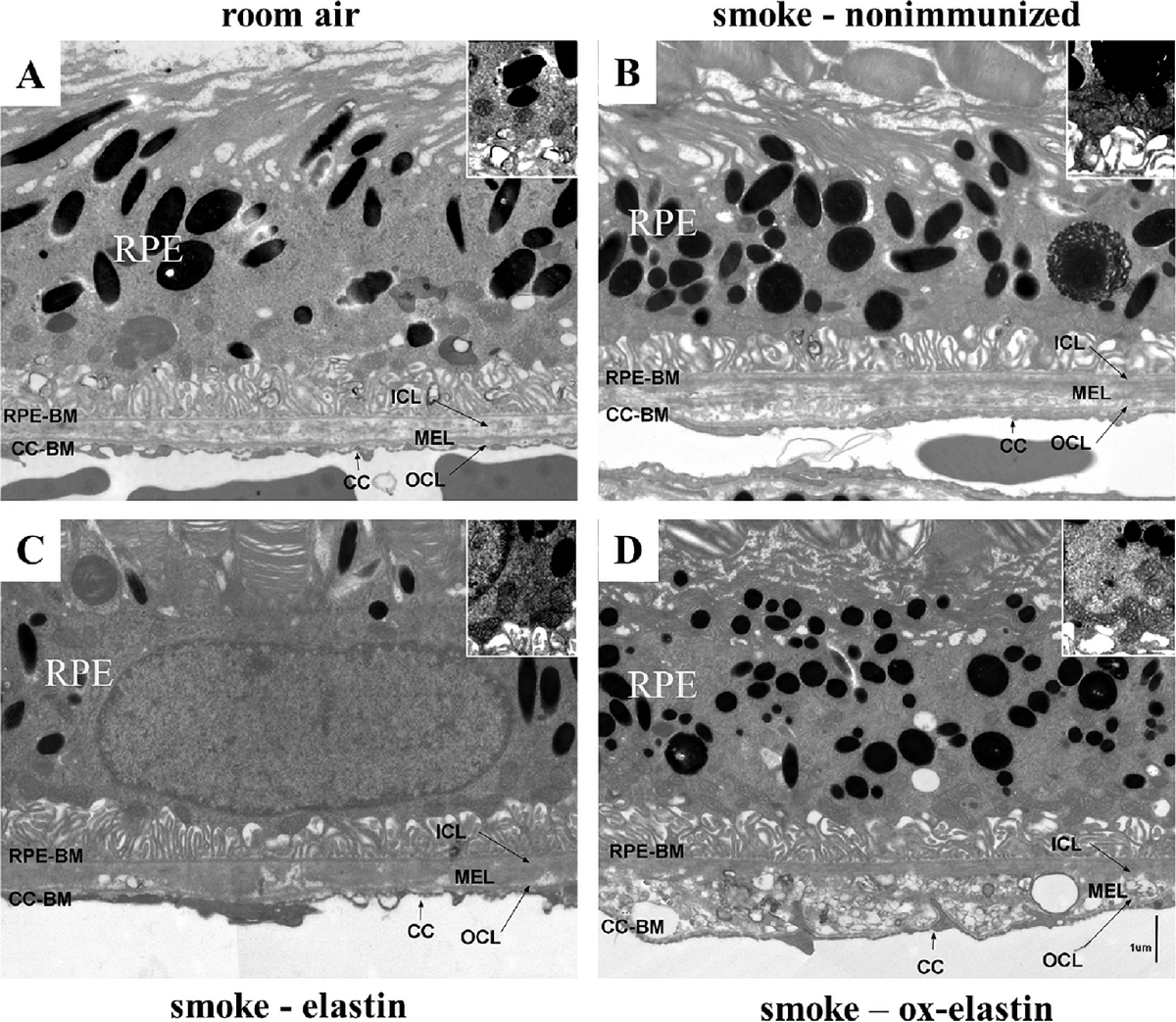
Methods: C57BL/6J mice were immunized with elastin or elastin peptide oxidatively modified by cigarette smoke (ox-elastin). Mice were then exposed to cigarette smoke or air for 6 months. Visual function was assessed by optokinetic response, retinal morphology by spectral-domain optical coherence tomography and electron microscopy, and complement activation and antibody deposition by Western blot.

Results: Ox-elastin IgG and IgM antibodies were elevated in ox-elastin immunized mice following 6 months of smoke, whereas elastin immunization had a smaller effect. Ox-elastin immunization exacerbated smoke-induced vision loss, with thicker BrM and more damaged retinal pigment epithelium (RPE) mitochondria compared with mice immunized with elastin or nonimmunized controls. These changes were correlated with increased levels of IgM, IgG2, IgG3, and complement activation products in RPE/choroid.

Conclusions: These data demonstrate that SIOP mice generate elastin-specific antibodies and that immunization with ox-elastin exacerbates ocular pathology. Elastin antibodies represented complement fixing isotypes that, together with the increased presence of complement activation seen in immunized mice, suggest that elastin antibodies exert pathogenic effects through mediating complement activation.

Teleworking In Response to COVID-19

The Marclab for Connectomics is immediately implementing teleworking strategies, moving many functions off campus in response to the COVID-19 outbreak/pandemic.

I am feeling fortunate in the much of our work is computational, but we do have a substantial wet lab component in addition to the ultrastructural data capture component.

We are not shutting down completely, yet. All students and postdocs will be working from home.  We have limited capacity to process tissue as only one person at a time will be in the laboratory proper, sectioning, performing immuno procedures, or capturing ultrastructural data.

I will be working intermittently from home and the laboratory office and working to determine where we are at from a personnel standpoint and how we will be impacted by NIH funding decisions. The good news is that NIH, I believe will honor charging salaries to grants, even if we cannot maintain our planned effort.  This means no job losses for the time being.

The COVID-19 outbreak is getting bigger and bigger.  I have cancelled all of my travel for the year to date, including a trip with friends to Lisbon in two weeks to walk around with cameras that I was planning on being the highlight of my year.  Trips to California, Italy, Sweden, and Germany are also off the table, at least through May/June.  ARVO has been cancelled. FASEB has been cancelled.

Why?

– COVID-19 is far more infectious than the flu.

– COVID-19 is far more deadly than the flu. On the order of 300-1000x more deadly. The difference is that it is not as common as the flu… yet.

-Our current Surgeon General just said on the 9th that the outbreak was already “contained”. I don’t even know where to start with this one as it is gross misinformation.

-I am seeing a level of buildup in the outbreak community and in the military that I’ve never seen before. This buildup is stymied by an administration that keeps talking it down. But I’ve never before seen a military response (calling out National Guard units specialized in biowarfare management) for an outbreak. At the same time, there is no coordination between the services that I can see. Navy is not talking with Army for instance, which is bizarre.

-The President has not invoked the Defense Production Act yet… I have no idea why.

-There is nobody at CDC that is coordinating all the information from the individual states as of today. This infrastructure *used* to exist. I don’t know why it is not there and that scares me, and makes me more angry than ever with the Trump administration.

HHS is estimating that a “moderate” outbreak will have 200,000 hospitalizations required for respiratory distress from COVID-19. We only have 100,000 ICU beds total in the United States, and some percentage of those are already in use, being optimized for markets. That looks to me like Italy’s situation right now, and their advanced medical system (among the best in Europe) is starting to fail which is why Italy has just extended their quarantine to the entire country.

-Per the Johns Hopkins dashboard, there is effectively no screening being done in Africa. No screening being done in Russia, with one backchannel report to me of at least 300 active cases in Moscow, but the doctors are not allowed to report it.

-No screening being done in the former Russian republics either.

-In all of India, there are only 43 reported cases. That does not make any sense to me.

-The lack of reporting for an infectious disease that is far more infectious than the flu indicates to me that COVID-19 is already in the population and is already spreading.

-Because it is relatively asymptomatic amongst much of the younger populace, it is not being seen/found. And as such, is a potent vector for spreading rapidly in communities.

-Universities around the country (not just my institution, the University of Utah) are prohibiting travel.

All of this means that if this is in fact as bad as it sounds, then we need to take active measures to limit the spread and the impact upon our healthcare infrastructure that is already going to be strained and we need to flatten the curve.
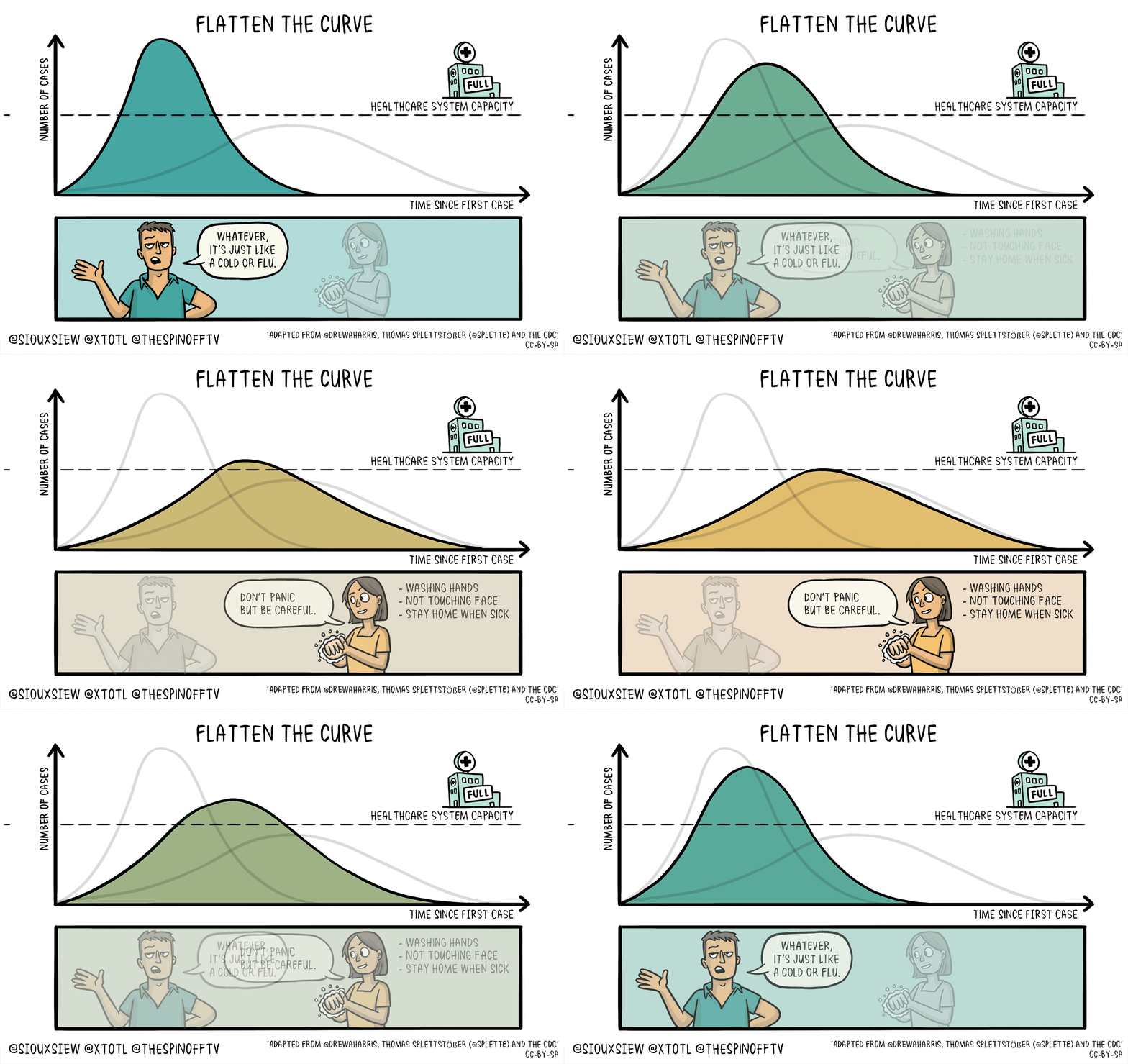
 

This *feels* very different from previous outbreaks, and the lack of coordination from the White House to CDC and .mil folks is astoundingly disturbing. Don’t forget, back in 2018, the Trump administration shut down the CDC foreign disease outbreak monitoring program.  This was the program that helped monitor foreign disease outbreaks and help define what sequences vaccine makers should use.

We have thus been relying upon WHO for information, and they even offered us primers for COVID-19 to make test kits.  But someone in the Trump administration told the CDC not to take them and we’d develop our own. My information says that those US developed primers had problems recognizing the epitope, and now people want to know *why* we rejected WHO primers. Speculation is that they wanted to use IP from the US made primers to capitalize on those markets.

This is infuriating and another example of why we should not be using economic models to address disease outbreaks, a position I’ve been taking on Twitter for some time now.

The WHO certified this outbreak as a pandemic on the 11th.  I have some real concerns, as some, like the former CDC director are advising that in a worst case scenario, up to 1.6 million Americans could be killed by COVID-19.  My concern is that from his numbers, even if this outbreak is just 1% infectious, and just moderately severe, that means 16,350 possible deaths in America.  That is not the bad part though. More importantly, 16,350 deaths will likely mean ICU beds are needed, and we only *have* 100k beds in the US.  Additionally, optimal occupancy for intensive care units (ICU) in the United States is accepted to be 70-75%. This means that at 1% infection rate, and moderately severe mortality rates, ALL of our ICU bed capacity could be occupied by COVID-19.  If it is worse, and the numbers from Italy’s example suggest it is, this could reach crisis levels quickly.

For all these reasons, I am taking active measures to reduce the exposure of people in my laboratory and help to reduce pressure on our healthcare systems.


Bryan William Jones, PhD


Müller Cell Metabolic Signatures: Evolutionary Conservation and Disruption in Disease

We have a new manuscript out in Trends in Endocrinology & Metabolism, Müller Cell Metabolic Signatures: Evolutionary Conservation and Disruption in Disease.

Authors: Rebecca L. Pfeiffer @BeccaPfeiffer19, Robert E. Marc @robertmarc60, and Bryan William Jones @BWJones.

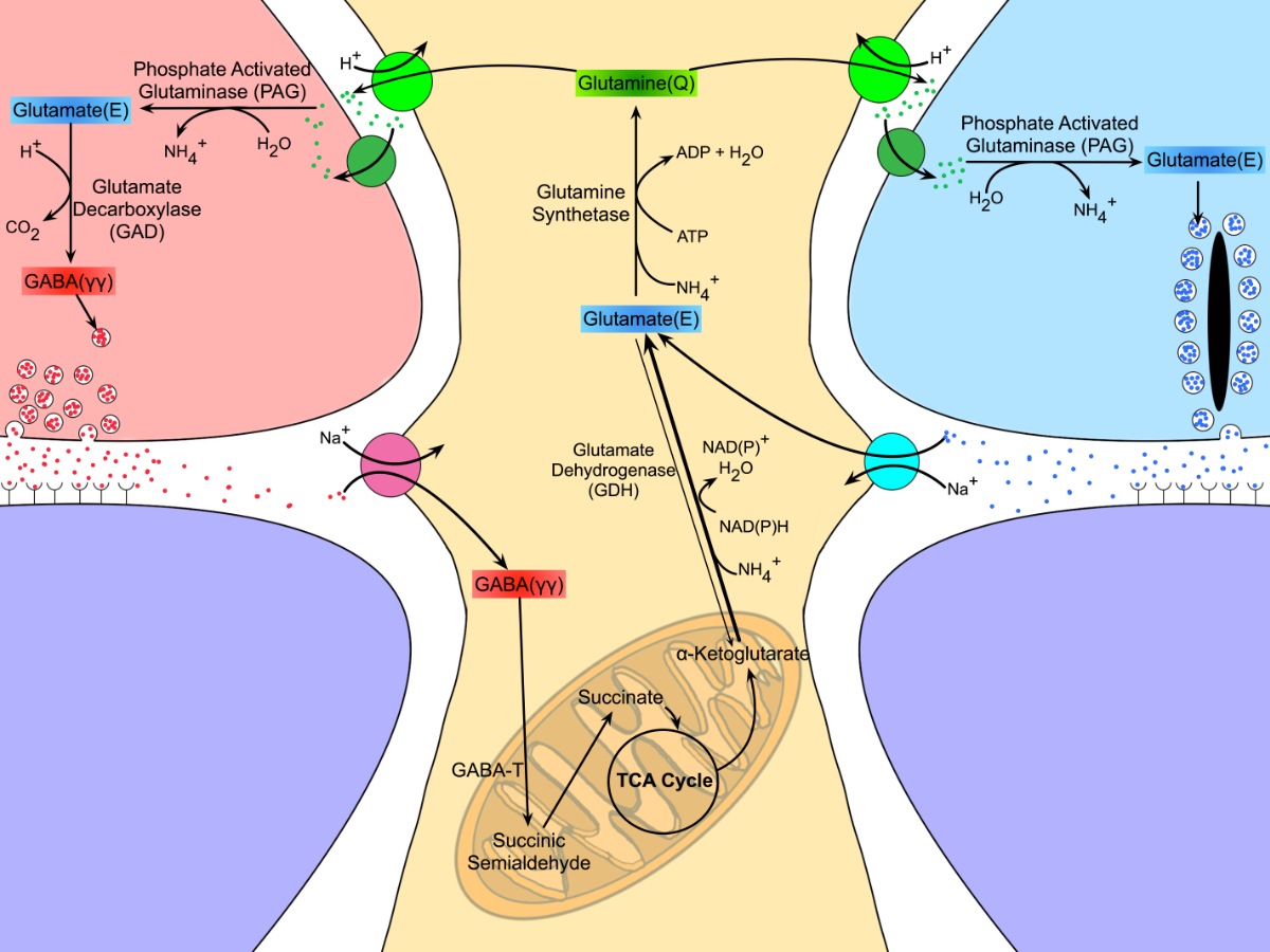
This manuscript functions as both a review and presents some exciting new data demonstrating how the glutamate cycle is disrupted during retinal degenerative disease.

Abstract: Müller cells are glia that play important regulatory roles in retinal metabolism. These roles have been evolutionarily conserved across at least 300 million years. Müller cells have a tightly locked metabolic signature in the healthy retina, which rapidly degrades in response to insult and disease. This variation in metabolic signature occurs in a chaotic fashion, involving some central metabolic pathways. The cause of this divergence of Müller cells, from a single class with a unique metabolic signature to numerous separable metabolic classes, is currently unknown and illuminates potential alternative metabolic pathways that may be revealed in disease. Understanding the impacts of this heterogeneity on degenerate retinas and the implications for the metabolic support of surrounding neurons will be critical to long-term integration of retinal therapeutics for the restoration of visual perception following photoreceptor degeneration.

Optic Cup Morphogenesis Requires Neural Crest-Mediated Basement Membrane Assembly

We have a new manuscript out in Development, Optic cup morphogenesis requires neural crest-mediated basement membrane assembly.

Authors: Chase D. Bryan @CDBE30, Macaulie A. Casey, Rebecca L. Pfeiffer @BeccaPfeiffer19, Bryan W. Jones @BWJones, and Kristen M. Kwan @BlockInTheBack

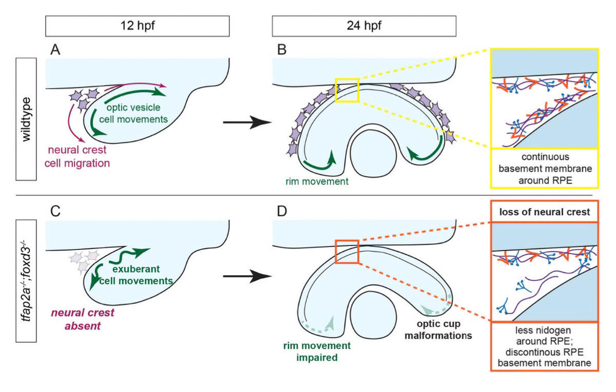
This is a collaborative project out of the Kwan lab that we helped out with some of the ultrastructural work and analysis.

Abstract: Organogenesis requires precise interactions between a developing tissue and its environment. In vertebrates, the developing eye is surrounded by a complex extracellular matrix as well as multiple mesenchymal cell populations. Disruptions to either the matrix or periocular mesenchyme can cause defects in early eye development, yet in many cases, the underlying mechanism is unknown. Here, using multidimensional imaging and computational analyses in zebrafish, we establish that cell movements in the developing optic cup require neural crest. Ultrastructural analysis reveals that basement membrane formation around the developing eye is also dependent on neural crest, but only specifically around the retinal pigment epithelium. Neural crest cells produce the extracellular matrix protein nidogen: impairing nidogen function disrupts eye development, and strikingly, expression of nidogen in the absence of neural crest partially restores optic cup morphogenesis. These results demonstrate that eye formation is regulated in part by extrinsic control of extracellular matrix assembly.