Tag Archives: Bryan Jones

Retinal Prosthetics, Optogenetics and Photoswitches

Retinal-Prosthetics-Optogenetics-and-Photoswitches

We have a new publication, Retinal Prosthetics, Optogenetics and Photoswitches in ACS Chemical Neuroscience.  Authors are:  Robert E. MarcRebecca L. Pfeiffer, and Bryan W. Jones.

Abstract:

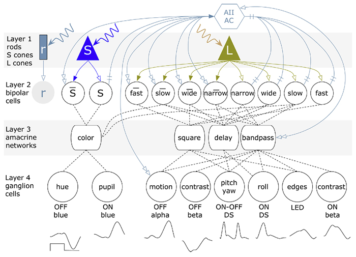
Three technologies have emerged as therapies to restore light sensing to profoundly blind patients suffering from late-stage retinal degenerations: (1) retinal prosthetics, (2) optogenetics, and (3) chemical photoswitches. Prosthetics are the most mature and the only approach in clinical practice. Prosthetic implants require complex surgical intervention and provide only limited visual resolution but can potentially restore navigational ability to many blind patients. Optogenetics uses viral delivery of type 1 opsin genes from prokaryotes or eukaryote algae to restore light responses in survivor neurons. Targeting and expression remain major problems, but are potentially soluble. Importantly, optogenetics could provide the ultimate in high-resolution vision due to the long persistence of gene expression achieved in animal models. Nevertheless, optogenetics remains challenging to implement in human eyes with large volumes, complex disease progression, and physical barriers to viral penetration. Now, a new generation of photochromic ligands or chemical photoswitches (azobenzene-quaternary ammonium derivatives) can be injected into a degenerated mouse eye and, in minutes to hours, activate light responses in neurons. These photoswitches offer the potential for rapidly and reversibly screening the vision restoration expected in an individual patient. Chemical photoswitch variants that persist in the cell membrane could make them a simple therapy of choice, with resolution and sensitivity equivalent to optogenetics approaches. A major complexity in treating retinal degenerations is retinal remodeling: pathologic network rewiring, molecular reprogramming, and cell death that compromise signaling in the surviving retina. Remodeling forces a choice between upstream and downstream targeting, each engaging different benefits and defects. Prosthetics and optogenetics can be implemented in either mode, but the use of chemical photoswitches is currently limited to downstream implementations. Even so, given the high density of human foveal ganglion cells, the ultimate chemical photoswitch treatment could deliver cost-effective, high-resolution vision for the blind.
Synapse Classification And Localization In Electron Micrographs

Synapse-classification_

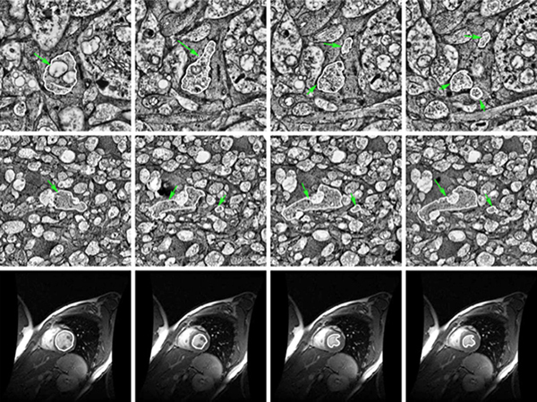
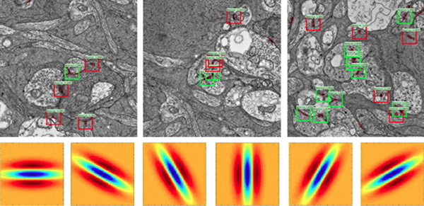
We have a new publication, Synapse Classification And Localization In Electron Micrographs in Pattern Recognition Letters.  Authors are: Vignesh JagadeeshJames Anderson, Bryan W. JonesRobert MarcSteven Fisher and B.S. Manjunath.

Abstract:  Classification and detection of biological structures in Electron Micrographs (EM) is a relatively new large scale image analysis problem. The primary challenges are in modeling diverse visual characteristics and development of scalable techniques. In this paper we propose novel methods for synapse detection and localization, an important problem in connectomics. We first propose an attribute based descriptor for characterizing synaptic junctions. These descriptors are task specific, low dimensional and can be scaled across large image sizes. Subsequently, techniques for fast localization of these junctions are proposed. Experimental results on images acquired from a mammalian retinal tissue compare favorably with state of the art descriptors used for object detection.

Robust Segmentation based Tracking using an Adaptive Wrapper for Inducing Priors

Robust-Segmentation-based-Tracing-using-an-Adaptive-Wrapper-for-Inducing-Priors

We have published another manuscript, Robust Segmentation based Tracking using an Adaptive Wrapper for Inducing Priors.  This manuscript describes the work on adaptive tracing and proposes an algorithm that adapts a generic tracing algorithm to an application of interest.  In our specific case, it is boundaries of cells in high frequency space in transmission electron microscopy images.  But the approach in this paper is applicable to biological, medical, remote sensing and surveillance data as well utilizing priors specific to the application.  The co-authors on the paper are: Vignesh JagadeeshJames Anderson, Bryan W. JonesRobert E. MarcSteven K Fisher and B.S Manjunath.

FASEB Bio-Art Competition Winner 2013

1254_40X_GBY_72dpi-1

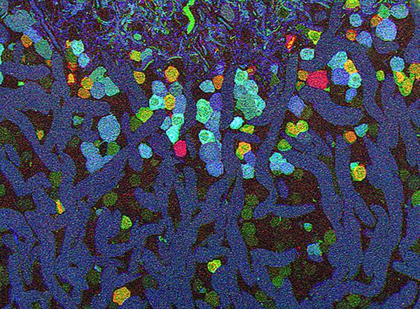
Bryan W. Jones and Robert E. Marc and were selected as 2013 FASEB BioArt Winners (Press release here).  This image shows a region of an amazingly complex retina from a goldfish (Carassius auratus auratus) analyzed using tools called Computational Molecular Phenotyping (CMP) that reveal the metabolic state of the all cell types in tissues.  These cells were labeled with antibodies for the presence of two fundamental amino acid metabolites (anti-glycine in red, anti-GABA in blue) and an amino acid tracer of physiologic activity (anti-AGB in green).   These labels allow us to visualize the metabolic state and therefore, classes of bipolaramacrine and horizontal cells.  More details on the image here.


The Alternative Complement Pathway Deficiency Amerliorates Chronic Smoked-Induced Functional And Morphological Ocular Injury

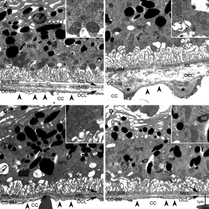
Alex Woodell, Beth Coughlin, Kannan Kunchithapautham, Sarah Casey, Tucker Williamson, W. Drew Ferrell,  Carl Atkinson, Bryan Jones and Baerbel Rohrer have a new manuscript out, The Alternative Complement Pathway Deficiency Amerliorates Chronic Smoked-Induced Functional And Morphological Ocular Injury in PLOS One.

The short story is: Don’t smoke.  But then you knew that.  Where this paper contributes is that it provides clear findings that show ocular pathologies generated by cigarette smoke are dependent upon activation of the immune system, in particular complement and the alternative pathway which are critical findings in the treatment of AMD.