Tag Archives: Bryan Jones

The Rod-Cone Crossover Connectome of Mammalian Bipolar Cells

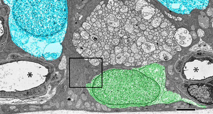
We have a new publication out (direct link), The rod-cone crossover connectome of mammalian bipolar cells authored by Scott Lauritzen, Crystal Sigulinsky, James Anderson, Michael Kalloniatis, Noah Nelson, Danny Emrich, Chris Rapp, Nicolas McCarthy, Ethan Kerzner, Mariah Meyer, Bryan W. Jones, and Robert Marc.

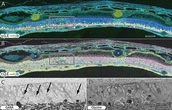
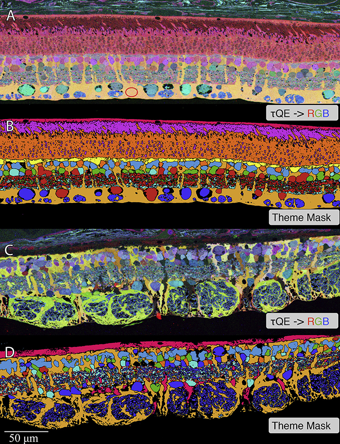
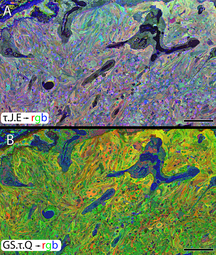
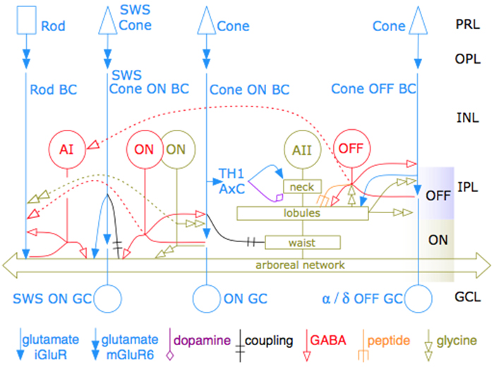
Abstract: The basis of cross-suppression between rod and cone channels has long been an enigma. Using rabbit retinal connectome RC1, we show that all cone bipolar cell (BC) classes inhibit rod BCs via amacrine cell (AC) motifs (C1-6); that all cone BC classes are themselves inhibited by AC motifs (R1-5, R25) driven by rod BCs. A sparse symmetric AC motif (CR) is presynaptic and postsynaptic to both rod and cone BCs. ON cone BCs of all classes drive inhibition of rod BCs via motif C1 wide-field GABAergic ACs (γACs) and motif C2 narrow field glycinergic ON ACs (GACs). Each rod BC receives ≈ 10 crossover AC synapses and each ON cone BC can target ≈ 10 or more rod BCs via separate AC processes. OFF cone BCs mediate monosynaptic inhibition of rod BCs via motif C3 driven by OFF γACs and GACs and disynaptic inhibition via motifs C4 and C5 driven by OFF wide-field γACs and narrow-field GACs, respectively. Motifs C4 and C5 form halos of 60-100 inhibitory synapses on proximal dendrites of AI γACs. Rod BCs inhibit surrounding arrays of cone BCs through AII GAC networks that access ON and OFF cone BC patches via motifs R1, R2, R4 R5 and a unique ON AC motif R3 that collects rod BC inputs and targets ON cone BCs. Crossover synapses for motifs C1, C4, C5 and R3 are 3-4x larger than typical feedback synapses, which may be a signature for synaptic winner-take-all switches.

Retinal Remodeling And Metabolic Alterations in Human AMD

We have a new publication out (direct link, open access), Müller Cell Metabolic Chaos During Retinal Degeneration authored by Bryan W. JonesRebecca Pfeiffer, William Ferrell, Carl Watt, James Tucker, and Robert Marc.

Abstract:

Age-related macular degeneration (AMD) is a progressive retinal degeneration resulting in central visual field loss, ultimately causing debilitating blindness. AMD affects 18% of Americans from 65 to 74, 30% older than 74 years of age and is the leading cause of severe vision loss and blindness in Western populations. While many genetic and environmental risk factors are known for AMD, we currently know less about the mechanisms mediating disease progression. The pathways and mechanisms through which genetic and non-genetic risk factors modulate development of AMD pathogenesis remain largely unexplored. Moreover, current treatment for AMD is palliative and limited to wet/exudative forms. Retina is a complex, heterocellular tissue and most retinal cell classes are impacted or altered in AMD. Defining disease and stage-specific cytoarchitectural and metabolic responses in AMD is critical for highlighting targets for intervention. The goal of this article is to illustrate cell types impacted in AMD and demonstrate the implications of those changes, likely beginning in the retinal pigment epithelium (RPE), for remodeling of the the neural retina. Tracking heterocellular responses in disease progression is best achieved with computational molecular phenotyping (CMP), a tool that enables acquisition of a small molecule fingerprint for every cell in the retina. CMP uncovered critical cellular and molecular pathologies (remodeling and reprogramming) in progressive retinal degenerations such as retinitis pigmentosa (RP). We now applied these approaches to normal human and AMD tissues mapping progression of cellular and molecular changes in AMD retinas, including late-stage forms of the disease.

Müller Cell Metabolic Chaos During Retinal Degeneration

We have a new publication out (direct link, open access), Müller Cell Metabolic Chaos During Retinal Degeneration authored by Rebecca PfeifferRobert Marc, Mineo Kondo, Hiroko Terasaki and Bryan W. Jones.

Abstract:

Müller cells play a critical role in retinal metabolism and are among the first cells to demonstrate metabolic changes in retinal stress or disease. The timing, extent, regulation, and impacts of these changes are not yet known. We evaluated metabolic phenotypes of Müller cells in the degenerating retina.

Retinas harvested from wild-type (WT) and rhodopsin Tg P347L rabbits were fixed in mixed aldehydes and resin embedded for computational molecular phenotyping (CMP). CMP facilitates small molecule fingerprinting of every cell in the retina, allowing evaluation of metabolite levels in single cells.

CMP revealed signature variations in metabolite levels across Müller cells from TgP347L retina. In brief, neighboring Müller cells demonstrated variability in taurine, glutamate, glutamine, glutathione, glutamine synthetase (GS), and CRALBP. This variability showed no correlation across metabolites, implying the changes are functionally chaotic rather than simply heterogeneous. The inability of any clustering algorithm to classify Müller cell as a single class in the TgP347L retina is a formal proof of metabolic variability in the present in degenerating retina.

Although retinal degeneration is certainly the trigger, Müller cell metabolic alterations are not a coherent response to the microenvironment. And while GS is believed to be the primary enzyme responsible for the conversion of glutamate to glutamine in the retina, alternative pathways appear to be unmasked in degenerating retina. Somehow, long term remodeling involves loss of Müller cell coordination and identity, which has negative implications for therapeutic interventions that target neurons alone.

2-nm Resolution Anatomy of Retinal Neuro-Glial-Vascular Architecture

This abstract was presented today, May 2th at the 2016 Association for Research in Vision and Opthalmology (ARVO) meetings in Seattle, Washington by Jefferson R. Brown, Rebecca L. Pfeiffer, Crystal Sigulinsky, Felix Vazquez-Chona, Daniel Emrich, Bryan W. Jones, Robert E. Marc.

Abstract Number: 995

Author Block: Jefferson R. Brown, Rebecca L. Pfeiffer, Crystal Sigulinsky, Felix Vazquez-Chona, Daniel Emrich, Bryan W. Jones, Robert E. Marc
1 Dept of Ophthalmology, University of Utah, Salt Lake City, Utah, United States

Disclosure Block:Jefferson R. Brown, None; Rebecca L. Pfeiffer, None; Crystal Sigulinsky, None; Felix Vazquez-Chona, None; Daniel Emrich, None; Bryan W. Jones, None; Robert E. Marc, Signature Immunologics (Code I (Personal Financial Interest) )

Purpose:Retinal vasculature is strongly affected by degenerative pathologies and in turn, may also contribute to their progression. However, much of what we understand about the normal, healthy interaction between neurons, glia, and blood vessels at the ultrastructural level is limited to single section electron microscopy. The technology of serial section transmission electron microscopy (ssTEM) extends the high definition of TEM imaging into three dimensions to create volumes, allowing for more thorough visualization and analysis of the vascular-glial-neuronal complex.

Methods:RC2 is a 40TB ssTEM volume of over 1,400 horizontal sections of retinal tissue derived from an adult female C57BL/6J mouse. The tissue sample is 250 um in diameter and spans the outer nuclear layer to the vitreal surface. Baseline resolution is 2.18nm per pixel. Visualization, navigation and metadata annotations of the database are made via the Viking software suite.

Results:Much of the retinal vascular basement membrane directly contacts Muller cells. In the ganglion cell layer, direct basement membrane contact with astrocytes is frequent. Microglia commonly contact the basement membrane, and occasionally direct contact of neurons onto basement membrane was observed. Full 3D reconstruction of all vascular pathways with associated endothelia and pericytes within the volume was completed, demonstrating that all the retinal capillary layers are continuous with one another [Figure].

Conclusions:The presence of occasional direct neuronal contact onto vascular basement membrane supports earlier work by Ochs and colleagues (2000) and suggests the blood-retina barrier does not universally involve retinal glia. However, since such contacts are extremely sparse, it remains to be seen whether this finding has biologic significance, though their existence suggests significance. The RC2 volume is a valuable resource to aid in discovery of defining characteristics of wild type neurovascular architecture.


The intro figure is a side view of reconstruction of all vasculature within the RC2 volume. Vessels at the top of the figure correspond to the outer plexiform layer, while those at the bottom correspond to the ganglion cell layer. This capillary plexus is one continuous structure. Visualization by VikingView software.

Retinal Remodeling in Human Retinitis Pigmentosa

We have a new publication out (Direct Link, Free Open Access), Retinal Remodeling in Human Retinitis Pigmentosa authored by Bryan W. Jones, Rebecca Pfeiffer, Drew Ferrell, Carl Watt, Michael Marmor and Robert Marc.

Abstract: Retinitis Pigmentosa (RP) in the human is a progressive, currently irreversible neural degenerative disease usually caused by gene defects that disrupt the function or architecture of the photoreceptors. While RP can initially be a disease of photoreceptors, there is increasing evidence that the inner retina becomes progressively disorganized as the outer retina degenerates. These alterations have been extensively described in animal models, but remodeling in humans has not been as well characterized. This study, using computational molecular phenotyping (CMP) seeks to advance our understanding of the retinal remodeling process in humans. We describe cone mediated preservation of overall topology, retinal reprogramming in the earliest stages of the disease in retinal bipolar cells, and alterations in both small molecule and protein signatures of neurons and glia. Furthermore, while Müller glia appear to be some of the last cells left in the degenerate retina, they are also one of the first cell classes in the neural retina to respond to stress which may reveal mechanisms related to remodeling and cell death in other retinal cell classes. Also fundamentally important is the finding that retinal network topologies are altered. Our results suggest interventions that presume substantial preservation of the neural retina will likely fail in late stages of the disease. Even early intervention offers no guarantee that the interventions will be immune to progressive remodeling. Fundamental work in the biology and mechanisms of disease progression are needed to support vision rescue strategies.

Store-Operated Calcium Entry In Müller Glia Is Controlled By Synergistic Activation Of TRPC And Orai Channels

We have a new publication out as collaborators with colleages, Store-Operated Calcium Entry In Müller Glia Is Controlled By Synergistic Activation Of TRPC And Orai Channels authored by Tünde Molnár, Oleg Yarishkin, Peter Barabas, Anthony Iuso, Bryan W. Jones, Robert Marc, Tam Phuong, and David Krizaj.

Bonus, we got the cover!  The image was created by Tam Phuong.


Significance: Store-operated Ca2+ signaling represents a major signaling pathway and source of cytosolic Ca2+ in astrocytes. Here, we show that the store-operated response in Müller cells, radial glia that perform key structural, signaling, osmoregulatory and mechanosensory functions within the retina, is mediated through synergistic activation of TRPC and Orai channels. The endfoot disproportionately expresses the depletion sensor STIM1, contains an extraordinarily high density of ER cisternae that shadow neuronal, astrocyte, vascular and axonal structures, interface with mitochondria but also originates SOCE-induced transcellular Ca2+ waves that propagate glial excitation into the proximal retina. These results identify a molecular mechanism that underlies complex interactions between the plasma membrane and calcium stores and contributes to radial glial function, regulation and response to mechanical stress.

Abstract: The endoplasmic reticulum (ER) is at the epicenter of astrocyte Ca2+ signaling. We sought to identify the molecular mechanism underlying store-operated calcium entry (SOCE) that repletes ER Ca2+ stores in mouse Müller cells. Store depletion, induced through blockade of sequestration transporters in Ca2+-free saline, induced synergistic activation of canonical transient receptor potential (TRPC1) and Orai channels. Store-operated TRPC1 channels were identified by their electrophysiological properties, pharmacological blockers and ablation of the Trpc1 gene. ICRAC (Ca2+ release-activated) currents were identified by ion permeability, voltage-dependence and sensitivity to selective Orai antagonists Synta66 and GSK7975A. Depletion-evoked calcium influx was initiated at the Müller endfoot and apical process, triggering centrifugal propagation of Ca2+ waves into the cell body. EM analysis of the endfoot compartment showed high-density ER cisternae that shadow retinal ganglion cell (RGC) somata and axons, protoplasmic astrocytes, vascular endothelial cells and ER-mitochondrial contacts at the vitreal surface of the endfoot. The mouse retina expresses transcripts encoding both Stim and all known Orai genes; Müller glia predominantly express STIM1 whereas STIM2 is mainly confined to the outer plexiform and retinal ganglion cell layers. Elimination of TRPC1 facilitated Müller gliosis induced by the elevation of intraocular pressure (IOP), suggesting that TRPC channels might play a neuroprotective role during mechanical stress. These findings expand the current knowledge about store-operated signaling in astroglia, as well as calcium signaling pathways in Müller astroglia and functional roles these cells play in retinal physiology and pathology.

Seasonal And Post-Trauma Remodeling Of The Ground Squirrel Retina

We have a new publication out, Seasonal and post-trauma remodeling in cone-dominant ground squirrel retina authored by Dana Merriman, Ben Sajdak, Wei Li and Bryan W. Jones.

Abstract:

With a photoreceptor mosaic containing ∼85% cones, the ground squirrel is one of the richest known mammalian sources of these important retinal cells. It also has a visual ecology much like the human’s. While the ground squirrel retina is understandably prominent in the cone biochemistry, physiology, and circuitry literature, far less is known about the remodeling potential of its retinal pigment epithelium, neurons, macroglia, or microglia. This review aims to summarize the data from ground squirrel retina to this point in time, and to relate them to data from other brain areas where appropriate. We begin with a survey of the ground squirrel visual system, making comparisons with traditional rodent models and with human. Because this animal’s status as a hibernator often goes unnoticed in the vision literature, we then present a brief primer on hibernation biology. Next we review what is known about ground squirrel retinal remodeling concurrent with deep torpor and with rapid recovery upon re-warming. Notable here is rapidly-reversible, temperature-dependent structural plasticity of cone ribbon synapses, as well as pre- and post-synaptic plasticity throughout diverse brain regions. It is not yet clear if retinal cell types other than cones engage in torpor-associated synaptic remodeling. We end with the small but intriguing literature on the ground squirrel retina’s remodeling responses to insult by retinal detachment. Notable for widespread loss of (cone) photoreceptors, there is surprisingly little remodeling of the RPE or Müller cells. Microglial activation appears minimal, and remodeling of surviving second- and third-order neurons seems absent, but both require further study. In contrast, traumatic brain injury in the ground squirrel elicits typical macroglial and microglial responses. Overall, the data to date strongly suggest a heretofore unrecognized, natural checkpoint between retinal deafferentiation and RPE and Müller cell remodeling events. As we continue to discover them, the unique ways by which ground squirrel retina responds to hibernation or injury may be adaptable to therapeutic use.

Development Of Animal Models Of Local Retinal Degeneration

Henri Lorach, Jennifer Kung, Corinne Beier, Yossi Mandel, Roopa Dalal, Philip Huie, Jenny Wang, Sengjun Lee, Alexander Sher, Bryan W. Jones, and Daniel Palanker have a new publication, Development of Animal Models of Local Retinal Degeneration (the IOVS direct link is here).

Many of the models of retinal degeneration we explore are genetic.  This project was designed to explore two other alternative approaches to retinal degeneration that are non-genetic and capable of producing highly localized retinal degeneration with precise onset time.


The AII Amacrine Cell Connectome: A Dense Network Hub

AII-connectome

We have a new publication in Frontiers in Neuroscience, The AII Amacrine Cell Connectome: A Dense Network Hub.  Authors are Robert E. MarcJames R. Anderson, Bryan W. Jones, Crystal Sigulinsky and J. Scott Lauritzen.

Abstract:  The mammalian AII retinal amacrine cell is a narrow-field, multistratified glycinergic neuron best known for its role in collecting scotopic signals from rod bipolar cells and distributing them to ON and OFF cone pathways in a crossover network via a combination of inhibitory synapses and heterocellular AII::ON cone bipolar cell gap junctions. Long considered a simple cell, a full connectomics analysis shows that AII cells possess the most complex interaction repertoire of any known vertebrate neuron, contacting at least 28 different cell classes, including every class of retinal bipolar cell. Beyond its basic role in distributing rod signals to cone pathways, the AII cell may also mediate narrow-field feedback and feedforward inhibition for the photopic OFF channel, photopic ON-OFF inhibitory crossover signaling, and serves as a nexus for a collection of inhibitory networks arising from cone pathways that likely negotiate fast switching between cone and rod vision. Further analysis of the complete synaptic counts for five AII cells shows that (1) synaptic sampling is normalized for anatomic target encounter rates; (2) qualitative targeting is specific and apparently errorless; and (3) that AII cells strongly differentiate partner cohorts by synaptic and/or coupling weights. The AII network is a dense hub connecting all primary retinal excitatory channels via precisely weighted drive and specific polarities. Homologs of AII amacrine cells have yet to be identified in non-mammalians, but we propose that such homologs should be narrow-field glycinergic amacrine cells driving photopic ON-OFF crossover via heterocellular coupling with ON cone bipolar cells and glycinergic synapses on OFF cone bipolar cells. The specific evolutionary event creating the mammalian AII scotopic-photopic hub would then simply be the emergence of large numbers of pure rod bipolar cells.


A Multi-Scale Computational Model For The Study Of Retinal Prosthetic Stimulation

Multiscale-model-of-retina

We have a new publication in IEEE, A Multi-Scale Computational Model For The Study Of Retinal Prosthetic Stimulation.  Authors are: Kyle LoizosGianluca Lazzi, J. Scott Lauritzen, James R. Anderson, Bryan W. Jones and Robert E. Marc.

Abstract: An implantable retinal prosthesis has been developed to restore vision to patients who have been blinded by degenerative diseases that destroy photoreceptors. By electrically stimulating the surviving retinal cells, the damaged photoreceptors may be bypassed and limited vision can be restored. While this has been shown to restore partial vision, the understanding of how cells react to this systematic electrical stimulation is largely unknown. Better predictive models and a deeper understanding of neural responses to electrical stimulation is necessary for designing a successful prosthesis. In this work, a computational model of an epi-retinal implant was built and simulated, spanning multiple spatial scales, including a large-scale model of the retina and implant electronics, as well as underlying neuronal networks.