Author Archives: bwjones

Comparative Anatomy And Connectivity Of The AII Amacrine Cell In Mouse And Rabbit Retina

This abstract was presented today, April 8th at the 2019 Association for Research in Vision and Opthalmology (ARVO) meetings in Vancouver, Canada by Selena Wirthlin, Crystal L. SigulinskyJames R. Anderson, Daniel P. Emrich, Christopher Rapp, Jeebika Dahal, Rebecca L. Pfeiffer, Kevin D. Rapp, Jia-Hui Yang, Carl B. Watt, Robert E. Marc and Bryan W. Jones.

Full resolution version here.

Purpose
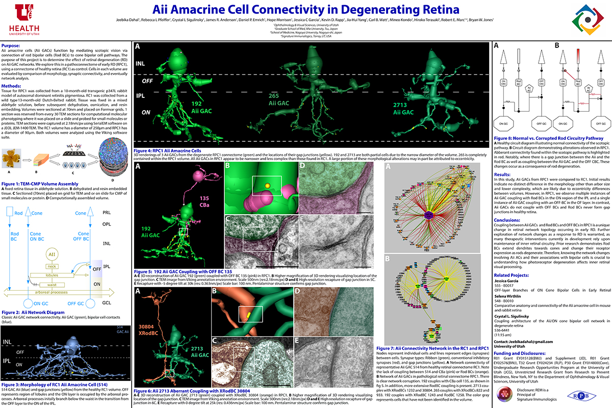
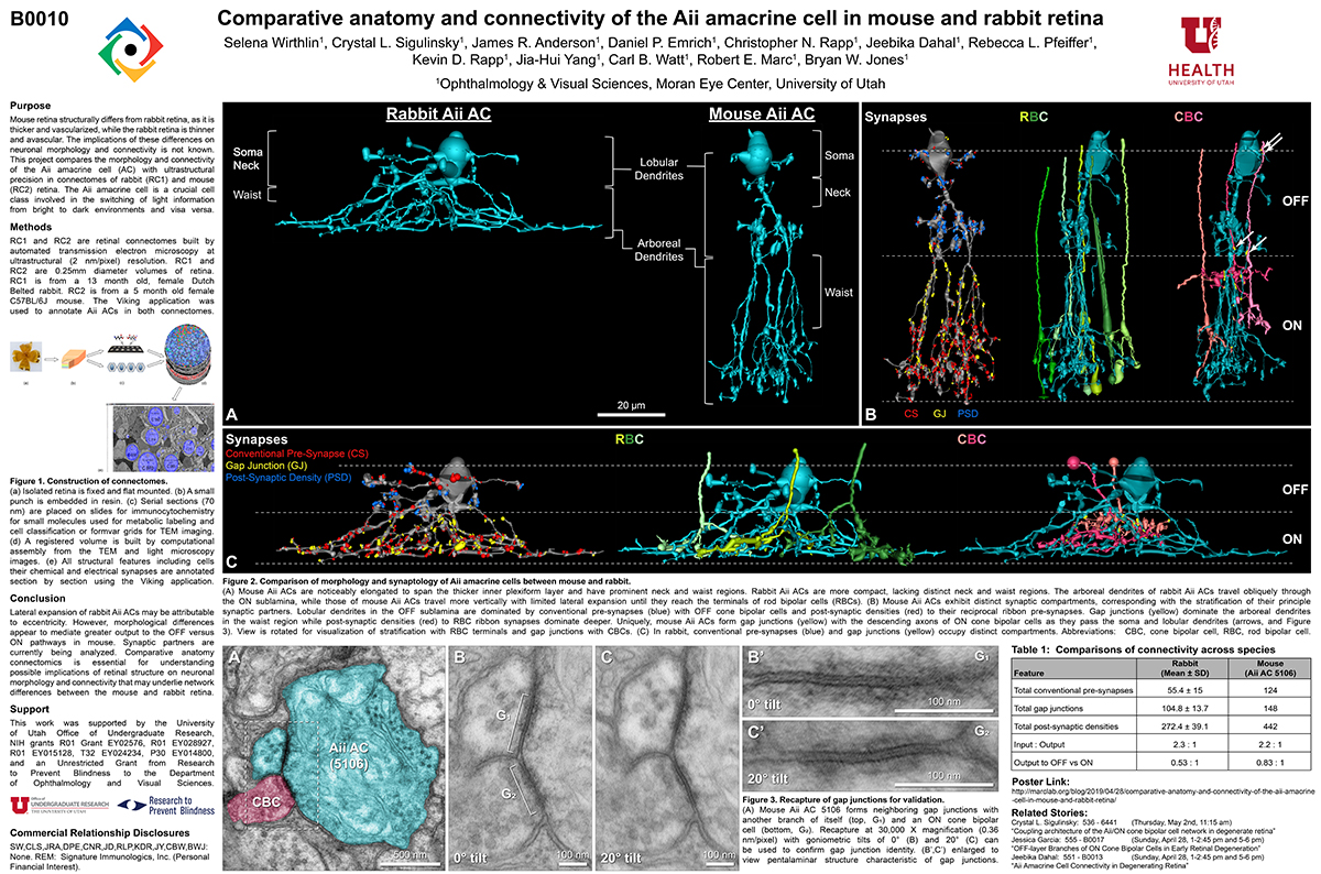
Mouse retina structurally differs from rabbit retina, as it is thicker and vascularized, while the rabbit retina is thinner and avascular. The implications of these differences on neuronal morphology and connectivity is not known. This project compares the morphology and connectivity of the Aii amacrine cell (AC) with ultrastructural precision in connectomes of mouse (RC2) and rabbit (RC1) retina.

Methods
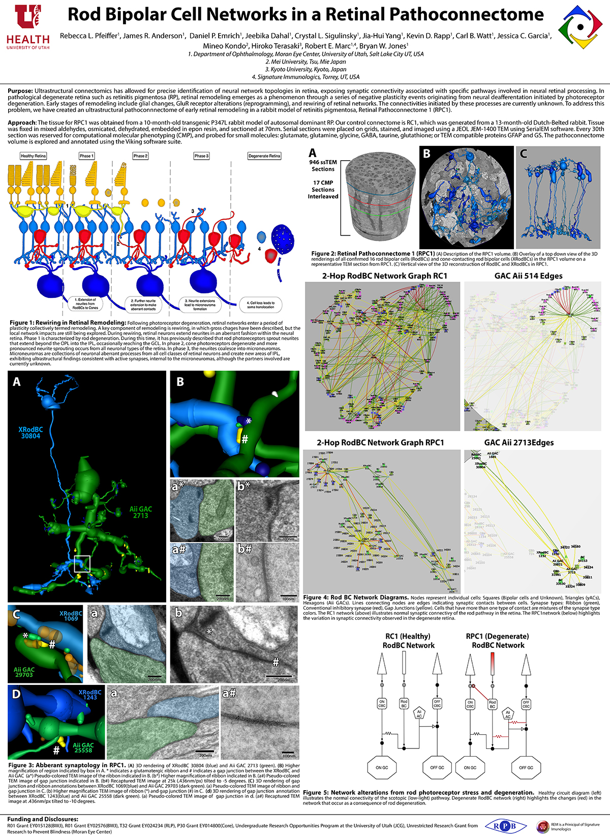
RC1 and RC2 are connectomes built by automated transmission electron microscopy at ultrastructural (2 nm/pixel) resolution. RC1 and RC2 are 0.25mm diameter volumes of retina. RC1 is from a 13 month old, female Dutch Belted rabbit. RC2 is from a 5 month old female C57BL/6J mouse. The Viking application was used to annotate Aii ACs in both connectomes.

Results
Mouse Aii ACs are noticeably elongated to span the thicker inner plexiform layer (IPL) and have a prominent neck region. Lobular appendages of Aii ACs in both species extend thin stalks from the soma, neck and proximal arboreal dendrites in the OFF sublamina, predominantly forming reciprocal synapses with OFF cone bipolar cells (BCs). In rabbits, multiple arboreal dendrites emerge from the base of the neck, branch and travel obliquely through the ON sublamina, and form gap junctions with ON cone BCs, neighbor Aii ACs, and itself. They extend laterally at the base of the IPL, collecting ribbon input from rod BCs. In contrast, mouse arboreal dendrites stem from a single primary dendrite that branches as it travels vertically through the IPL without self-branch interaction, terminating at variable depths that align with the more broadly ramified axon terminals of rod BCs. Conventional synapse to gap junction ratios reveal greater output in the OFF vs ON layer in mouse compared to rabbit. Notably, mouse Aii ACs form gap junctions with the descending axons of ON cone BCs as they pass its soma, in contrast to rabbit, where gap junctions do not form at contacts proximal to ON cone BC axon terminals.

Conclusions
Lateral expansion of rabbit Aii ACs may be attributable to eccentricity. However, morphological differences appear to mediate greater output to the OFF versus ON pathway in mouse. Synaptic partners are currently being analyzed. Comparative anatomy connectomics is essential for understanding possible implications of retinal structure on neuronal morphology and connectivity that may underlie network differences between the mouse and rabbit retina.

Aii Amacrine Cell Connectivity in Degenerating Retina

This abstract was presented today, April 8th at the 2019 Association for Research in Vision and Opthalmology (ARVO) meetings in Vancouver, Canada by Jeebika Dahal, Rebecca L. Pfeiffer, Crystal L. Sigulinsky, James R. Anderson, Daniel P. Emrich, Hope Morrison, Jessica C. Garcia, Kevin D. Rapp, Jia-Hui Yang, Carl B. Watt, Mineo Kondo, Hiroko Terasaki, Robert E. Marc and Bryan W. Jones.

Full resolution version here.

Purpose
Aii amacrine cells (Aii ACs) function in mediating scotopic vision via connection of rod bipolar cells (Rod BCs) to cone bipolar cell pathways. The purpose of this project is to determine the effect of retinal degeneration (RD) on Aii AC networks. We explore this in a pathoconnectome of early RD (RPC1), using a connectome of healthy retina (RC1) as control. Cells in each volume are evaluated by comparison of morphology, synaptic connectivity, and eventually network analysis.

Methods
Tissue for RPC1 was collected from a 10 month old transgenic p347L rabbit model of autosomal dominant retinitis pigmentosa. RC1 was collected from a 13 month old Dutch-Belted rabbit, with no indications of degeneration. Tissue was fixed in a mixed aldehyde solution, before subsequent dehydration, osmication, and resin embedding. Volumes were sectioned at 70nm (RPC1) and 90nm (RC1) and placed on formvar grids. 1 section was reserved from every 30 TEM sections for computational molecular phenotyping where it was placed on a slide and probed for small molecules or proteins. TEM sections were captured at 2.18nm/px using SerialEM software on a JEOL JEM-1400 TEM. The RC1 volume has a diameter of 250µm and RPC1 has a diameter of 90µm. Both volumes were analyzed using the Viking software suite.

Results
In this study, Aii ACs from RPC1 were compared to RC1. Initial results indicate no distinct difference in the morphology other than arbor size, which are likely due to eccentricity differences between volumes. However, in RPC1, we observe multiple instances of Aii AC coupling with Rod BCs in the ON region of the IPL. In contrast, Rod BCs never form gap junctions in healthy retina.

Conclusions
Coupling between Aii ACs and Rod BCs in RPC1 is a unique change in retinal network topology occurring in early RD. Further exploration of network changes as a response to RD is warranted, as many therapeutic interventions currently in development rely upon maintenance of inner retinal circuitry. Prior research demonstrates Rod BCs extend dendrites towards cones and change their receptor expression as rods degenerate. Therefore, knowing the network changes involving Aii ACs and their associations with bipolar cells is crucial to understanding how photoreceptor degeneration affects inner retinal visual processing.

OFF-layer Branches of ON Cone Bipolar Cells in Early Retinal Degeneration

This abstract was presented today, April 8th at the 2019 Association for Research in Vision and Opthalmology (ARVO) meetings in Vancouver, Canada by Jessica C. Garcia, Rebecca L. Pfeiffer, Crystal L. Sigulinsky, James R. Anderson, Daniel P. Emrich, Jeebika Dahal, Hope Morrison, Kevin D. Rapp, Jia-Hui Yang, Carl B. Watt, Mineo Kondo, Hiroko Terasaki, Robert E. Marc and Bryan W. Jones.

Full resolution version here.

Purpose: Cone bipolar cells are customarily classified into superclasses of ON (ON-BCs) and OFF (OFF-BCs). ON or OFF specialization is further segregated by stratification within the inner plexiform layer. Retinal degeneration induces negative plasticity termed remodeling, that includes aberrant neurite extension from multiple cell types (rewiring) and ON-BCs switching their glutamate receptors to match that of OFF-BCs (reprogramming). Previous analysis in healthy retina shows that ON-BCs can make small ribbon synapses in the descending axon or, less frequently, simple single branch projections in the OFF layer. What impact remodeling has on these OFF-layer branches is unknown. In this study, we compare OFF branches from ON-BCs in a connectome of early retinal degeneration (RPC1) to our healthy retinal connectome (RC1).

Methods: Retinal tissues selected for RC1 and RPC1 were collected post-mortem from a 13 month old Dutch-belted healthy female rabbit and a male 10 month old transgenic P347L rabbit model of autosomal dominant retinitis pigmentosa, respectively. RPC1 shows signs of remodeling including rod outer-segment degeneration and aberrant neurite extension. Tissues were fixed in mixed aldehydes and subsequently osmicated, dehydrated, resin embedded, and sectioned at 90 nm (RC1) or 70 nm (RPC1). Sections were placed on formvar grids, stained, and imaged at 2nm/px on a JEOL JEM-1400 TEM using SerialEM software. 1 section was reserved from every 30 for Computational Molecular Phenotyping, and probed for small molecules. Both volumes were evaluated using the Viking software suite.

Results: Ribbons of ON-BCs formed in the OFF layer branches have been previously described to contact glycinergic amacrine cells (ACs), GABAergic ACs, ON ganglion cells, and intrinsically photosensitive ganglion cells. Initial analysis of OFF branches of ON-BCs in RPC1 demonstrate more complex branching than in RC1 and increased number of synapses on these branches. In contrast to the inconsistent OFF layer branch stratification observed in RC1, the OFF branches in RPC1 appear to stratify at a similar level. Evaluation of synaptic partners is ongoing.

Conclusions: Increased complexity and number of synapses found in the OFF branches of some ON-BCs ultimately may represent ON network corruption. Exploring synaptic partners will reveal potential network alterations in retinal degenerative disease.

Marclab Off To ARVO2019

The Marclab is off to ARVO 2019 and eager to share some of what we’ve been up to over the past year.  We have undergraduate Jeebika Dahal presenting her work on the AII Amacrine Cell Connectivity Changes In Degenerating Retina on Sunday (see poster B0013 Abstract Number: 551 – B0013).  Undergraduate Selena Wirthlin will present her work on the Comparative Anatomy and Connectivity Of The AII Amacrine Cell In Mouse And Rabbit Retina on Sunday (poster B0010 Abstract Number: 548 – B0010). Undergraduate and US Navy veteran Jessica Garcia will present her work Sunday on OFF-layer Branches Of ON Cone Bipolar Cells In Early Retinal Degeneration (B0017 Abstract Number: 555 – B0017). And postdoc Crystal Sigulinsky will present her work on Coupling Architecture Of The Aii/ON Cone Bipolar Cell Network In Degenerate Retina in a platform presentation on Thursday at 11:15am (Abstract Number: 6441).

We hope to see you there!

Hypoxia Tolerance In The Norrin-Deficient Retina And The Chronically Hypoxic Brain Studied At Single-Cell Resolution

We have a new paper out in PNAS, Hypoxia tolerance in the Norrin-deficient retina and the chronically hypoxic brain studied at single-cell resolution.

Authors: Jacob S. Heng, Amir Rattner, Genevieve L. Stein-O’Brien, Briana L. Winer, Bryan W. Jones, Hilary J. Vernon, Loyal A. Goff, and Jeremy Nathans.

Abstract: The mammalian CNS is capable of tolerating chronic hypoxia, but cell type-specific responses to this stress have not been systematically characterized. In the Norrin KO (NdpKO) mouse, a model of familial exudative vitreoretinopathy (FEVR), developmental hypovascularization of the retina produces chronic hypoxia of inner nuclear-layer (INL) neurons and Muller glia. We used single-cell RNA sequencing, untargeted metabolomics, and metabolite labeling from 13C-glucose to compare WT and NdpKO retinas. In NdpKO retinas, we observe gene expression responses consistent with hypoxia in Muller glia and retinal neurons, and we find a metabolic shift that combines reduced flux through the TCA cycle with increased synthesis of serine, glycine, and glutathione. We also used single-cell RNA sequencing to compare the responses of individual cell types in NdpKO retinas with those in the hypoxic cerebral cortex of mice that were housed for 1 week in a reduced oxygen environment (7.5% oxygen). In the hypoxic cerebral cortex, glial transcriptome responses most closely resemble the response of Muller glia in the NdpKO retina. In both retina and brain, vascular endothelial cells activate a previously dormant tip cell gene expression program, which likely underlies the adaptive neoangiogenic response to chronic hypoxia. These analyses of retina and brain transcriptomes at single-cell resolution reveal both shared and cell type-specific changes in gene expression in response to chronic hypoxia, implying both shared and distinct cell type-specific physiologic responses.

Rod Bipolar Cell Networks In A Retinal Pathoconnectome

We presented a poster on Rod Bipolar Cell Networks In A Retinal Pathoconnectome at the 2019 HHMI Connectomics meeting in Berlin today. Downsampled PDF of poster here.

Authors: Rebecca L. Pfeiffer, James R. Anderson, Daniel P. Emrich, Jeebika Dahal, Crystal L. Sigulinsky, Jia-Hui Yang, Kevin D. Rapp, Carl B. Watt, Jessica C. Garcia, Mineo Kondo, Hiroko Terasaki, Robert E. Marc, and Bryan W. Jones.

Abstract: Ultrastructural connectomics has allowed for precise identification of neural network topologies in retina, exposing synaptic connectivity associated with specific pathways involved in neural retinal processing. In pathological degenerate retina such as retinitis pigmentosa (RP), retinal remodeling emerges as a phenomenon through a series of negative plasticity events originating from neural deafferentation initiated by photoreceptor degeneration. Early stages of remodeling include glial changes, GluR receptor alterations (reprogramming), and rewiring of retinal networks. The connectivities initiated by these processes are currently unknown. To address this problem, we have created an ultrastructural pathoconnnectome of early retinal remodeling in a rabbit model of retinitis pigmentosa, Retinal Pathoconnectome 1 (RPC1).

Mapping the network architecture of gap junctional coupling among parallel processing channels in the mammalian retina

We presented a poster on Mapping the network architecture of gap junctional coupling among parallel processing channels in the mammalian retina at the 2019 HHMI Connectomics meeting in Berlintoday. Downsampled PDF of poster here.

Authors: Crystal L. Sigulinsky, James R. Anderson, Ethan Kerzner, Christopher N. Rapp, Rebecca L. Pfeiffer, Daniel P. Emrich, Kevin D. Rapp, Noah T. Nelson, J. Scott Lauritzen, Miriah Meyer, Robert E. Marc, and Bryan W. Jones.

Abstract: Electrical synapses are fundamental components of neural networks. Gap junctions provide the anatomical basis for electrical synapses and are prevalent throughout the neural retina with essential roles in signal transmission. Gap junctions within and between the parallel processing channels afforded by retinal bipolar cells have been reported or predicted, but their roles, partners, and patterns remain largely unknown. Here, we took advantage of the high resolution of Retinal Connectome 1 (RC1) to reconstruct ON cone bipolar cells (CBCs) and map their coupling topologies.

Selena Sullivan Wirthlin Annotating Connectomes

One of the undergraduates in the lab, Selena Sullivan has been working on an exciting project in the lab in the realm of comparative anatomy.  She will be presenting this work at this year’s ARVO conference, exploring how different cells of a single type are between species in terms of morphology and synaptic and gap junctional connectivity.

via: Jonesblog