Development Of Animal Models Of Local Retinal Degeneration

Henri Lorach, Jennifer Kung, Corinne Beier, Yossi Mandel, Roopa Dalal, Philip Huie, Jenny Wang, Sengjun Lee, Alexander Sher, Bryan W. Jones, and Daniel Palanker have a new publication, Development of Animal Models of Local Retinal Degeneration (the IOVS direct link is here).

Many of the models of retinal degeneration we explore are genetic.  This project was designed to explore two other alternative approaches to retinal degeneration that are non-genetic and capable of producing highly localized retinal degeneration with precise onset time.


In Situ Metabolomic Signatures Of Neuroprotection, Apoptosis, And Microglial Phagocytosis

This abstract was presented July 1st at the 11th International Converence of the Metabolomics Society at the University of California, Davis by Felix Vazquez-Chona, Drew Ferrell, Bryan W. Jones and Robert E. Marc.


Metabolic dysregulation is an early hallmark of neurodegenerative diseases including Alzheimer’s disease and age-related macular degeneration. Mapping metabolic adaptation with cellular resolution and tissue- wide context is crucial to define networks regulating neuronal survival, cell death progression, and immune cell response.

Computational Molecular Phenotyping (CMP) explores the amine metabolome (amino acids and amines). Technically, CMP metabolomics combines amine metabolite trapping, ultrathin microscopy (50-200 nm), immunodetection, pattern recognition, and clustering algorithms. Here we mapped the in situ distribution of over 30 core amine metabolites in retinal cells challenged by light-induced oxidative stress. Metabolomic profiles were phenotyped using ultrastructural, biochemical, and proteomic indices of oxidative stress.

CMP enabled precise visualization of >30 metabolites in every retinal cell. CMP resolved and phenotyped metabolomic profiles to specific degeneration and microglial functional states in the light-damaged retina. Cone photoreceptor survival correlated with enhanced antioxidant glutathione content. Rod photoreceptor apoptosis coincided with rapid depletion of organic osmolytes followed by nuclear import of cationic arginine metabolites. Delay in cell death increased necrosis and DNA damage-induced apoptosis. Microglial chemotaxis enhanced distinct signatures of glutamate and glutathione metabolism; whereas, phagocytosis coinduced classic (M1) and alternative (M2) arginine metabolites of macrophage activation.

CMP discovers and phenotypes cell classes, tracks cell state, and maps disease with single-cell resolution in any tissue or organism.

Webvision Chapter: Retinal Degeneration, Remodeling and Plasticity

We have published a new chapter in Webvision, Retinal Degeneration, Remodeling and Plasticity that covers the history of the study of retinal degenerations and some of the implications for vision rescue.  Authors are Bryan W. Jones, Rebecca L. Pfeiffer and Robert E. Marc.  It will, like other Webvision chapters evolve over time, which is the whole point of Webvision, but we hope it will generate some discussion.

Ultrastructural Connectomics Reveals The Entire Chemical And Electrical Synaptic Cohort Of An ON Cone Bipolar Cell In The Inner Plexiform Layer Of The Rabbit Retina

593-Horizontal

This abstract was presented at the 2014 Society for Neuroscience meeting in Washington D.C. by J. Scott Lauritzen, Crystal L. Sigulinsky, Danny P. Emrich, Joshua M. Dudleston, Noah T. Nelson, Rebecca L. Pfeiffer, Nathan R. Sherbotie, John V. Hoang, Jefferson R. Brown, Carl B. WattJames R. Anderson, Bryan W. Jones and Robert E. Marc.

Purpose: Despite large-scale efforts aimed at mapping the mammalian nervous system, the entire synaptic cohort of a single mammalian neuron of any class has never been mapped. To this end we reconstructed all chemical and electrical synaptic partners of a single ON cone bipolar cell (ON CBC) in the inner plexiform layer (IPL) of the rabbit retina. We then searched all members of the same cell class for repeating network motifs and explored postsynaptic cell sampling topologies from this bipolar cell (BC).

Methods: Cells in retinal connectome 1 (RC1) were annotated with Viking viewer, and explored via graph visualization of connectivity and 3D rendering (Anderson et al., 2011 J Microscopy). Small molecule signals in RC1, e.g. GABA, glycine, and L-glutamate, combined with morphological reconstruction and connectivity analysis allow robust cell classification. The default resolution of RC1 is 2.18nm/pixel, however goniometric recapture at 0.273 nm/pixel was performed as needed for synapse verification.

Results: ON CBC 593 is one of 20 BCs of this class in RC1, the axonal arbors of which tile with gap junctions between nearest neighbors at their distal axonal tips. ON CBC 593 contains 194 ribbons, 274 postsynaptic densities, 20 gap junctions, and 66 conventional synapses, for a total of 554 synaptic connections. Twenty ganglion cells sample the glutamatergic output. ON CBC 593 is presynaptic to 262 amacrine cell (AC) processes, and is postsynaptic to 228 AC processes. Of these, 33% form reciprocal connections. We approximate that ON CBC 593 forms synapses with 50 distinct ACs. ON CBC 593 is routinely pre- and postsynaptic to within-class, cross-class, feedback, and feedforward inhibition motifs, including 1 instance of OFF-ON crossover inhibition. ON CBC 593 forms 12 gap junctions with at least 2 AII ACs, 7 with 5 ON CBCs, and 1 with itself. We searched for repeating network motifs across all ON CBCs of this class in RC1. Thus far, 80% of these form in-class inhibitory motifs, and 75% form cross-class inhibitory motifs. All ACs and GCs discovered to contact multiple branches of ON CBC 593 form synapses on every branch.

Conclusions: An individual bipolar cell is inherently multi-kinetic, receiving inhibition driven by all ON CBC classes, sharing these signals via gap junctions with ON CBCs of the same class, and driving inhibition of all ON CBC classes. This constitutes a substrate for multi-channel coordination throughout the IPL, and predicts multi-kinetic BC responses. The results establish a normative framework against which members of the same and different classes may be compared, and foster interpretation of BC physiological behavior under different stimulus regimes.

The AII Amacrine Cell Connectome: A Dense Network Hub

AII-connectome

We have a new publication in Frontiers in Neuroscience, The AII Amacrine Cell Connectome: A Dense Network Hub.  Authors are Robert E. MarcJames R. Anderson, Bryan W. Jones, Crystal Sigulinsky and J. Scott Lauritzen.

Abstract:  The mammalian AII retinal amacrine cell is a narrow-field, multistratified glycinergic neuron best known for its role in collecting scotopic signals from rod bipolar cells and distributing them to ON and OFF cone pathways in a crossover network via a combination of inhibitory synapses and heterocellular AII::ON cone bipolar cell gap junctions. Long considered a simple cell, a full connectomics analysis shows that AII cells possess the most complex interaction repertoire of any known vertebrate neuron, contacting at least 28 different cell classes, including every class of retinal bipolar cell. Beyond its basic role in distributing rod signals to cone pathways, the AII cell may also mediate narrow-field feedback and feedforward inhibition for the photopic OFF channel, photopic ON-OFF inhibitory crossover signaling, and serves as a nexus for a collection of inhibitory networks arising from cone pathways that likely negotiate fast switching between cone and rod vision. Further analysis of the complete synaptic counts for five AII cells shows that (1) synaptic sampling is normalized for anatomic target encounter rates; (2) qualitative targeting is specific and apparently errorless; and (3) that AII cells strongly differentiate partner cohorts by synaptic and/or coupling weights. The AII network is a dense hub connecting all primary retinal excitatory channels via precisely weighted drive and specific polarities. Homologs of AII amacrine cells have yet to be identified in non-mammalians, but we propose that such homologs should be narrow-field glycinergic amacrine cells driving photopic ON-OFF crossover via heterocellular coupling with ON cone bipolar cells and glycinergic synapses on OFF cone bipolar cells. The specific evolutionary event creating the mammalian AII scotopic-photopic hub would then simply be the emergence of large numbers of pure rod bipolar cells.


A Multi-Scale Computational Model For The Study Of Retinal Prosthetic Stimulation

Multiscale-model-of-retina

We have a new publication in IEEE, A Multi-Scale Computational Model For The Study Of Retinal Prosthetic Stimulation.  Authors are: Kyle LoizosGianluca Lazzi, J. Scott Lauritzen, James R. Anderson, Bryan W. Jones and Robert E. Marc.

Abstract: An implantable retinal prosthesis has been developed to restore vision to patients who have been blinded by degenerative diseases that destroy photoreceptors. By electrically stimulating the surviving retinal cells, the damaged photoreceptors may be bypassed and limited vision can be restored. While this has been shown to restore partial vision, the understanding of how cells react to this systematic electrical stimulation is largely unknown. Better predictive models and a deeper understanding of neural responses to electrical stimulation is necessary for designing a successful prosthesis. In this work, a computational model of an epi-retinal implant was built and simulated, spanning multiple spatial scales, including a large-scale model of the retina and implant electronics, as well as underlying neuronal networks.


Retinal Prosthetics, Optogenetics and Photoswitches

Retinal-Prosthetics-Optogenetics-and-Photoswitches

We have a new publication, Retinal Prosthetics, Optogenetics and Photoswitches in ACS Chemical Neuroscience.  Authors are:  Robert E. MarcRebecca L. Pfeiffer, and Bryan W. Jones.

Abstract:

Three technologies have emerged as therapies to restore light sensing to profoundly blind patients suffering from late-stage retinal degenerations: (1) retinal prosthetics, (2) optogenetics, and (3) chemical photoswitches. Prosthetics are the most mature and the only approach in clinical practice. Prosthetic implants require complex surgical intervention and provide only limited visual resolution but can potentially restore navigational ability to many blind patients. Optogenetics uses viral delivery of type 1 opsin genes from prokaryotes or eukaryote algae to restore light responses in survivor neurons. Targeting and expression remain major problems, but are potentially soluble. Importantly, optogenetics could provide the ultimate in high-resolution vision due to the long persistence of gene expression achieved in animal models. Nevertheless, optogenetics remains challenging to implement in human eyes with large volumes, complex disease progression, and physical barriers to viral penetration. Now, a new generation of photochromic ligands or chemical photoswitches (azobenzene-quaternary ammonium derivatives) can be injected into a degenerated mouse eye and, in minutes to hours, activate light responses in neurons. These photoswitches offer the potential for rapidly and reversibly screening the vision restoration expected in an individual patient. Chemical photoswitch variants that persist in the cell membrane could make them a simple therapy of choice, with resolution and sensitivity equivalent to optogenetics approaches. A major complexity in treating retinal degenerations is retinal remodeling: pathologic network rewiring, molecular reprogramming, and cell death that compromise signaling in the surviving retina. Remodeling forces a choice between upstream and downstream targeting, each engaging different benefits and defects. Prosthetics and optogenetics can be implemented in either mode, but the use of chemical photoswitches is currently limited to downstream implementations. Even so, given the high density of human foveal ganglion cells, the ultimate chemical photoswitch treatment could deliver cost-effective, high-resolution vision for the blind.

Ultrastructural Reconstruction of ON Cone Bipolar Cell Projective Fields In The Innter Plexiform Layer of The Rabbit Retina

retina reconstruction

This abstract was presented at the 2014 FASEB Summer Research Conference in Saxtons River, Vermont by J. Scott Lauritzen, Crystal L. Sigulinsky, Noah T. Nelson, Nathan R. Sherbotie, Danny P. Emrich, Rebecca L. Pfeiffer, Jefferson R. Brown, John V. Hoang, Joshua M. Dudleston, Carl B. Watt, Kevin Rapp, Marguerite V. Shaw, Jia-Hui Yang, James R. Anderson, Bryan W. Jones and Robert E. Marc.

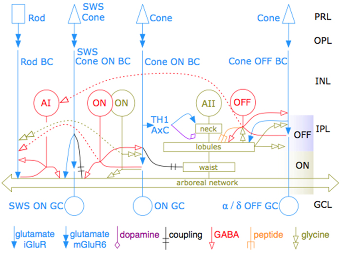
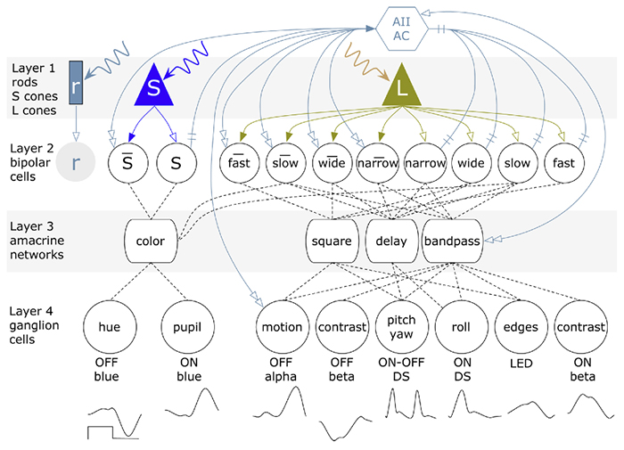
Purpose: Functional mapping in tiger salamander shows that bipolar cell (BC) projective fields far exceed their axonal fields, and directly implicates wide-field GABAergic amacrine cells (wf γACs) and gap junctions (Asari & Meister, 2014). Strikingly, single BCs exert differential effects on functionally distinct ganglion cells (GCs), likely achieved by privatized amacrine cell (AC) presynaptic inhibition to specific BC-GC synaptic pairs (Asari & Meister, 2012). To address whether BC projective fields in the mammal are equally broad, wf γAC- and gap junction-dependent, and GC type unselective, we reconstructed all electrical and chemical synaptic partners of a single ON cone BC in the inner plexiform layer of the rabbit retina, and searched BC-GC synaptic pairs for differential synaptic inhibition.

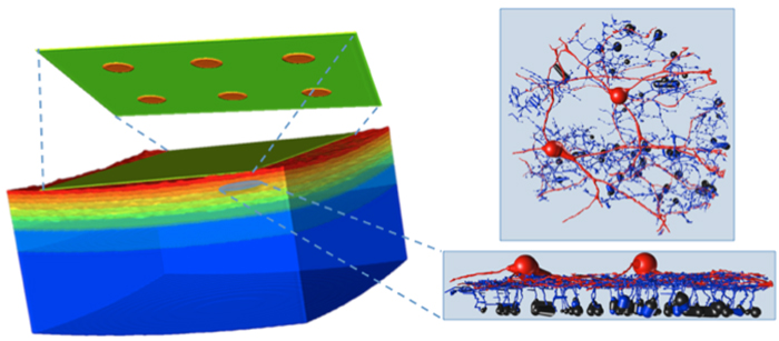
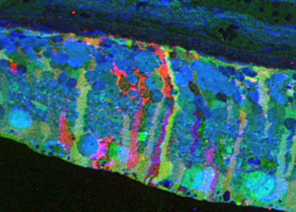
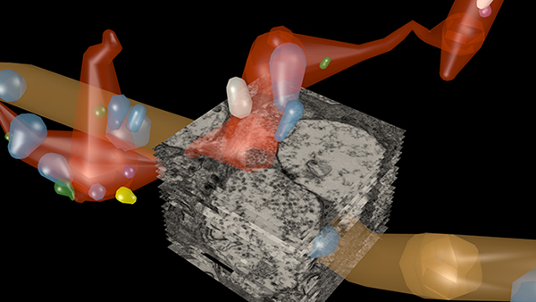
Methods: Cells in retinal connectome 1 (RC1) were annotated with Viking viewer, and explored via connectivity visualizations and 3D rendering (Anderson et al., 2011). Small molecule signals embedded in RC1, e.g. GABA, glycine, and L-glutamate, combined with morphological reconstruction and connectivity analysis allow robust cell classification. We used the MacNeil et al. (2004) rabbit BC classification scheme.

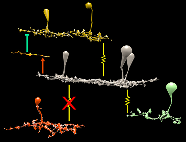
Results: CBb5w 593 is one of 20 ON cone BCs of this class in RC1. This CBb5w is presynaptic to 17 distinct GCs and 262 AC processes, and postsynaptic to 228 AC processes. The majority of these ACs are wf γACs. We estimate this BC forms synapses with 50 unique ACs. Asari & Meister (2014) found that single bipolar cell projective fields range up to 1 mm, far beyond a BC axonal field, and differentially drive multiple classes of GC. We discovered BC-BC within- and cross-class coupling and lateral inhibition that construct sign-conserving and sign-inverting projective fields to many distinct ganglion cell classes across the entire 0.25 mm diameter of RC1, much greater than a 60 µm BC axonal field. Cross-class projections access a broader set of GCs than expected from in-class projections alone. The BC-BC coupling is independent of BC-AII AC coupling. 94% of the CBb5w 593 BC-GC synaptic pairs receive feedback inhibition within the varicosity of the ribbon, but the number of feedback synapses is highly variable (coefficient of variation = 0.81). 35% of the BC-GC pairs receive feedforward inhibition within 2 microns of the postsynaptic density.

Conclusions: Mammalian BCs use novel cross-class topologies to distribute signals to a wide range of GCs and establish projective fields similar to those discovered in non-mammalian species. BC-BC within- and cross-class coupling and lateral inhibition via wf γACs establish sign-conserving and sign-inverting projective fields, respectively, up to 1 mm diameters. BC-GC synaptic pairs overwhelmingly employ feedback vs. feedforward inhibition to modulate signaling, and the numbers of feedback synapses are highly variable across these pairs, accounting for privatized and differential GC responses to the same BC drive.

A Synaptic Basis for Small World Network Design in the ON Inner Plexiform Layer of the Rabbit Retina

Bipolar cells_

This abstract was presented today at the 2014 Association for Research in Vision and Opthalmology (ARVO) meetings in Orlando, Florida by J Scott Lauritzen, Noah T. Nelson, Crystal L. Sigulinsky, Nathan Sherbotie, John Hoang, Rebecca L. PfeifferJames R. Anderson, Carl B. Watt, Bryan W. Jones and Robert E. Marc.

Purpose: Converging evidence suggests that large- and intermediate-scale neural networks throughout the nervous system exhibit small world’ design characterized by high local clustering of connections yet short path length between neuronal modules (Watts & Strogatz 1998 Nature; Sporns et al.2004 Trends in Cog Sci). It is suspected that this organizing principle scales to local networks (Ganmor et al. 2011 J Neurosci; Sporns 2006 BioSystems) but direct observation of synapses and local network topologies mediating small world design has not been achieved in any neuronal tissue. We sought direct evidence for synaptic and topological substrates that instantiate small world network architectures in the ON inner plexiform layer (IPL) of the rabbit retina. To test this we mined ≈ 200 ON cone bipolar cells (BCs) and ≈ 500 inhibitory amacrine cell (AC) processes in the ultrastructural rabbit retinal connectome (RC1).

Methods: BC networks in RC1 were annotated with the Viking viewer and explored via graph visualization of connectivity and 3D rendering (Anderson et al. 2011 J Microscopy). Small molecule signals embedded in RC1 e.g. GABA glycine and L-glutamate combined with morphological reconstruction and connectivity analysis allow for robust cell classification. MacNeil et al. (2004 J Comp Neurol) BC classification scheme used for clarity.

Results: Homocellular BC coupling (CBb3::CBb3 CBb4::CBb4 CBb5::CBb5) and within-class BC inhibitory networks (CBb3 → AC –| CBb3 CBb4 → AC –| CBb4 CBb5 → AC –| CBb5) in each ON IPL strata form laminar-specific functional sheets with high clustering coefficients. Heterocellular BC coupling (CBb3::CBb4 CBb4::CBb5 CBb3::CBb5) and cross-class BC inhibitory networks (CBb3 → AC –| CBb4 CBb4 → AC –| CBb3 CBb4 → AC –| CBb5 CBb5 → AC –| CBb4 CBb3 → AC –| CBb5 CBb5 → AC –| CBb3) establish short synaptic path lengths across all ON IPL laminae.

Conclusions: The retina contains a greater than expected number of synaptic hubs that multiplex parallel channels presynaptic to ganglion cells. The results validate a synaptic basis (ie. direct synaptic connectivity) and local network topology for the small world architecture indicated at larger scales providing neuroanatomical plausibility of this organization for local networks and are consistent with small world design as a fundamental organizing principle of neural networks on multiple spatial scales.

Support:  NIH EY02576 (RM), NIH EY015128 (RM), NSF 0941717 (RM), NIH EY014800 Vision Core (RM), RPB CDA (BWJ), Thome AMD Grant (BWJ).

Metabolic Changes Associated With Müller Cells In A Transgenic Rabbit Model Of Retinal Degeneration

Retina-RLP

This abstract was presented today at the 2014 Association for Research in Vision and Opthalmology (ARVO) meetings in Orlando, Florida by  Rebecca L. PfeifferBryan W. Jones and Robert E. Marc.

Purpose: Müller cells play a central role in retinal metabolism via the glutamate cycle. During retinal degeneration Müller cells are among the first to demonstrate changes, reflected in alterations of metabolic signatures and morphology. The timing, extent and regulation of these changes is not fully characterized. To address this issue, we evaluated Müller cell metabolic phenotypes at multiple stages of retinal remodeling.

Methods: Samples were collected post-mortem from both WT and P347L rabbits. The retinas were then divided into fragments, fixed in buffered aldehydes, and embedded in epoxy resins. Tissues were sectioned at 200nm followed by classification with computational molecular phenotyping (CMP) using an array of small and macromolecular signatures (aspartate (D), glutamate (E), glycine (G), glutamine (Q), glutathione (J), GABA (yy), taurine (T), CRALBP, Glutamine Synthetase (GS), and GFAP). Levels of amino acid or protein were quantified by selecting a region of interest either within the Müller cell population or surrounding neurons and evaluating the intensity of the signal within that region.

Results: CMP reveals overall decreases in GS levels over the course of degeneration. Of notable importance, we saw that in regions of near complete photoreceptor loss neighboring Müller cells may express independent variation in metabolic signatures of E, Q, and GS. Also observed in these Müller cells, ratios of GS:E and GS:Q are not consistent with the ratios seen in WT retina. These results are inconsistent with the current models of both E to Q metabolism and microenvironment regulation of Müller cell phenotypes.

Conclusions: These observations indicate two conclusions. First, although the degenerate state of the retina is the likely trigger inducing Müller cells to express altered metabolic signatures, the rate at which the metabolic state changes is not purely a product of the surrounding environment, but also a stochastic change within individual Müller cells. Second, although it is commonly accepted that GS is the primary enzyme which converts Q to E as part of the glutamate cycle, in degenerate retina alternative pathways may be utilized following decrease in GS.

Support: NIH EY02576 (RM), NIH EY015128 (RM), NSF 0941717 (RM), NIH EY014800 Vision Core (RM), RPB CDA (BWJ), Thome AMD Grant (BWJ).