Category Archives: Connectomics

Aii Amacrine Cell Connectivity in Degenerating Retina

This abstract was presented today, April 8th at the 2019 Association for Research in Vision and Opthalmology (ARVO) meetings in Vancouver, Canada by Jeebika Dahal, Rebecca L. Pfeiffer, Crystal L. Sigulinsky, James R. Anderson, Daniel P. Emrich, Hope Morrison, Jessica C. Garcia, Kevin D. Rapp, Jia-Hui Yang, Carl B. Watt, Mineo Kondo, Hiroko Terasaki, Robert E. Marc and Bryan W. Jones.

Full resolution version here.

Purpose
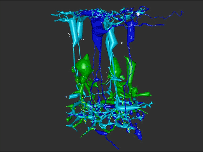
Aii amacrine cells (Aii ACs) function in mediating scotopic vision via connection of rod bipolar cells (Rod BCs) to cone bipolar cell pathways. The purpose of this project is to determine the effect of retinal degeneration (RD) on Aii AC networks. We explore this in a pathoconnectome of early RD (RPC1), using a connectome of healthy retina (RC1) as control. Cells in each volume are evaluated by comparison of morphology, synaptic connectivity, and eventually network analysis.

Methods
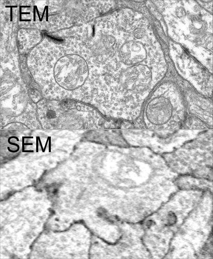
Tissue for RPC1 was collected from a 10 month old transgenic p347L rabbit model of autosomal dominant retinitis pigmentosa. RC1 was collected from a 13 month old Dutch-Belted rabbit, with no indications of degeneration. Tissue was fixed in a mixed aldehyde solution, before subsequent dehydration, osmication, and resin embedding. Volumes were sectioned at 70nm (RPC1) and 90nm (RC1) and placed on formvar grids. 1 section was reserved from every 30 TEM sections for computational molecular phenotyping where it was placed on a slide and probed for small molecules or proteins. TEM sections were captured at 2.18nm/px using SerialEM software on a JEOL JEM-1400 TEM. The RC1 volume has a diameter of 250µm and RPC1 has a diameter of 90µm. Both volumes were analyzed using the Viking software suite.

Results
In this study, Aii ACs from RPC1 were compared to RC1. Initial results indicate no distinct difference in the morphology other than arbor size, which are likely due to eccentricity differences between volumes. However, in RPC1, we observe multiple instances of Aii AC coupling with Rod BCs in the ON region of the IPL. In contrast, Rod BCs never form gap junctions in healthy retina.

Conclusions
Coupling between Aii ACs and Rod BCs in RPC1 is a unique change in retinal network topology occurring in early RD. Further exploration of network changes as a response to RD is warranted, as many therapeutic interventions currently in development rely upon maintenance of inner retinal circuitry. Prior research demonstrates Rod BCs extend dendrites towards cones and change their receptor expression as rods degenerate. Therefore, knowing the network changes involving Aii ACs and their associations with bipolar cells is crucial to understanding how photoreceptor degeneration affects inner retinal visual processing.

OFF-layer Branches of ON Cone Bipolar Cells in Early Retinal Degeneration

This abstract was presented today, April 8th at the 2019 Association for Research in Vision and Opthalmology (ARVO) meetings in Vancouver, Canada by Jessica C. Garcia, Rebecca L. Pfeiffer, Crystal L. Sigulinsky, James R. Anderson, Daniel P. Emrich, Jeebika Dahal, Hope Morrison, Kevin D. Rapp, Jia-Hui Yang, Carl B. Watt, Mineo Kondo, Hiroko Terasaki, Robert E. Marc and Bryan W. Jones.

Full resolution version here.

Purpose: Cone bipolar cells are customarily classified into superclasses of ON (ON-BCs) and OFF (OFF-BCs). ON or OFF specialization is further segregated by stratification within the inner plexiform layer. Retinal degeneration induces negative plasticity termed remodeling, that includes aberrant neurite extension from multiple cell types (rewiring) and ON-BCs switching their glutamate receptors to match that of OFF-BCs (reprogramming). Previous analysis in healthy retina shows that ON-BCs can make small ribbon synapses in the descending axon or, less frequently, simple single branch projections in the OFF layer. What impact remodeling has on these OFF-layer branches is unknown. In this study, we compare OFF branches from ON-BCs in a connectome of early retinal degeneration (RPC1) to our healthy retinal connectome (RC1).

Methods: Retinal tissues selected for RC1 and RPC1 were collected post-mortem from a 13 month old Dutch-belted healthy female rabbit and a male 10 month old transgenic P347L rabbit model of autosomal dominant retinitis pigmentosa, respectively. RPC1 shows signs of remodeling including rod outer-segment degeneration and aberrant neurite extension. Tissues were fixed in mixed aldehydes and subsequently osmicated, dehydrated, resin embedded, and sectioned at 90 nm (RC1) or 70 nm (RPC1). Sections were placed on formvar grids, stained, and imaged at 2nm/px on a JEOL JEM-1400 TEM using SerialEM software. 1 section was reserved from every 30 for Computational Molecular Phenotyping, and probed for small molecules. Both volumes were evaluated using the Viking software suite.

Results: Ribbons of ON-BCs formed in the OFF layer branches have been previously described to contact glycinergic amacrine cells (ACs), GABAergic ACs, ON ganglion cells, and intrinsically photosensitive ganglion cells. Initial analysis of OFF branches of ON-BCs in RPC1 demonstrate more complex branching than in RC1 and increased number of synapses on these branches. In contrast to the inconsistent OFF layer branch stratification observed in RC1, the OFF branches in RPC1 appear to stratify at a similar level. Evaluation of synaptic partners is ongoing.

Conclusions: Increased complexity and number of synapses found in the OFF branches of some ON-BCs ultimately may represent ON network corruption. Exploring synaptic partners will reveal potential network alterations in retinal degenerative disease.

Rod Bipolar Cell Networks In A Retinal Pathoconnectome

We presented a poster on Rod Bipolar Cell Networks In A Retinal Pathoconnectome at the 2019 HHMI Connectomics meeting in Berlin today. Downsampled PDF of poster here.

Authors: Rebecca L. Pfeiffer, James R. Anderson, Daniel P. Emrich, Jeebika Dahal, Crystal L. Sigulinsky, Jia-Hui Yang, Kevin D. Rapp, Carl B. Watt, Jessica C. Garcia, Mineo Kondo, Hiroko Terasaki, Robert E. Marc, and Bryan W. Jones.

Abstract: Ultrastructural connectomics has allowed for precise identification of neural network topologies in retina, exposing synaptic connectivity associated with specific pathways involved in neural retinal processing. In pathological degenerate retina such as retinitis pigmentosa (RP), retinal remodeling emerges as a phenomenon through a series of negative plasticity events originating from neural deafferentation initiated by photoreceptor degeneration. Early stages of remodeling include glial changes, GluR receptor alterations (reprogramming), and rewiring of retinal networks. The connectivities initiated by these processes are currently unknown. To address this problem, we have created an ultrastructural pathoconnnectome of early retinal remodeling in a rabbit model of retinitis pigmentosa, Retinal Pathoconnectome 1 (RPC1).

Heterocellular Coupling Between Amacrine Cells and Ganglion Cells

We have a new paper out In Frontiers in Neural Circuits, Heterocellular Coupling Between Amacrine Cells and Ganglion Cells. This manuscript preprint was published in BioRxiv.

Authors: Robert E. Marc, Crystal Lynn Sigulinsky, Rebecca L. Pfeiffer, Daniel Emrich, James Russel Anderson and Bryan William Jones.

Abstract: All superclasses of retinal neurons, including bipolar cells (BCs), amacrine cells (ACs) and ganglion cells (GCs), display gap junctional coupling. However, coupling varies extensively by class. Heterocellular AC coupling is common in many mammalian GC classes. Yet, the topology and functions of coupling networks remains largely undefined. GCs are the least frequent superclass in the inner plexiform layer and the gap junctions mediating GC-to-AC coupling (GC::AC) are sparsely arrayed amidst large cohorts of homocellular AC::AC, BC::BC, GC::GC and heterocellular AC::BC gap junctions. Here, we report quantitative coupling for identified GCs in retinal connectome 1 (RC1), a high resolution (2 nm) transmission electron microscopy-based volume of rabbit retina. These reveal that most GC gap junctions in RC1 are suboptical. GC classes lack direct cross-class homocellular coupling with other GCs, despite opportunities via direct membrane contact, while OFF alpha GCs and transient ON directionally selective (DS) GCs are strongly coupled to distinct AC cohorts. Integrated small molecule immunocytochemistry identifies these as GABAergic ACs (γ+ ACs). Multi-hop synaptic queries of RC1 connectome further profile these coupled γ+ ACs. Notably, OFF alpha GCs couple to OFF γ+ ACs and transient ON DS GCs couple to ON γ+ ACs, including a large interstitial amacrine cell, revealing matched ON/OFF photic drive polarities within coupled networks. Furthermore, BC input to these γ+ ACs is tightly matched to the GCs with which they couple. Evaluation of the coupled versus inhibitory targets of the γ+ ACs reveals that in both ON and OFF coupled GC networks these ACs are presynaptic to GC classes that are different than the classes with which they couple. These heterocellular coupling patterns provide a potential mechanism for an excited GC to indirectly inhibit nearby GCs of different classes. Similarly, coupled γ+ ACs engaged in feedback networks can leverage the additional gain of BC synapses in shaping the signaling of downstream targets based on their own selective coupling with GCs. A consequence of coupling is intercellular fluxes of small molecules. GC::AC coupling involves primarily γ+ cells, likely resulting in GABA diffusion into GCs. Surveying GABA signatures in the GC layer across diverse species suggests the majority of vertebrate retinas engage in GC::γ+ AC coupling.

Rod Bipolar Cell Networks in Early Retinal Remodeling

Rebecca Pfeiffer, a post-doc in the laboratory presented her work on “Rod Bipolar Cell Networks in Early Retinal Remodeling” as a platform presentation at the ISER 2018 meeting in Belfast, Northern Ireland.

Authors: Rebecca Pfeiffer, James R. Anderson, Daniel P. Emrich, Jeebika Dahal, Crystal L Sigulinsky, Hope AB Morrison, Jia-Hui Yang, Carl B. Watt, Kevin D. Rapp, Jessica C Garcia, Mineo Kondo, Hiroko Terasaki, Robert E. Marc, and Bryan W. Jones.

Abstract: Retinal remodeling is a form of negative plasticity that occurs as a consequence of retinal degenerative diseases. Part of retinal remodeling involves anomalous sprouting of processes, termed neurites. The synaptic structures and partners of the neurites are not yet defined, leading to uncertainty about the consistency of network motifs between healthy and degenerate retina. Our goal is to map out the identities and network relationships of bipolar cell networks using a connectomics strategy. Retinal connectomes or ultrastructural maps of neuronal connectivity have substantially contributed to our understanding of retinal network topology, providing ground truth against which pathological network topologies can be evaluated. We have generated the first pathoconnectome (RPC1), or connectome of pathological tissues, of early retinal remodeling at 2nm/pixel, and are currently investigating the impact of remodeling on network architecture.
The tissue for RPC1 was obtained from a 10mo transgenic P347L rabbit model of autosomal dominant retinitis pigmentosa. Tissue was fixed in mixed aldehydes, osmicated, dehydrated, embedded in epon resin, and sectioned at 70nm. Serial sections were placed on grids, stained, and imaged using a JEOL JEM-1400 TEM using SerialEM software. Every 30th section was reserved for computational molecular phenotyping (CMP), and probed for small molecules: glutamate, glutamine, glycine, GABA, taurine, glutathione; or TEM compatible proteins GFAP and GS. The pathoconnectome volume is explored and annotated using the Viking software suite.
RPC1 was selected as an example of early retinal remodeling, demonstrating Muller cell hypertrophy, metabolic dysregulation, and degeneration of rod outer segments, indicating phase 1 remodeling and neuronal sprouting. We have observed the presence of both cone pedicles and rod spherules within the OPL to be synaptically active with neurites from some rod bipolar cells forming functional synapses with both rod spherules and cone pedicles. These rod bipolar cells also exhibit structurally altered ribbon synapses. We are currently evaluating network motifs and comparing them to networks established from our previous connectome, RC1, generated from a healthy rabbit.
These findings allow us to evaluate and analyze the impact of retinal remodeling on retinal networks which may have important implications for therapeutic interventions being developed which rely on inner retina network integrity.

Coupling Architecture Of The Aii/ON Cone Bipolar Cell Network In The Degenerate Retina

Crystal Sigulinsky, a post-doc in the lab, presented her work on “coupling architecture of the
Aii/ON cone bipolar cell network in the degenerate retina” at the RD2018 meeting in Killarney, Ireland today.  Authors are: Crystal L Sigulinsky, Rebecca L Pfeiffer, James R Anderson, Jeebika Dahal, Hope Morrison, Daniel P. Emrich, Jessica C Garcia, Jia-Hui Yang, Carl B. Watt, Kevin D. Rapp, Mineo Kondo, Hiroko Terasaki, Robert E. Marc, and Bryan W. Jones.

Purpose: Retinal network hyperactivity within degenerative retinal networks is a component of the disease process with implications for therapeutic interventions for blinding diseases that depend upon the surviving retinal network. Connexin36-containing gap junctions centered on the Aii amacrine cell network appear to mediate the aberrant signaling observed in mouse models of retinal degeneration. However, it remains unclear whether this hyperactivity reflects changes in the underlying circuitry or dysfunction/dysregulation of the normative circuitry. Mapping retinal circuitry in the ultrastructural rabbit Retinal Connectome, RC1, has revealed Aii network topologies explicitly involving gap junctions. In addition to canonical Aii-to-Aii and Aii-to-ON cone bipolar cell (CBC) coupling, we describe pervasive in- and cross-class coupling motifs among ON CBCs that extend and dramatically expand the coupled Aii network topologies. Since virtually every gap junction in the inner plexiform layer contains Connexin36, these circuits likely participate in the aberrant signaling of degenerate retinas. This study examines these Aii and ON CBC coupling motifs in Retinal PathoConnectome 1 (RPC1), an ultrastructural pathoconnectome of a rabbit model of retinitis pigmentosa.

Approach: RPC1 is a 2nm/pixel resolution volume of retina from a 10 month old, transgenic P347L rabbit model of autosomal dominant retinitis pigmentosa in early phase 1 retinal remodeling, a time point where cone and rod photoreceptors are still present, albeit going through cell stress. RPC1 spans the vitreous to basal outer nuclear layer and was built by automated transmission electron microscopy and computational assembly. ON CBCs, Aii amacrine cells, and their coupling partners were annotated using the Viking application and explored with 3D rendering and graph visualization of connectivity. Gap junctions were validated by 0.25 nm resolution recapture with goniometric tilt when necessary. Motifs were compared to those discovered in RC1. RC1 is a 2 nm resolution, 0.25 mm diameter volume of a light-adapted adult female Dutch Belted rabbit retina spanning the ganglion cell through inner nuclear layers.

Conclusions: RPC1 shows degeneration of rod outer segments, Müller cell hypertrophy and neuronal sprouting, characteristic of early stage retinal degeneration and phase 1 remodeling, when retinal hyperactivity and its reliance on gap junctional coupling has likely already initiated and human patients would still have some vision. All major coupling motifs (Aii-to-Aii, Aii-to-ON CBC, and ON CBC-to-ON CBC) were observed. Preliminary examinations indicate that several ON CBC classes retained their class-specific coupling profiles, accepting and rejecting specific combinations of Aii and ON CBC class partnerships. However, recent findings reveal aberrant partnerships in the coupled network, including both loss of prominent motifs and acquisition of novel ones. Thus, clear aberrant morphological and synaptic changes have been identified in RPC1, including changes in the coupling specificity and gap junction distributions of both Aii amacrine cells and ON CBCs (Figure 6). This suggests that the Aii/ON CBC circuit topology is already altered during early phase 1 remodeling, with substantial implications for therapeutic interventions in human subjects. The full coupling network is actively being examined and progress has begun on RPC2, a second pathoconnectome for examining later, phase 2 remodeling in this same model.

An almost full size poster available here in pdf format.

SEM vs. TEM

SEM vs. TEM is a tradeoff of convenience, resolution, cost and speed. The very physics of SEM signal integration means that the fundamental acquisition time for large canonical volume datasets are incompatible with 5 year grant cycles. SEM based approaches can potentially rival TEM, but dwell time/pixel increases logarithmically with resolution.

To give you some idea for the resolution differences at routine capture speeds, both of these above images capture a region within the inner plexiform layer of retina, looking at bipolar cell terminals. The TEM image was captured at a standard operating resolution of 2nm/pixel. The SEM image was captured at 16nm/pixel. You cannot see any gap junctions that might be present in the SEM image and you can only infer or guess at synaptic ribbons. And look at the texture!

You *can* get better resolution with SEM, but as I said before, the capture time increases logarithmically. To accomplish what we perform in 8-10 hours with a TEM, would take 108-115 hours on a current, cutting edge multi beam SEM. There are many other advantages of TEM including the ability to capture higher resolution images faster, be able to re-image in goniometric tilt series, be able to integrate molecular markers inside connectome volumes, and a TEM is about 1/3rd the cost of an SEM. Also, SEM images tend to be texturally poor as they are made from capturing electron backscatter of surfaces rather than made by projection of electrons through a small volume, and there is tremendous value in the texture of ultrastructural images. Ergo, this is why we use TEM.

This is not to say that SEM is not a great tool. It is just not the best tool for large scale connectomics where you have to have the resolution to capture all synapses and gap junctions, over large areas. For smaller volumes that do not require a canonical sampling of cell classes, SEM is absolutely an appropriate tool.

This content was originally published on Jonesblog.

Nornir listed on NITRC

Nornir is now registered with NeuroImaging Tools & Resources Collaboratory (NITRC).

Nornir’s takes large sets of overlapping images in 2D and produces registered (a.k.a. aligned) 2D mosaics and 3D volumes of any size and scale. Registered slices may be exported as a single large images or viewed/annoted with our Viking viewer.

Nornir has been used successfully on transmission electron microscopy, scanning electron microscopy images, and light microscopy images. Nornir supports interleaving different imaging methods into the same volume. Support for SerialEM, Objective Imaging, and Digital Micrograph (DM4) raw data is available. Adding formats is not complicated and the author will consider requests.

Nornir runs on fairly humble hardware for the task. A 32-core 64GB Xeon system built a ~60 TB 250um diameter 2.12nm/pixel volume from roughly 1400 slices. Nornir works incrementally, only updating data that has changed.

Installation is fairly simple and primarily uses Python’s PIP installer.

For further information: http://nornir.github.io/

A Pathoconnectome of Early Retinal Remodeling

This abstract was presented today, Monday, April 30th at the 2018 Association for Research in Vision and Opthalmology (ARVO) meetings in Honolulu, Hawaii by Rebecca Pfeiffer, Robert E. Marc, James R. Anderson, Daniel P. Emrich, Carl B. Watt, Jia-Hui Yang, Kevin D. Rapp, Jeebika Dahal, Mineo Kondo, Hiroko Terasaki, and Bryan W. Jones.

Purpose:
Retinal remodeling is a consequence of retinal degenerative disease, during which neurons sprout new neurites whose synaptic structures and partners are not yet defined. Simultaneously during remodeling, Müller cells (MCs) undergo structural and metabolic changes, whose impact on surrounding neurons is an active area of research. Retinal connectomes have elucidated and validated fundamental networks. These data provide further classification of neuronal types and subtypes and a precise framework for modeling of retinal function, based on ground truth networks. The creation of the first pathoconnectome (RPC1), a connectome from pathological retinal tissue, provides the opportunity to determine connectivites between neurons, while simultaneously evaluating glial remodeling. Computational Molecular Phenotyping (CMP) embedded within the ultrastructure provides metabolic factors of pathologies.

Methods:
RPC1 was collected post-mortem from a 10mo TgP347L rabbit model of adRP, fixed in 1% FA, 2.5% GA, 3% sucrose, and 1mM MgSO4 in cacodylate buffer (pH 7.4). The tissue was osmicated, dehydrated, resin embedded, and sectioned at 70nm. Sections were placed on formvar grids, stained, and imaged on a JEOL JEM-1400 TEM using SerialEM. 1 section was reserved from every 30 section for CMP, where it was probed for small molecules: glutamate, glutamine, glycine, GABA, taurine, glutathione; or proteins GFAP and GS. RPC1 was evaluated using the Viking software suite.

Results:
RPC1 was chosen based on early features of retinal degeneration/remodeling: degeneration of rod OS, MC hypertrophy, and neuronal sprouting. RPC1 consists of 948 serial sections spanning the ONL to the vitreous, with a diameter of 90µm. We find dendrites extending from rod bipolar cells to cone pedicles, originally described in light microscopy, and active synaptic contacts. We also see alterations of synaptic structure in the IPL, and MC morphological changes affecting surface to volume and neuron/glial relationships. Network motifs are being actively investigated.

Conclusions:
We observe many features of remodeling previously described using light microscopy, and confirm active synaptic contact. We also find synaptic structural features, not previously described. In addition, early evaluation of MC morphology demonstrates marked changes in MC shape and associations with nearby neurons and glia, which, combined with CMP, will be instrumental in understanding how MCs affect retinal remodeling.