Category Archives: Publications

An Update On Retinal Prostheses

We have a new manuscript out in Clinical Neurophysiology, An Update on Retinal Prostheses. PubMedDirect Link PDF here.

Authors: Lauren N. Ayton, Nick Barnes, Gislin Dagnelie, Takashi Fujikado, Georges Goetz, Ralf Hornig, Bryan W. Jones, Mahiul M.K. Muqit, Daniel L. Rathbun, Katarina Stingl, James D. Weiland, Matthew A. Petoe.

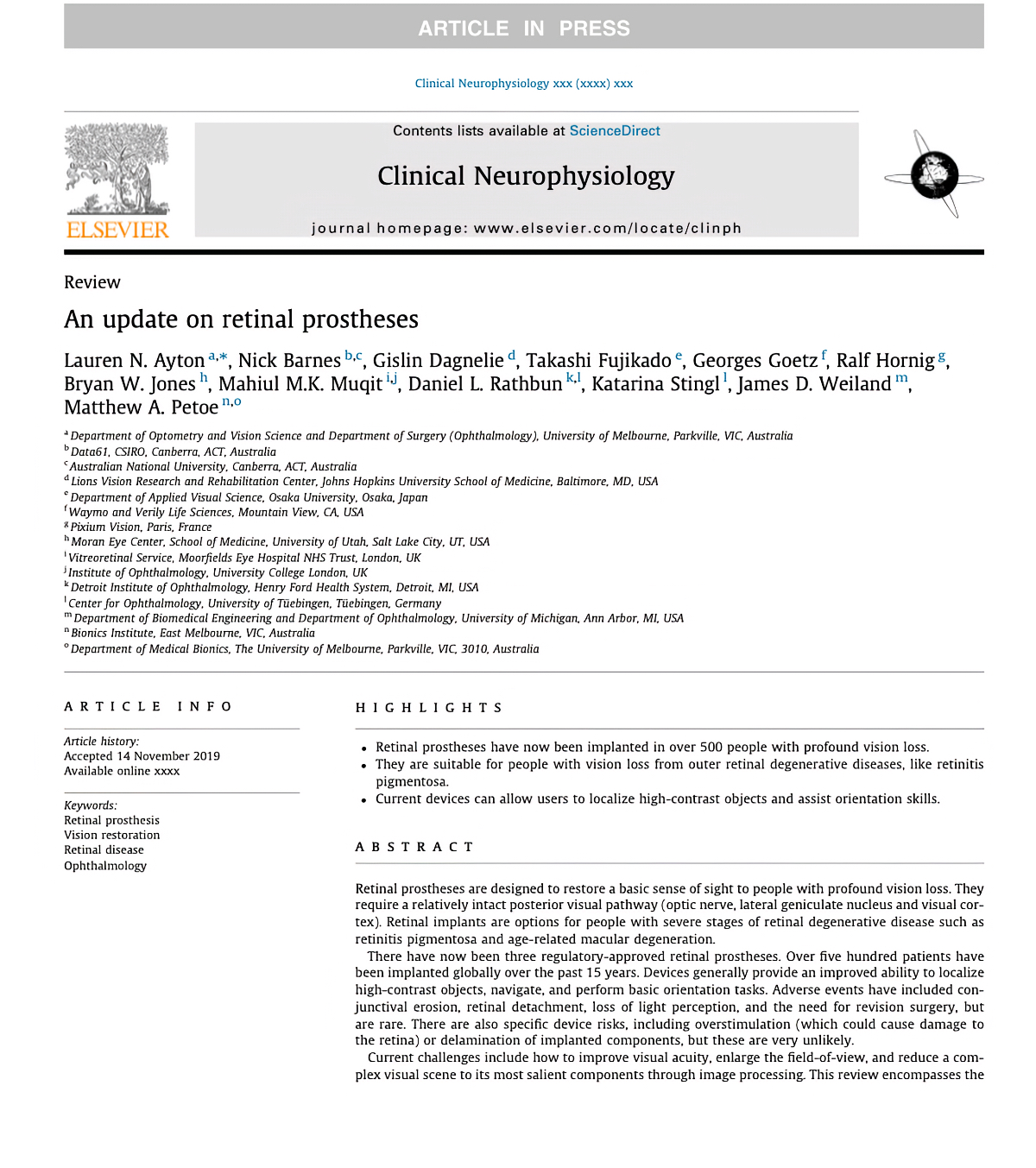
Abstract: Retinal prostheses are designed to restore a basic sense of sight to people with profound vision loss. They require a relatively intact posterior visual pathway (optic nerve, lateral geniculate nucleus and visual cor- tex). Retinal implants are options for people with severe stages of retinal degenerative disease such as retinitis pigmentosa and age-related macular degeneration.

There have now been three regulatory-approved retinal prostheses. Over five hundred patients have been implanted globally over the past 15 years. Devices generally provide an improved ability to localize high-contrast objects, navigate, and perform basic orientation tasks. Adverse events have included con- junctival erosion, retinal detachment, loss of light perception, and the need for revision surgery, but are rare. There are also specific device risks, including overstimulation (which could cause damage to the retina) or delamination of implanted components, but these are very unlikely.
Current challenges include how to improve visual acuity, enlarge the field-of-view, and reduce a com- plex visual scene to its most salient components through image processing. This review encompasses the work of over 40 individual research groups who have built devices, developed stimulation strategies, or investigated the basic physiology underpinning retinal prostheses. Current technologies are summarized, along with future challenges that face the field.


Contrast Sensitivity Isocontours Of The Central Visual Field

We have a new manuscript out in Nature Scientific Reports, Contrast Sensitivity Isocontours Of The Central Visual Field.

Authors: Agnes Y. J. Choi, Lisa Nivison-Smith, Jack Phu, Barbara Zangerl, Sieu K. Khuu, Bryan W. Jones, Rebecca L. Pfeiffer, Robert E. Marc & Michael Kalloniatis.

Abstract: Standard automated perimetry (SAP), the most common form of perimetry used in clinical practice, is associated with high test variability, impacting clinical decision making and efficiency. Contrast sensitivity isocontours (CSIs) may reduce test variability in SAP by identifying regions of the visual field with statistically similar patterns of change that can be analysed collectively and allow a point (disease)-to-CSI (normal) comparison in disease assessment as opposed to a point (disease)-to-point (normal) comparison. CSIs in the central visual field however have limited applicability as they have only been described using visual field test patterns with low, 6° spatial sampling. In this study, CSIs were determined within the central 20° visual field using the 10-2 test grid paradigm of the Humphrey Field Analyzer which has a high 2° sampling frequency. The number of CSIs detected in the central 20° visual field was greater than previously reported with low spatial sampling and stimulus size dependent: 6 CSIs for GI, 4 CSIs for GII and GIII, and 3 CSIs for GIV and GV. CSI number and distribution were preserved with age. Use of CSIs to assess visual function in age-related macular degeneration (AMD) found CSI guided analysis detected a significantly greater deviation in sensitivity of AMD eyes from normal compared to a standard clinical pointwise comparison (−1.40 ± 0.15 dB vs −0.96 ± 0.15 dB; p < 0.05). This work suggests detection of CSIs within the central 20° is dependent on sampling strategy and stimulus size and normative distribution limits of CSIs can indicate significant functional deficits in diseases affecting the central visual field such as AMD.

Development Of A Spatial Model Of Age-Related Change In The Macular Ganglion Cell Layer To Predict Function From Structural Changes

We have a new paper out in the Americal Journal of Ophthalmology, Development of a spatial model of age-related change in the macular ganglion cell layer to predict function from structural changes.

Authors: Janelle Tong, Jack Phu, Sieu K. Khuu, Nayuta Yoshioka, Agnes Y. Choi, Lisa Nivison-Smith, Robert E. Marc, Bryan W. Jones, Rebecca L. Pfeiffer, Michael Kalloniatis, and Barbara Zangerl.

Purpose: To develop location specific models of normal, age-related changes in the macular ganglion cell layer (GCL) from optical coherence tomography (OCT). Using these OCT-derived models, we predicted visual field (VF) sensitivity and compared these results to actual VF sensitivities.

Design: Retrospective cohort study

Methods: Single eyes of 254 normal participants were retrospectively enrolled from the Centre for Eye Health (Sydney, Australia). Macular GCL measurements were obtained using Spectralis OCT. Cluster algorithms were performed to identify spatial patterns demonstrating similar age- related change. Quadratic and linear regression models were subsequently utilized to characterize age-related GCL decline. 40 participants underwent additional testing with Humphrey VFs, and 95% prediction intervals were calculated to measure the predictive ability of structure-function models incorporating cluster-based pooling, age-correction and consideration of spatial summation.

Results: Quadratic GCL regression models provided a superior fit (p = <0.0001-0.0066), establishing that GCL decline commences in the late 30’s across the macula. The equivalent linear rates of GCL decline showed eccentricity-dependent variation (0.13μm/year centrally versus 0.06μm/year peripherally), however average, normalized GCL loss per year was consistent across the 64 macular measurement locations at 0.26%. The 95% prediction intervals describing predicted VF sensitivities were significantly narrower across all cluster- based structure-function models (3.79-4.99dB) compared with models without clustering applied (5.66-6.73dB, p <0.0001).

Conclusions: Combining spatial clustering with age-dependent regression allowed the development of robust models describing GCL changes with age. The resultant superior predictive ability of VF sensitivity from ganglion cell measurements may be applied to future models of disease development to improve detection of early macular GCL pathology.

Hypoxia Tolerance In The Norrin-Deficient Retina And The Chronically Hypoxic Brain Studied At Single-Cell Resolution

We have a new paper out in PNAS, Hypoxia tolerance in the Norrin-deficient retina and the chronically hypoxic brain studied at single-cell resolution.

Authors: Jacob S. Heng, Amir Rattner, Genevieve L. Stein-O’Brien, Briana L. Winer, Bryan W. Jones, Hilary J. Vernon, Loyal A. Goff, and Jeremy Nathans.

Abstract: The mammalian CNS is capable of tolerating chronic hypoxia, but cell type-specific responses to this stress have not been systematically characterized. In the Norrin KO (NdpKO) mouse, a model of familial exudative vitreoretinopathy (FEVR), developmental hypovascularization of the retina produces chronic hypoxia of inner nuclear-layer (INL) neurons and Muller glia. We used single-cell RNA sequencing, untargeted metabolomics, and metabolite labeling from 13C-glucose to compare WT and NdpKO retinas. In NdpKO retinas, we observe gene expression responses consistent with hypoxia in Muller glia and retinal neurons, and we find a metabolic shift that combines reduced flux through the TCA cycle with increased synthesis of serine, glycine, and glutathione. We also used single-cell RNA sequencing to compare the responses of individual cell types in NdpKO retinas with those in the hypoxic cerebral cortex of mice that were housed for 1 week in a reduced oxygen environment (7.5% oxygen). In the hypoxic cerebral cortex, glial transcriptome responses most closely resemble the response of Muller glia in the NdpKO retina. In both retina and brain, vascular endothelial cells activate a previously dormant tip cell gene expression program, which likely underlies the adaptive neoangiogenic response to chronic hypoxia. These analyses of retina and brain transcriptomes at single-cell resolution reveal both shared and cell type-specific changes in gene expression in response to chronic hypoxia, implying both shared and distinct cell type-specific physiologic responses.

Rod Bipolar Cell Networks In A Retinal Pathoconnectome

We presented a poster on Rod Bipolar Cell Networks In A Retinal Pathoconnectome at the 2019 HHMI Connectomics meeting in Berlin today. Downsampled PDF of poster here.

Authors: Rebecca L. Pfeiffer, James R. Anderson, Daniel P. Emrich, Jeebika Dahal, Crystal L. Sigulinsky, Jia-Hui Yang, Kevin D. Rapp, Carl B. Watt, Jessica C. Garcia, Mineo Kondo, Hiroko Terasaki, Robert E. Marc, and Bryan W. Jones.

Abstract: Ultrastructural connectomics has allowed for precise identification of neural network topologies in retina, exposing synaptic connectivity associated with specific pathways involved in neural retinal processing. In pathological degenerate retina such as retinitis pigmentosa (RP), retinal remodeling emerges as a phenomenon through a series of negative plasticity events originating from neural deafferentation initiated by photoreceptor degeneration. Early stages of remodeling include glial changes, GluR receptor alterations (reprogramming), and rewiring of retinal networks. The connectivities initiated by these processes are currently unknown. To address this problem, we have created an ultrastructural pathoconnnectome of early retinal remodeling in a rabbit model of retinitis pigmentosa, Retinal Pathoconnectome 1 (RPC1).

Heterocellular Coupling Between Amacrine Cells and Ganglion Cells

We have a new paper out In Frontiers in Neural Circuits, Heterocellular Coupling Between Amacrine Cells and Ganglion Cells. This manuscript preprint was published in BioRxiv.

Authors: Robert E. Marc, Crystal Lynn Sigulinsky, Rebecca L. Pfeiffer, Daniel Emrich, James Russel Anderson and Bryan William Jones.

Abstract: All superclasses of retinal neurons, including bipolar cells (BCs), amacrine cells (ACs) and ganglion cells (GCs), display gap junctional coupling. However, coupling varies extensively by class. Heterocellular AC coupling is common in many mammalian GC classes. Yet, the topology and functions of coupling networks remains largely undefined. GCs are the least frequent superclass in the inner plexiform layer and the gap junctions mediating GC-to-AC coupling (GC::AC) are sparsely arrayed amidst large cohorts of homocellular AC::AC, BC::BC, GC::GC and heterocellular AC::BC gap junctions. Here, we report quantitative coupling for identified GCs in retinal connectome 1 (RC1), a high resolution (2 nm) transmission electron microscopy-based volume of rabbit retina. These reveal that most GC gap junctions in RC1 are suboptical. GC classes lack direct cross-class homocellular coupling with other GCs, despite opportunities via direct membrane contact, while OFF alpha GCs and transient ON directionally selective (DS) GCs are strongly coupled to distinct AC cohorts. Integrated small molecule immunocytochemistry identifies these as GABAergic ACs (γ+ ACs). Multi-hop synaptic queries of RC1 connectome further profile these coupled γ+ ACs. Notably, OFF alpha GCs couple to OFF γ+ ACs and transient ON DS GCs couple to ON γ+ ACs, including a large interstitial amacrine cell, revealing matched ON/OFF photic drive polarities within coupled networks. Furthermore, BC input to these γ+ ACs is tightly matched to the GCs with which they couple. Evaluation of the coupled versus inhibitory targets of the γ+ ACs reveals that in both ON and OFF coupled GC networks these ACs are presynaptic to GC classes that are different than the classes with which they couple. These heterocellular coupling patterns provide a potential mechanism for an excited GC to indirectly inhibit nearby GCs of different classes. Similarly, coupled γ+ ACs engaged in feedback networks can leverage the additional gain of BC synapses in shaping the signaling of downstream targets based on their own selective coupling with GCs. A consequence of coupling is intercellular fluxes of small molecules. GC::AC coupling involves primarily γ+ cells, likely resulting in GABA diffusion into GCs. Surveying GABA signatures in the GC layer across diverse species suggests the majority of vertebrate retinas engage in GC::γ+ AC coupling.

Increasing Electrical Stimulation Efficacy in Degenerated Retina: Stimulus Waveform Design in a Multiscale Computational Model

We have a new publication out (direct link), Increasing Electrical Stimulation Efficacy in Degenerated Retina: Stimulus Waveform Design in a Multiscale Computational Model authored by Kyle Loizos, Robert Marc, Mark Humayun, James R. Anderson, Bryan W. Jones and Gianluca Lazzi.

Abstract—A computational model of electrical stimulation of the retina is proposed for investigating current waveforms used in prosthetic devices for restoring partial vision lost to retina degen- erative diseases. The model framework combines a connectome- based neural network model characterized by accurate mor- phological and synaptic properties with an Admittance Method model of bulk tissue and prosthetic electronics. In this model, the retina was computationally “degenerated,” considering cellular death and anatomical changes that occur early in disease, as well as altered neural behavior that develops throughout the neurodegeneration and is likely interfering with current attempts at restoring vision. A resulting analysis of stimulation range and threshold of ON ganglion cells within retina that are either healthy or in beginning stages of degeneration is presented for currently-used stimulation waveforms, and an asymmetric biphasic current stimulation for subduing spontaneous firing to allow increased control over ganglion cell firing patterns in degenerated retina is proposed. Results show that stimulation thresholds of retinal ganglion cells do not notably vary after beginning stages of retina degeneration. In addition, simulation of proposed asymmetric waveforms showed the ability to enhance the control of ganglion cell firing via electrical stimulation.

Corticostriatal Circuit Defects in Hoxb8 Mutant Mice

We have a new publication in Molecular Psychiatry, Corticostriatal circuit defects in Hoxb8 mutant mice. (Direct link here).  Authors are:  Naveen Nagarajan, Bryan W. Jones, Peter West, Robert Marc, and Mario R. Capecchi.

Abstract: Hoxb8 mutant mice exhibit compulsive grooming and hair removal dysfunction similar to humans with the obsessive-compulsive disorder (OCD)-spectrum disorder, trichotillomania. As, in the mouse brain, the only detectable cells that label with Hoxb8 cell lineage appear to be microglia, we suggested that defective microglia cause the neuropsychiatric disorder. Does the Hoxb8 mutation in microglia lead to neural circuit dysfunctions? We demonstrate that Hoxb8 mutants contain corticostriatal circuit defects. Golgi staining, ultra-structural and electrophysiological studies of mutants reveal excess dendritic spines, pre- and postsynaptic structural defects, long-term potentiation and miniature postsynaptic current defects. Hoxb8 mutants also exhibit hyperanxiety and social behavioral deficits similar to mice with neuronal mutations in Sapap3, Slitrk5 and Shank3, reported models of OCD and autism spectrum disorders (ASDs). Long-term treatment of Hoxb8 mutants with fluoxetine, a serotonin reuptake inhibitor, reduces excessive grooming, hyperanxiety and social behavioral impairments. These studies provide linkage between the neuronal defects induced by defective Hoxb8-microglia and neuronal dysfunctions directly generated by mutations in synaptic components that result in mice, which display similar pathological grooming, hyperanxiety and social impairment deficits. Our results shed light on Hoxb8 microglia-driven circuit-specific defects and therapeutic approaches that will become essential to developing novel
therapies for neuropsychiatric diseases such as OCD and ASDs with Hoxb8-microglia being the central target.

Pattern Recognition Analysis Reveals Unique Contrast Sensitivity Isocontours Using Static Perimetry Thresholds Across The Visual Field

We have a new publication in IOVS, Pattern Recognition Analysis Reveals Unique Contrast Sensitivity Isocontours Using Static Perimetry Thresholds Across The Visual Field (Direct link here).  Authors are:  Jack Phu, Sieu Khuu, Lisa Nivison-Smith, Barbara Zangerl, Agnes Yiu, Jeung Choi, Bryan W. JonesRebecca Pfeiffer, Robert Marc, and Michael Kalloniatis.

Purpose
To determine the locus of test locations that exhibit statistically similar age-related decline in sensitivity to light increments and age-corrected contrast sensitivity isocontours (CSIs) across the central visual field (VF). We compared these CSIs with test point clusters used by the Glaucoma Hemifield Test (GHT).

Methods
Sixty healthy observers underwent testing on the Humphrey Field Analyzer 30-2 test grid using Goldmann (G) stimulus sizes I-V. Age-correction factors for GI-V were determined using linear regression analysis. Pattern recognition analysis was used to cluster test locations across the VF exhibiting equal age-related sensitivity decline (age-related CSIs), and points of equal age-corrected sensitivity (age-corrected CSIs) for GI-V.

Results
There was a small but significant test size–dependent sensitivity decline with age, with smaller stimuli declining more rapidly. Age-related decline in sensitivity was more rapid in the periphery. A greater number of unique age-related CSIs was revealed when using smaller stimuli, particularly in the mid-periphery. Cluster analysis of age-corrected sensitivity thresholds revealed unique CSIs for GI-V, with smaller stimuli having a greater number of unique clusters. Zones examined by the GHT consisted of test locations that did not necessarily belong to the same CSI, particularly in the periphery.

Conclusions
Cluster analysis reveals statistically significant groups of test locations within the 30-2 test grid exhibiting the same age-related decline. CSIs facilitate pooling of sensitivities to reduce the variability of individual test locations. These CSIs could guide future structure-function and alternate hemifield asymmetry analyses by comparing matched areas of similar sensitivity signatures.