Tag Archives: Bryan W. Jones

Coupling Architecture Of The Aii/ON Cone Bipolar Cell Network In The Degenerate Retina

Crystal Sigulinsky, a post-doc in the lab, presented her work on “coupling architecture of the
Aii/ON cone bipolar cell network in the degenerate retina” at the RD2018 meeting in Killarney, Ireland today.  Authors are: Crystal L Sigulinsky, Rebecca L Pfeiffer, James R Anderson, Jeebika Dahal, Hope Morrison, Daniel P. Emrich, Jessica C Garcia, Jia-Hui Yang, Carl B. Watt, Kevin D. Rapp, Mineo Kondo, Hiroko Terasaki, Robert E. Marc, and Bryan W. Jones.

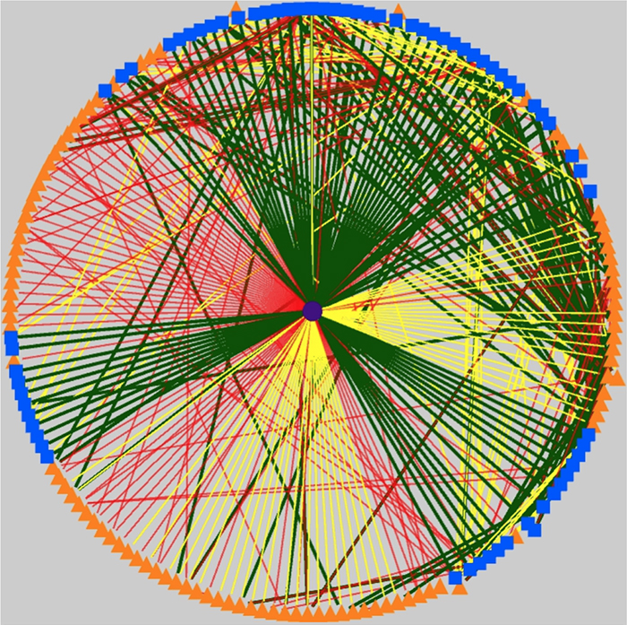
Purpose: Retinal network hyperactivity within degenerative retinal networks is a component of the disease process with implications for therapeutic interventions for blinding diseases that depend upon the surviving retinal network. Connexin36-containing gap junctions centered on the Aii amacrine cell network appear to mediate the aberrant signaling observed in mouse models of retinal degeneration. However, it remains unclear whether this hyperactivity reflects changes in the underlying circuitry or dysfunction/dysregulation of the normative circuitry. Mapping retinal circuitry in the ultrastructural rabbit Retinal Connectome, RC1, has revealed Aii network topologies explicitly involving gap junctions. In addition to canonical Aii-to-Aii and Aii-to-ON cone bipolar cell (CBC) coupling, we describe pervasive in- and cross-class coupling motifs among ON CBCs that extend and dramatically expand the coupled Aii network topologies. Since virtually every gap junction in the inner plexiform layer contains Connexin36, these circuits likely participate in the aberrant signaling of degenerate retinas. This study examines these Aii and ON CBC coupling motifs in Retinal PathoConnectome 1 (RPC1), an ultrastructural pathoconnectome of a rabbit model of retinitis pigmentosa.

Approach: RPC1 is a 2nm/pixel resolution volume of retina from a 10 month old, transgenic P347L rabbit model of autosomal dominant retinitis pigmentosa in early phase 1 retinal remodeling, a time point where cone and rod photoreceptors are still present, albeit going through cell stress. RPC1 spans the vitreous to basal outer nuclear layer and was built by automated transmission electron microscopy and computational assembly. ON CBCs, Aii amacrine cells, and their coupling partners were annotated using the Viking application and explored with 3D rendering and graph visualization of connectivity. Gap junctions were validated by 0.25 nm resolution recapture with goniometric tilt when necessary. Motifs were compared to those discovered in RC1. RC1 is a 2 nm resolution, 0.25 mm diameter volume of a light-adapted adult female Dutch Belted rabbit retina spanning the ganglion cell through inner nuclear layers.

Conclusions: RPC1 shows degeneration of rod outer segments, Müller cell hypertrophy and neuronal sprouting, characteristic of early stage retinal degeneration and phase 1 remodeling, when retinal hyperactivity and its reliance on gap junctional coupling has likely already initiated and human patients would still have some vision. All major coupling motifs (Aii-to-Aii, Aii-to-ON CBC, and ON CBC-to-ON CBC) were observed. Preliminary examinations indicate that several ON CBC classes retained their class-specific coupling profiles, accepting and rejecting specific combinations of Aii and ON CBC class partnerships. However, recent findings reveal aberrant partnerships in the coupled network, including both loss of prominent motifs and acquisition of novel ones. Thus, clear aberrant morphological and synaptic changes have been identified in RPC1, including changes in the coupling specificity and gap junction distributions of both Aii amacrine cells and ON CBCs (Figure 6). This suggests that the Aii/ON CBC circuit topology is already altered during early phase 1 remodeling, with substantial implications for therapeutic interventions in human subjects. The full coupling network is actively being examined and progress has begun on RPC2, a second pathoconnectome for examining later, phase 2 remodeling in this same model.

An almost full size poster available here in pdf format.

Increasing Electrical Stimulation Efficacy in Degenerated Retina: Stimulus Waveform Design in a Multiscale Computational Model

We have a new publication out (direct link), Increasing Electrical Stimulation Efficacy in Degenerated Retina: Stimulus Waveform Design in a Multiscale Computational Model authored by Kyle Loizos, Robert Marc, Mark Humayun, James R. Anderson, Bryan W. Jones and Gianluca Lazzi.

Abstract—A computational model of electrical stimulation of the retina is proposed for investigating current waveforms used in prosthetic devices for restoring partial vision lost to retina degen- erative diseases. The model framework combines a connectome- based neural network model characterized by accurate mor- phological and synaptic properties with an Admittance Method model of bulk tissue and prosthetic electronics. In this model, the retina was computationally “degenerated,” considering cellular death and anatomical changes that occur early in disease, as well as altered neural behavior that develops throughout the neurodegeneration and is likely interfering with current attempts at restoring vision. A resulting analysis of stimulation range and threshold of ON ganglion cells within retina that are either healthy or in beginning stages of degeneration is presented for currently-used stimulation waveforms, and an asymmetric biphasic current stimulation for subduing spontaneous firing to allow increased control over ganglion cell firing patterns in degenerated retina is proposed. Results show that stimulation thresholds of retinal ganglion cells do not notably vary after beginning stages of retina degeneration. In addition, simulation of proposed asymmetric waveforms showed the ability to enhance the control of ganglion cell firing via electrical stimulation.

A Pathoconnectome of Early Retinal Remodeling

This abstract was presented today, Monday, April 30th at the 2018 Association for Research in Vision and Opthalmology (ARVO) meetings in Honolulu, Hawaii by Rebecca Pfeiffer, Robert E. Marc, James R. Anderson, Daniel P. Emrich, Carl B. Watt, Jia-Hui Yang, Kevin D. Rapp, Jeebika Dahal, Mineo Kondo, Hiroko Terasaki, and Bryan W. Jones.

Purpose:
Retinal remodeling is a consequence of retinal degenerative disease, during which neurons sprout new neurites whose synaptic structures and partners are not yet defined. Simultaneously during remodeling, Müller cells (MCs) undergo structural and metabolic changes, whose impact on surrounding neurons is an active area of research. Retinal connectomes have elucidated and validated fundamental networks. These data provide further classification of neuronal types and subtypes and a precise framework for modeling of retinal function, based on ground truth networks. The creation of the first pathoconnectome (RPC1), a connectome from pathological retinal tissue, provides the opportunity to determine connectivites between neurons, while simultaneously evaluating glial remodeling. Computational Molecular Phenotyping (CMP) embedded within the ultrastructure provides metabolic factors of pathologies.

Methods:
RPC1 was collected post-mortem from a 10mo TgP347L rabbit model of adRP, fixed in 1% FA, 2.5% GA, 3% sucrose, and 1mM MgSO4 in cacodylate buffer (pH 7.4). The tissue was osmicated, dehydrated, resin embedded, and sectioned at 70nm. Sections were placed on formvar grids, stained, and imaged on a JEOL JEM-1400 TEM using SerialEM. 1 section was reserved from every 30 section for CMP, where it was probed for small molecules: glutamate, glutamine, glycine, GABA, taurine, glutathione; or proteins GFAP and GS. RPC1 was evaluated using the Viking software suite.

Results:
RPC1 was chosen based on early features of retinal degeneration/remodeling: degeneration of rod OS, MC hypertrophy, and neuronal sprouting. RPC1 consists of 948 serial sections spanning the ONL to the vitreous, with a diameter of 90µm. We find dendrites extending from rod bipolar cells to cone pedicles, originally described in light microscopy, and active synaptic contacts. We also see alterations of synaptic structure in the IPL, and MC morphological changes affecting surface to volume and neuron/glial relationships. Network motifs are being actively investigated.

Conclusions:
We observe many features of remodeling previously described using light microscopy, and confirm active synaptic contact. We also find synaptic structural features, not previously described. In addition, early evaluation of MC morphology demonstrates marked changes in MC shape and associations with nearby neurons and glia, which, combined with CMP, will be instrumental in understanding how MCs affect retinal remodeling.

Impact of Glaucoma On Retinal Ganglion Cell Subtypes: A Single-Cell RNA-seq Analysis of the DBA/2J Mouse

This abstract was presented today, May 1st at the 2018 Association for Research in Vision and Opthalmology (ARVO) meetings in Honolulu, Hawaii by Siamak Yousefi, Hao Chen, Jesse Ingels, Sumana R. Chintalapudi, Megan Mulligan, Bryan W. Jones, Vanessa Marie Morales-Tirado, Pete Williams, Simon W. John, Felix Struebing, Eldon E. Geisert, Monica Jablonski, Lu Lu, Robert Williams

Purpose
We are developing methods to define molecular signatures of cellular stress during early stages of glaucoma for major subtypes of retinal ganglion cells (RGCs). Our first aim is to develop reliable mRNA biomarkers for RGC subtypes in the DBA/2J (D2) mouse model prior to disease onset. Our second objective is to quantify cellular stress in RGC subtypes at early stages of disease using known sets of stress-responsive transcripts (e.g. Struebing et al, 2016 PMID:27733864; Williams et al. 2017, PMID:28209901; Lu et al, ARVO 2018).

Methods
Whole retinas from D2 or D2.Cg-Tg(Thy1-CFP)23Jrs/SjJ at 130 to 150 days-of-age were dissociated gently and size selected (>10 µm). RGCs were enriched using THY1 antibody-coated beads. Fluidigm HT microfluidics plates were used to isolate and generate scRNA-seq libraries of full length polyA-positive mRNAs using SMART-Seq v4. Libraries were sequenced using HiSeq3000, PE151. Following alignment using STAR, expression was normalized to log2(FPKM+1) across ~25,000 unique transcript models. Cells with fewer than 1000 detected genes and genes expressed in fewer than 1% of RGCs were excluded. Sets of genes with high variance and/or high expression were used for principal component analysis (PCA). Twenty PCs were used for graph-based unsupervised clustering and visualized using t-distributed stochastic neighbor embedding (tSNE). Gene specificity was computed for all transcripts across all clusters. The top transcripts per cluster with expression >1 in 1% or more of cells, were used to diagnose cellular identify of clusters. The top 30 genes per cluster were searched in PubMed against a panel of cell and tissue specific terms using Chilibot.

Results
The scRNA-seq protocol generates 150,000 – 200,000 uniquely mapped mRNA reads/cell and ~5000 genes/cells. We currently have 1600 cells, of which over half are RGCs. Around 75% of cells are positive for two or more of the following RGC markers: Thy1, Rbpms, Rbpms2, Jam2, G3bp1, and Ywhaz. This set of cells and different subsets of genes are now being used for RGC clustering. We have identified at least 17 clusters in initial datasets using these protocols and are now linking clusters to major classes of RGCs.

Conclusions
Molecular signatures of cellular stress and RGC subtypes in early stage of glaucoma should now be identifiable using unsupervised learning techniques.

Metabolic, Excitation and Functional Mapping of Diabetic Retinopathy

This abstract was presented today, Monday, April 30th at the 2018 Association for Research in Vision and Opthalmology (ARVO) meetings in Honolulu, Hawaii by Felix R. Vazquez-Chona, Tam T.T. Phuong, Oleg Yarishkin, Bryan W. Jones, and David Krizaj

Purpose:
Loss of vision in diabetic retinopathy is associated with extensive shifts in retinal metabolic and synaptic function yet the general principles that govern the metabolic remodeling remain unknown. To define the metabolic signature in hyperglycemic retina we took advantage of in situ metabolomics, excitation mapping and gene knockdown. Specifically, we investigated whether manipulation of the swelling-activated calcium-permeable TRPV4 (transient receptor potential isoform 4) channel contributes to the metabolic program of the degenerating neurogliovascular subunit in diabetic mice.

Methods:
Type I diabetes in wild type (WT) and TRPV4-/- mice was induced with streptozotocin (STZ). We visualized glutamate (NMDA)-gated excitation and glucose transport using the organic cation agmatine (AGB2+) and the glucose analog glucosamine (GCN). Retinas were fixed in glutaraldehyde, sectioned, and incubated with antibodies targeting GCN and AGB. Cell classification and metabolic status were interrogated using Computational Metabolic Profiling (CMP) and probes against ADP, alanine, arginine, aspartate, citrulline, GABA, glutamate, glycine, glutathione, glutamine, isoleucine, taurine, glutamine synthetase, CRALBP, GFAP, and tomato lectin.

Results:
Amacrine and ganglion cells in control retinas responded to NMDA activation with a large elevations in AGB and GCN signals. Diabetic amacrine cells maintained a robust dynamic range of AGB and GCN signals which however were markedly diminished in RGCs. Metabolomic maps of diabetic WT retinas showed that the outer retina remains metabolically quiescent whereas the ganglion cell layer displayed cells with lower glutamate and GABA signals. Diabetic TRPV4-deficient retinas displayed metabolomic, excitation, and glucose transport maps that were comparable to control retinas. ERG analysis showed modest STZ-induced changes in scotopic a- and b-waves of WT and KO eyes.

Conclusions:
Our preliminary electrophysiological and metabolomic findings suggest that STZ-induced diabetes spares the inner retina but alters amacrine-ganglion cell signaling, the neurogliovascular unit organization together with RGC metabolism. TRPV4 inactivation partially rescues the metabolic, excitation, physiologic phenotypes imposed by hyperglycemia. These results suggest that ambient sensing through polymodal TRP channels links retinal neuronal, glial and endothelial signaling to cellular metabolism and visual function.

Corticostriatal Circuit Defects in Hoxb8 Mutant Mice

We have a new publication in Molecular Psychiatry, Corticostriatal circuit defects in Hoxb8 mutant mice. (Direct link here).  Authors are:  Naveen Nagarajan, Bryan W. Jones, Peter West, Robert Marc, and Mario R. Capecchi.

Abstract: Hoxb8 mutant mice exhibit compulsive grooming and hair removal dysfunction similar to humans with the obsessive-compulsive disorder (OCD)-spectrum disorder, trichotillomania. As, in the mouse brain, the only detectable cells that label with Hoxb8 cell lineage appear to be microglia, we suggested that defective microglia cause the neuropsychiatric disorder. Does the Hoxb8 mutation in microglia lead to neural circuit dysfunctions? We demonstrate that Hoxb8 mutants contain corticostriatal circuit defects. Golgi staining, ultra-structural and electrophysiological studies of mutants reveal excess dendritic spines, pre- and postsynaptic structural defects, long-term potentiation and miniature postsynaptic current defects. Hoxb8 mutants also exhibit hyperanxiety and social behavioral deficits similar to mice with neuronal mutations in Sapap3, Slitrk5 and Shank3, reported models of OCD and autism spectrum disorders (ASDs). Long-term treatment of Hoxb8 mutants with fluoxetine, a serotonin reuptake inhibitor, reduces excessive grooming, hyperanxiety and social behavioral impairments. These studies provide linkage between the neuronal defects induced by defective Hoxb8-microglia and neuronal dysfunctions directly generated by mutations in synaptic components that result in mice, which display similar pathological grooming, hyperanxiety and social impairment deficits. Our results shed light on Hoxb8 microglia-driven circuit-specific defects and therapeutic approaches that will become essential to developing novel
therapies for neuropsychiatric diseases such as OCD and ASDs with Hoxb8-microglia being the central target.

Pattern Recognition Analysis of Age-Related Retinal Ganglion Cell Signatures In The Human Eye

We have a new publication in IOVS, Pattern Recognition Analysis of Age-Related Retinal Ganglion Cell Signatures In The Human Eye (Direct link here).  Authors are:  Nayuta Yoshioka, Barbara Zangerl, Lisa Nivison-Smith, Sieu Khuu, Bryan W. Jones, Rebecca Pfeiffer, Robert Marc, and Michael Kalloniatis.

Purpose: We recently used pattern recognition analysis to show macula areas can be classified into statistically distinct clusters in accordance to their age-related retinal ganglion cell layer (RGCL) thickness change in a normal population. The aim of this study was to perform a retrospective cross-sectional analysis utilizing a large cohort of patients to establish accuracy of this model and to develop a normative dataset using a 50-year-old equivalent cohort.

Methods: Data was collected from patients seen at the Centre for Eye Health for optic nerve assessment without posterior pole disease. The grid-wise RGCL thickness was obtained from a single eye of each patient via Spectralis OCT macular scan over an 8×8 measurement grid. Measurements for patients outside the 45-54 age range (training cohort) were converted to 50-year-old equivalent value utilizing pattern recognition derived regression model which, in brief, consists of 8×8 grid clustered into 8 distinct classes according to the pattern of RGCL thickness change with age. Accuracy of the predictions was assessed by comparing the training cohort’s measurements to the 45-54 year reference cohort using t-test and one-way ANOVA.

Results: Data were collected from a total 248 patients aged 20 to 78.1 years. 80 patients within this group were aged 45 – 54 and formed the reference cohort (average±SD 49.6±2.83) and the remaining 168 eyes formed the training cohort (average age±SD 50.7±17.34). Converted values for the training set matched those of the reference cohort (average disparity±SD 0.10±0.42µm, range -0.74-1.34µm) and were not significantly different (p > 0.9). Most variability was observed with patients above 70 years of age (average disparity±SD -0.09±1.73µm, range -3.67 to 6.16µm) and central grids corresponding to the fovea (average disparity±SD 0.47±0.72µm, range -0.22 to 1.34µm).

Conclusions: Our regression model for normal age-related RGCL change can accurately convert and/or predict RGCL thickness for individuals in comparison to 50-year-equivalent reference cohort and could allow for more accurate assessment of RGCL thickness and earlier detection of significant loss in the future. Caution may be needed when applying the model in the foveal area or for patients older than 70 years.

Synaptic Inputs To A Gamma Ganglion Cell In Rabbit Retina

This abstract was presented today, May 8th at the 2017 Association for Research in Vision and Opthalmology (ARVO) meetings in Baltimore, Maryland by Andrea Bordt, Diego Perez, Robert E. Marc, James R. Anderson, Carl B. Watt, Bryan W. Jones, Crystal Sigulinsky, James S. Lauritzen, Danny Emrich, Noah Nelson, Luke Tseng, Weiley Liu, and David W. Marshak. Full resolution version here.

Purpose: There are at least 30 distinct types of mammalian retinal ganglion cells, each sensitive to different features of the visual environment, and these can be grouped according to their morphology. One such group, the gamma cells, was identified more than 40 years ago, but their synaptic inputs have never been described. That was the goal of this study.

Methods: The synaptic inputs to a subtype of gamma cell with dendrites ramifying in the outer sublamina of the inner plexiform layer (IPL) of the rabbit retina were identified in a retinal connectome developed using automated transmission electron microscopy.

Results: The gamma cell was always postsynaptic in the IPL, confirming its identity as a ganglion cell. The local synaptic input should produce relatively weak OFF reposnses to stimuli confined to the center of the gamma cell’s receptive field. It typically received only one synapse per bipolar cell from at least 4 types of OFF bipolar cells. Because bipolar cells vary in their response kinetics and contrast sensitivity. each type would provide a small, asynchronous excitatory input. The amacrine cells at the dyad synapses provided only a small amount presynaptic inhibition; reciprocal synapses were observed in only 3 of the 18 ribbon synapses. There was no glycinergic crossover inhibition, another local interaction that would enhance light responses. Local postsynaptic inhibition was somewhat more common; in 6 instances, the bipolar cells presynaptic to the gamma cell or their electrically coupled neighbors also provided input to an amacrine cell that inhibited the gamma cell. The other amacrine cell inputs to the gamma cell should have a much greater impact on the light responses because they are far more numerous. These are from axons and long dendrites of GABAergic amacrine cells, and they provide 60% of all the input. This finding suggests that many types of stimuli in the receptive field surround or outside of the classical receptive field would provide potent inhibition to the gamma cell.

Conclusions: The synaptic inputs rsuggest that gamma cells in rabbit retina would have light responses like their homologs in mouse retina, OFF responses to small stimuli in the receptive field center that are suppressed by a variety of larger stimuli. Thus, they would signal object motion selectively.

Photoreceptor Outer Segment-like Structures in Long-Term 3D Retinas from Human Pluripotent Stem Cells

We have a new publication out (direct link), Photoreceptor Outer Segment-like Structures in Long-Term 3D Retinas from Human Pluripotent Stem Cells authored by Karl Wahlin, Julien Maruotti, Srinivasa Sripathi, John Ball, Juan Angueyra, Catherin Kim, Rhonda Grebe, Wei Li, Bryan W. Jones and Don Zack.

Abstract: The retinal degenerative diseases, which together constitute a leading cause of hereditary blindness worldwide, are largely untreatable. Development of reliable methods to culture complex retinal tissues from human pluripotent stem cells (hPSCs) could offer a means to study human retinal development, provide a platform to investigate the mechanisms of retinal degeneration and screen for neuroprotective compounds, and provide the basis for cell-based therapeutic strategies. In this study, we describe an in vitro method by which hPSCs can be differentiated into 3D retinas with at least some important features reminiscent of a mature retina, including exuberant outgrowth of outer segment-like structures and synaptic ribbons, photoreceptor neurotransmitter expression, and membrane conductances and synaptic vesicle release properties consistent with possible photoreceptor synaptic function. The advanced outer segment-like structures reported here support the notion that 3D retina cups could serve as a model for studying mature photoreceptor development and allow for more robust modeling of retinal degenerative disease in vitro.

Graffinity: Visualizing Connectivity in Large Graphs

We have a new publication out, (direct link)(Wiley link), Graffinity: Visualizing Connectivity in Large Graphs.  Authors are, Ethan Kerzner (@EthanKerzner), Alexander Lex, Crystal Sigulinsky, Timothy Urness, Bryan W. Jones,  Robert Marc, and Miriah Meyer.

Abstract:  Multivariate graphs are prolific across many fields, including transportation and neuroscience. A key task in graph analysis is the exploration of connectivity, to, for example, analyze how signals flow through neurons, or to explore how well different cities are connected by flights. While standard node-link diagrams are helpful in judging connectivity, they do not scale to large networks. Adjacency matrices also do not scale to large networks and are only suitable to judge connectivity of adjacent nodes. A key approach to realize scalable graph visualization are queries: instead of displaying the whole network, only a relevant subset is shown. Query-based techniques for analyzing connectivity in graphs, however, can also easily suffer from cluttering if the query result is big enough. To remedy this, we introduce techniques that provide an overview of the connectivity and reveal details on demand. We have two main contributions: (1) two novel visualization techniques that work in concert for summarizing graph connectivity; and (2) Graffinity, an open-source implementation of these visualizations supplemented by detail views to enable a complete analysis workflow. Graffinity was designed in a close collaboration with neuroscientists and is optimized for connectomics data analysis, yet the technique is applicable across domains. We validate the connectivity overview and our open-source tool with illustrative examples using flight and connectomics data.