Tag Archives: Robert E. Marc

A Synaptic Basis for Small World Network Design in the ON Inner Plexiform Layer of the Rabbit Retina

Bipolar cells_

This abstract was presented today at the 2014 Association for Research in Vision and Opthalmology (ARVO) meetings in Orlando, Florida by J Scott Lauritzen, Noah T. Nelson, Crystal L. Sigulinsky, Nathan Sherbotie, John Hoang, Rebecca L. PfeifferJames R. Anderson, Carl B. Watt, Bryan W. Jones and Robert E. Marc.

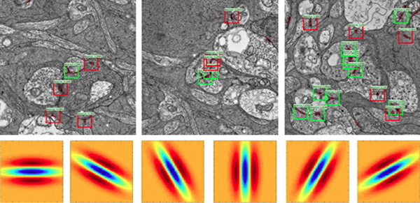
Purpose: Converging evidence suggests that large- and intermediate-scale neural networks throughout the nervous system exhibit small world’ design characterized by high local clustering of connections yet short path length between neuronal modules (Watts & Strogatz 1998 Nature; Sporns et al.2004 Trends in Cog Sci). It is suspected that this organizing principle scales to local networks (Ganmor et al. 2011 J Neurosci; Sporns 2006 BioSystems) but direct observation of synapses and local network topologies mediating small world design has not been achieved in any neuronal tissue. We sought direct evidence for synaptic and topological substrates that instantiate small world network architectures in the ON inner plexiform layer (IPL) of the rabbit retina. To test this we mined ≈ 200 ON cone bipolar cells (BCs) and ≈ 500 inhibitory amacrine cell (AC) processes in the ultrastructural rabbit retinal connectome (RC1).

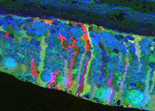
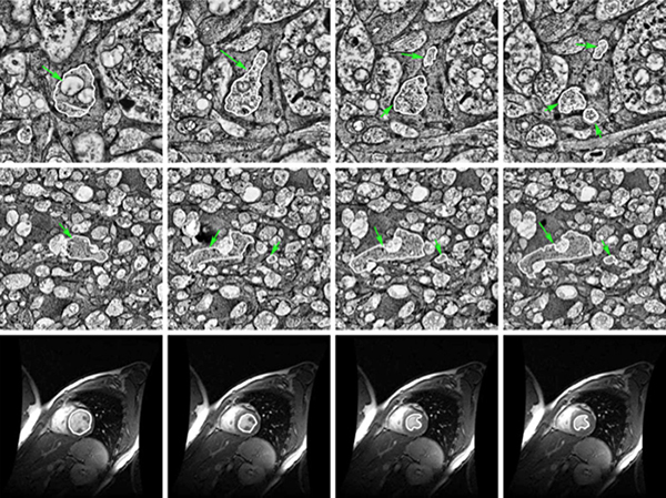
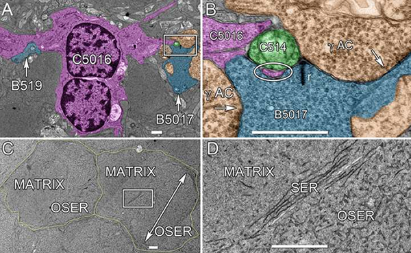
Methods: BC networks in RC1 were annotated with the Viking viewer and explored via graph visualization of connectivity and 3D rendering (Anderson et al. 2011 J Microscopy). Small molecule signals embedded in RC1 e.g. GABA glycine and L-glutamate combined with morphological reconstruction and connectivity analysis allow for robust cell classification. MacNeil et al. (2004 J Comp Neurol) BC classification scheme used for clarity.

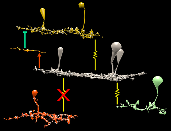
Results: Homocellular BC coupling (CBb3::CBb3 CBb4::CBb4 CBb5::CBb5) and within-class BC inhibitory networks (CBb3 → AC –| CBb3 CBb4 → AC –| CBb4 CBb5 → AC –| CBb5) in each ON IPL strata form laminar-specific functional sheets with high clustering coefficients. Heterocellular BC coupling (CBb3::CBb4 CBb4::CBb5 CBb3::CBb5) and cross-class BC inhibitory networks (CBb3 → AC –| CBb4 CBb4 → AC –| CBb3 CBb4 → AC –| CBb5 CBb5 → AC –| CBb4 CBb3 → AC –| CBb5 CBb5 → AC –| CBb3) establish short synaptic path lengths across all ON IPL laminae.

Conclusions: The retina contains a greater than expected number of synaptic hubs that multiplex parallel channels presynaptic to ganglion cells. The results validate a synaptic basis (ie. direct synaptic connectivity) and local network topology for the small world architecture indicated at larger scales providing neuroanatomical plausibility of this organization for local networks and are consistent with small world design as a fundamental organizing principle of neural networks on multiple spatial scales.

Support:  NIH EY02576 (RM), NIH EY015128 (RM), NSF 0941717 (RM), NIH EY014800 Vision Core (RM), RPB CDA (BWJ), Thome AMD Grant (BWJ).

Metabolic Changes Associated With Müller Cells In A Transgenic Rabbit Model Of Retinal Degeneration

Retina-RLP

This abstract was presented today at the 2014 Association for Research in Vision and Opthalmology (ARVO) meetings in Orlando, Florida by  Rebecca L. PfeifferBryan W. Jones and Robert E. Marc.

Purpose: Müller cells play a central role in retinal metabolism via the glutamate cycle. During retinal degeneration Müller cells are among the first to demonstrate changes, reflected in alterations of metabolic signatures and morphology. The timing, extent and regulation of these changes is not fully characterized. To address this issue, we evaluated Müller cell metabolic phenotypes at multiple stages of retinal remodeling.

Methods: Samples were collected post-mortem from both WT and P347L rabbits. The retinas were then divided into fragments, fixed in buffered aldehydes, and embedded in epoxy resins. Tissues were sectioned at 200nm followed by classification with computational molecular phenotyping (CMP) using an array of small and macromolecular signatures (aspartate (D), glutamate (E), glycine (G), glutamine (Q), glutathione (J), GABA (yy), taurine (T), CRALBP, Glutamine Synthetase (GS), and GFAP). Levels of amino acid or protein were quantified by selecting a region of interest either within the Müller cell population or surrounding neurons and evaluating the intensity of the signal within that region.

Results: CMP reveals overall decreases in GS levels over the course of degeneration. Of notable importance, we saw that in regions of near complete photoreceptor loss neighboring Müller cells may express independent variation in metabolic signatures of E, Q, and GS. Also observed in these Müller cells, ratios of GS:E and GS:Q are not consistent with the ratios seen in WT retina. These results are inconsistent with the current models of both E to Q metabolism and microenvironment regulation of Müller cell phenotypes.

Conclusions: These observations indicate two conclusions. First, although the degenerate state of the retina is the likely trigger inducing Müller cells to express altered metabolic signatures, the rate at which the metabolic state changes is not purely a product of the surrounding environment, but also a stochastic change within individual Müller cells. Second, although it is commonly accepted that GS is the primary enzyme which converts Q to E as part of the glutamate cycle, in degenerate retina alternative pathways may be utilized following decrease in GS.

Support: NIH EY02576 (RM), NIH EY015128 (RM), NSF 0941717 (RM), NIH EY014800 Vision Core (RM), RPB CDA (BWJ), Thome AMD Grant (BWJ).

Synapse Classification And Localization In Electron Micrographs

Synapse-classification_

We have a new publication, Synapse Classification And Localization In Electron Micrographs in Pattern Recognition Letters.  Authors are: Vignesh JagadeeshJames Anderson, Bryan W. JonesRobert MarcSteven Fisher and B.S. Manjunath.

Abstract:  Classification and detection of biological structures in Electron Micrographs (EM) is a relatively new large scale image analysis problem. The primary challenges are in modeling diverse visual characteristics and development of scalable techniques. In this paper we propose novel methods for synapse detection and localization, an important problem in connectomics. We first propose an attribute based descriptor for characterizing synaptic junctions. These descriptors are task specific, low dimensional and can be scaled across large image sizes. Subsequently, techniques for fast localization of these junctions are proposed. Experimental results on images acquired from a mammalian retinal tissue compare favorably with state of the art descriptors used for object detection.

Retinal connectomics: A New Era For Connectivity Analysis in The New Visual Neurosciences

New-Visual-Neurosciences

We have a new publication, this one a chapter titled: Retinal connectomics: A New Era For Connectivity Analysis in The New Visual Neurosciences (A little cheaper on Amazon here) textbook.  Authors are Robert E. Marc, Bryan W. Jones, James S. Lauritzen, Carl B. Watt and James R. Anderson.

Robust Segmentation based Tracking using an Adaptive Wrapper for Inducing Priors

Robust-Segmentation-based-Tracing-using-an-Adaptive-Wrapper-for-Inducing-Priors

We have published another manuscript, Robust Segmentation based Tracking using an Adaptive Wrapper for Inducing Priors.  This manuscript describes the work on adaptive tracing and proposes an algorithm that adapts a generic tracing algorithm to an application of interest.  In our specific case, it is boundaries of cells in high frequency space in transmission electron microscopy images.  But the approach in this paper is applicable to biological, medical, remote sensing and surveillance data as well utilizing priors specific to the application.  The co-authors on the paper are: Vignesh JagadeeshJames Anderson, Bryan W. JonesRobert E. MarcSteven K Fisher and B.S Manjunath.

Retinal Connectomics: Toward Complete, Accurate Networks

Retinal Connectomics_600

We have a new publication, Retinal connectomics: Toward complete, accurate networks in Progress in Retinal and Eye Research.  Authors are:  Robert E. Marc, Bryan W. JonesCarl B. Watt, Crystal Sigulinsky, James R. Anderson and J. Scott Lauritzen.

Abstract:
Connectomics is a strategy for mapping complex neural networks based on high-speed automated electron optical imaging, computational assembly of neural data volumes, web-based navigational tools to explore 1012-1015 byte (terabyte to petabyte) image volumes, and annotation and markup tools to convert images into rich networks with cellular metadata. These collections of network data and associated metadata, analyzed using tools from graph theory and classification theory, can be merged with classical systems theory, giving a more completely parameterized view of how biologic information processing systems are implemented in retina and brain. Networks have two separable features: topology and connection attributes. The first findings from connectomics strongly validate the idea that the topologies complete retinal networks are far more complex than the simple schematics that emerged from classical anatomy. In particular, connectomics has permitted an aggressive refactoring of the retinal inner plexiform layer, demonstrating that network function cannot be simply inferred from stratification; exposing the complex geometric rules for inserting different cells into a shared network; revealing unexpected bidirectional signaling pathways between mammalian rod and cone systems; documenting selective feedforward systems, novel candidate signaling architectures, new coupling motifs, and the highly complex architecture of the mammalian AII amacrine cell. This is but the beginning, as the underlying principles of connectomics are readily transferrable to non-neural cell complexes and provide new contexts for assessing intercellular communication.

Constructive Retinal Plasticity After Selective Ablation of the Photoreceptors

This abstract was presented today at the Association for Research in Vision and Opthalmology (ARVO) meetings in Seattle, Washington by Corinne N. Beier, Bryan W. Jones, Philip Huie, Yannis M. Paulus, Daniel Lavinsky, Loh-Shan B. Leung, Hiroyuki Nomoto,  Robert E. Marc, Daniel V. Palanker, and Alexander Sher.

Purpose: In the rabbit retina there is evidence of constructive retinal plasticity in response to focal ablation of a small patch of the photoreceptor layer by laser photocoagulation. After a two-month healing period, healthy photoreceptors migrate inwards filling the damaged area and restoring visual sensitivity to the lesion site. We investigated the integrity and function of the neural populations above the lesion, whether the migrating photoreceptors formed new connections with deafferented bipolar cells, and to what degree the new function resembled normal retinal function.

Methods: 
Retinal photocoagulation lesions of Moderate and Barely Visible clinical grades were produced in rabbits with a 532-nm laser, using beam diameter of 200 and 400 μm. Retinal ganglion cell (RGC) responses to spatio-temporal white noise stimulus were recorded on a 512-electrode array. Inner retinal neuron cell types were identified using Computational Molecular Phenotyping (CMP). Light evoked activity of the inner retinal neurons was measured through 1-amino-4-guanidobutane (AGB) labeling. Synaptic structure between photoreceptors and bipolar cells was characterized through transmission electron microscopy (TEM) imaging.

Results: 
The lesioned areas of the retina, after a two-month healing period, regained visual sensitivity. There was no significant difference between the response kinetics of RGCs with receptive fields covering the lesioned area and RGCs with receptive fields unaffected by the lesion. Furthermore, the average receptive field sizes of RGCs covering the lesion were consistent with the average receptive field sizes of RGCs unaffected by the lesion. CMP showed that all major inner retinal neuron cell types are present above both acute and healed lesions. Light evoked activity in the retina, as measured by AGB concentration levels, was diminished in the acute lesion but returned to within 10% of normal after two months. TEM images showed normal photoreceptor synaptic structure inside the healed lesion area.

Conclusions: 
Migrating photoreceptors establish new functional connectivity to deafferented bipolar cells and have normal synaptic structure. The new circuitry results in spatial and temporal properties of the RGC responses that resemble those of the healthy retina. In summary, the rewiring restores normal visual response in the lesioned area, indicating constructive retinal plasticity.

Support: Burroughs Wellcome Fund Career Award at the Scientific Interface; the Pew Charitable Trusts Scholarship in the Biomedical Sciences (A.S.), RPB CDA, Thome Foundation (BWJ), NIH EY02576, NIH EY015128, NSF 0941717, NIH EY014800 Vision Core (R.M.); NIH 5R01EY18608, AFOSR FA9550- 10-1-0503, DoD W81XWH-12-10575, Stanford University Bio-X (D.P.), Edward N. and Della L. Thome Memorial Foundation grant for Age-Related Macular Degeneration Research (BWJ) RPB unrestricted award (Moran Eye Center)

Retinal Metabolic Response to Cigarette Smoke

This abstract was presented today at the Association for Research in Vision and Opthalmology (ARVO) meetings in Seattle, Washington by Alexandra D. Butler, William D. Ferrell, Alex Woodell, Carl Atkinson, Baerbel Rohrer, Robert E. Marc and Bryan W. Jones.

Purpose:  Smoking is the single largest risk factor for age-related macular degeneration, aside from age. Several of the main genetic risk factors for AMD are polymorphisms occurring in complement genes involved in the alternative, classical and common terminal pathways. To better understand the metabolic impact of smoking on the retina, we used computational molecular phenotyping (CMP) and examined the effects of cigarette smoke on wild type (wt) retinas and mice in which either the alternative pathway (complement factor B, CfB) or the common terminal pathway (complement component 3, C3) was removed.

Methods:  Mice were exposed to either cigarette smoke or filtered air. Cigarette smoke (CS) was generated using an automated cigarette-smoking machine (Model TE-10, Teague Enterprises, Davis, CA) by burning 3R4F reference cigarettes (2.45 mg nicotine per cigarette; purchased from the Tobacco Health Research Institute, University of Kentucky, Lexington, KY). Mice were exposed to CS for 6 hours/day, 5 days/week for 6 months. Age matched room filtered air exposed mice were used as controls. Eyes were enucleated immediately post-mortem, fixed in 1% paraformaldehyde, 2.5% glutaraldehyde, dehydrated in graded methanols, embedded in eponates and histologically analyzed with CMP.

Results:  Alterations in retinal small molecule signatures from mice exposed to cigarette smoke were observed compared to retinas from non-smoked mice in wt, CfB and C3 knockout mice. Signal changes with arginine, glutamine and glutathione progressively increased in the retinas of smoked exposed wt, CfB and C3 knockout mice, indicating increased response profiles to cell stress. Both Müller cells and photoreceptors of wt smoked retinas demonstrated changes relative to non- smoked retinas.

Conclusions:  Arginine, glutamine and glutathione, amino acids known to be involved in cellular stress responses, were increased in retinal neurons and glial cells upon smoke exposure. Eliminating essential components of the complement system, a cascade required for the maintenance of the immune privilege of the eye, appears to exacerbate responses to cigarette smoke in oxidative damage response related pathways. Understanding complement-dependent alterations in the eye will aid in our understanding of AMD pathology and may open new avenues for novel treatment strategies.

Support:  RPB CDA (BWJ), Thome AMD Grant (BWJ), NIH EY02576 (RM), NIH EY015128 (RM), NSF 0941717 (RM), NIH EY014800 Vision Core (RM), NIH EY019320 (BR), VA merit award RX000444 (BR), grant to MUSC from RPB