Tag Archives: Robert E. Marc

Müller Cell Metabolic Chaos During Retinal Degeneration

We have a new publication out (direct link, open access), Müller Cell Metabolic Chaos During Retinal Degeneration authored by Rebecca PfeifferRobert Marc, Mineo Kondo, Hiroko Terasaki and Bryan W. Jones.

Abstract:

Müller cells play a critical role in retinal metabolism and are among the first cells to demonstrate metabolic changes in retinal stress or disease. The timing, extent, regulation, and impacts of these changes are not yet known. We evaluated metabolic phenotypes of Müller cells in the degenerating retina.

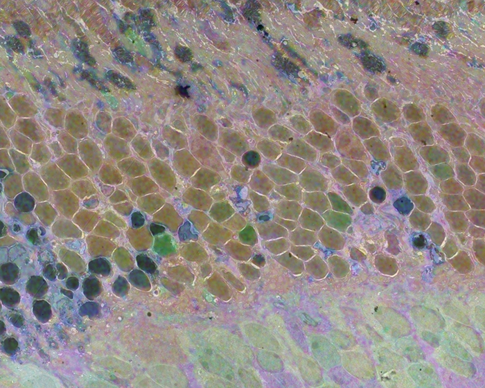
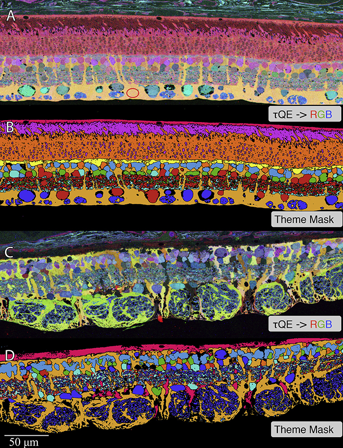
Retinas harvested from wild-type (WT) and rhodopsin Tg P347L rabbits were fixed in mixed aldehydes and resin embedded for computational molecular phenotyping (CMP). CMP facilitates small molecule fingerprinting of every cell in the retina, allowing evaluation of metabolite levels in single cells.

CMP revealed signature variations in metabolite levels across Müller cells from TgP347L retina. In brief, neighboring Müller cells demonstrated variability in taurine, glutamate, glutamine, glutathione, glutamine synthetase (GS), and CRALBP. This variability showed no correlation across metabolites, implying the changes are functionally chaotic rather than simply heterogeneous. The inability of any clustering algorithm to classify Müller cell as a single class in the TgP347L retina is a formal proof of metabolic variability in the present in degenerating retina.

Although retinal degeneration is certainly the trigger, Müller cell metabolic alterations are not a coherent response to the microenvironment. And while GS is believed to be the primary enzyme responsible for the conversion of glutamate to glutamine in the retina, alternative pathways appear to be unmasked in degenerating retina. Somehow, long term remodeling involves loss of Müller cell coordination and identity, which has negative implications for therapeutic interventions that target neurons alone.

Pattern Recognition Reveals Different Visual Field Signature Patterns When Using Spatially Equated Test Sizes Compared To Standard Goldmann III Alone

This abstract was presented today, May 2th at the 2016 Association for Research in Vision and Opthalmology (ARVO) meetings in Seattle, Washington by Michael Kalloniatis, Robert E. Marc, Sieu K. Khuu, Jack Phu, Barbara Zangerl, Lisa Nivison-Smith, Bryan W. Jones, and Rebecca L. Pfeiffer. 

Abstract Number: 4745

Author Block: Michael Kalloniatis, Robert E. Marc, Sieu K. Khuu, Jack Phu, Barbara Zangerl, Lisa Nivison-Smith, Bryan W. Jones, Rebecca L. Pfeiffer
1 Centre for Eye Health, SOVS, University of New South Wales, Kensington, New South Wales, Australia; 2 SOVS, UNSW, Sydney, New South Wales, Australia; 3 Univ of Utah/Moran Eye Center, Salt Lake City, Utah, United States

Disclosure Block:Michael Kalloniatis, 2014/094035 A1 (USA) and 13865419.9 (EU) (Code P (Patent) ); Robert E. Marc, None; Sieu K. Khuu, 2014/094035 A1 (USA) and 13865419.9 (EU) (Code P (Patent) ); Jack Phu, None; Barbara Zangerl, None; Lisa Nivison-Smith, None; Bryan W. Jones, None; Rebecca L. Pfeiffer, None

Purpose:To identify areas within the visual field with matching contrast sensitivity (CS) signature patterns as a function of age using pattern recognition and determine the discrimination of CS data when using spatially equated test stimuli compared to the single size Goldmann (G)III alone.

Methods:52 subjects (classified in decade age groups from 20-60+ years) were tested using the Humphrey Visual Field Analyser 30-2 paradigm in full threshold mode for GI to GV. At least two thresholds were obtained per size. Two visual field maps were analyzed: a spatially equated visual field where GI was used centrally, GII mid-peripherally and GIII in the outer rings to place the test size at or close to complete spatial summation and a second where a single GIII was used at all locations. Thresholds were expressed as dB* (Khuu & Kalloniatis, IOVS 2015), converted to pixel values and analyzed using an unsupervised classification using isodata clustering (PCI, Geomatica, Canada). Class separation was extracted across the ages to develop dot plots of decade measures of CS.

Results:The 77 data points across the central 60° visual field can be distilled into 6 functional classes using the spatially equated visual field (Class separation 1). The 6 classes reflect areas in visual space that change in a similar manner across the ages. The use of the single GIII target resulted in only 4 classes displaying a poorer discrimination over the central visual field (Class separation 2). Extracted dot plots from class separation illustrated average CS within each class could be assessed across the decades.

Conclusions:When using spatially equated visual field testing, concentric areas were separated into distinct CS signatures consistent with known visual field sensitivity. We confirmed these areas change systematically with age. GIII failed to discriminate central areas of the 30-2 that likely reflects the fact that this size operates outside complete spatial summation and thus may not be the optimal test size for assessing visual function in the central visual field. More importantly, we showed pattern recognition can be applied to complex visual field data sets to identify common features and age-related visual function changes. This analysis allows regions to be averaged as they are statistically identical: this approach will likely assist structure-function studies.

Progressive Retinal Remodeling In A Transgenic Rabbit Model Of Retinitis Pigmentosa

This poster was presented today, May 2th at the 2016 Association for Research in Vision and Opthalmology (ARVO) meetings in Seattle, Washington by Rebecca L. Pfeiffer, Bryan W. Jones, and Robert E. Marc.

Posterboard #: D0246

Abstract Number: 2256 – D0246

Author Block: Rebecca L. Pfeiffer1,2 , Bryan W. Jones1,2 , Robert E. Marc1,2 
1 Ophthalmology, University of Utah, Salt Lake City, Utah, United States; 2 Interdepartmental Program in Neuroscience, University of Utah, Salt Lake City, Utah, United States

Purpose:Retinal degenerations are a collection of neural disorders, usually precipitated by photoreceptor degeneration. All display progressive metabolic alterations and neural loss following the death of the photoreceptors. Although it has been demonstrated that the metabolism of Müller cells (MCs) is drastically altered in degeneration, the full impact of these changes on surrounding neurons and long-term characterization of remodeling was previously unavailable, due to short lifespans of model organisms.

Methods:Retinal samples were collected from WT and Tg P347L rabbits at ages ranging from 3 months to 6 years. Following enucleation, retinas were divided into fragments and incubated for 10 minutes at 35 degrees C in D-isomers of Glutamate (dE), Glutamine (dQ), or Aspartate (dD) and Ames-bicarbonate medium to explore retinal transport capabilities at each stage of degeneration. Retinas were then fixed in mixed aldehyde buffer and processed for transmission electron microscope connectomics, immunocytochemistry for a range of macromolecules, and computational molecular phenotyping for small molecules (CMP) (J Comp Neurol. 464:1, 2003).

Results:CMP reveals that single metabolic phenotype of MCs splits and diverges into many phenotypes continuously throughout degeneration. Further, all neuronal classes continue to die throughout degeneration. By 6 years, over 90% of neurons are lost, and the remaining glutamatergic neurons have altered metabolic signatures with a large increase in aspartate levels, at times exceeding glutamate. Transport of the D-isomers indicates that glutamate transport capabilities remain intact until the latest stages of degeneration. This may not be true of their GABA transporters.

Conclusions:These results suggest three main conclusions. First, retinal remodeling in degeneration is relentlessly progressive long after all photoreceptors have disappeared. Second, cell types previously thought to remain after degeneration onset, such as ganglion cells, will also ultimately die and the cells not lost often will change their metabolism. The consequence of this metabolic change in neurons is not yet fully explored. Third, the persistent robust glutamate transport capabilities of Müller cells indicate Müller cells can metabolize glutamate despite the massive loss of glutamine synthetase activity, likely unmasking alternate metabolic pathways.

2-nm Resolution Anatomy of Retinal Neuro-Glial-Vascular Architecture

This abstract was presented today, May 2th at the 2016 Association for Research in Vision and Opthalmology (ARVO) meetings in Seattle, Washington by Jefferson R. Brown, Rebecca L. Pfeiffer, Crystal Sigulinsky, Felix Vazquez-Chona, Daniel Emrich, Bryan W. Jones, Robert E. Marc.

Abstract Number: 995

Author Block: Jefferson R. Brown, Rebecca L. Pfeiffer, Crystal Sigulinsky, Felix Vazquez-Chona, Daniel Emrich, Bryan W. Jones, Robert E. Marc
1 Dept of Ophthalmology, University of Utah, Salt Lake City, Utah, United States

Disclosure Block:Jefferson R. Brown, None; Rebecca L. Pfeiffer, None; Crystal Sigulinsky, None; Felix Vazquez-Chona, None; Daniel Emrich, None; Bryan W. Jones, None; Robert E. Marc, Signature Immunologics (Code I (Personal Financial Interest) )

Purpose:Retinal vasculature is strongly affected by degenerative pathologies and in turn, may also contribute to their progression. However, much of what we understand about the normal, healthy interaction between neurons, glia, and blood vessels at the ultrastructural level is limited to single section electron microscopy. The technology of serial section transmission electron microscopy (ssTEM) extends the high definition of TEM imaging into three dimensions to create volumes, allowing for more thorough visualization and analysis of the vascular-glial-neuronal complex.

Methods:RC2 is a 40TB ssTEM volume of over 1,400 horizontal sections of retinal tissue derived from an adult female C57BL/6J mouse. The tissue sample is 250 um in diameter and spans the outer nuclear layer to the vitreal surface. Baseline resolution is 2.18nm per pixel. Visualization, navigation and metadata annotations of the database are made via the Viking software suite.

Results:Much of the retinal vascular basement membrane directly contacts Muller cells. In the ganglion cell layer, direct basement membrane contact with astrocytes is frequent. Microglia commonly contact the basement membrane, and occasionally direct contact of neurons onto basement membrane was observed. Full 3D reconstruction of all vascular pathways with associated endothelia and pericytes within the volume was completed, demonstrating that all the retinal capillary layers are continuous with one another [Figure].

Conclusions:The presence of occasional direct neuronal contact onto vascular basement membrane supports earlier work by Ochs and colleagues (2000) and suggests the blood-retina barrier does not universally involve retinal glia. However, since such contacts are extremely sparse, it remains to be seen whether this finding has biologic significance, though their existence suggests significance. The RC2 volume is a valuable resource to aid in discovery of defining characteristics of wild type neurovascular architecture.


The intro figure is a side view of reconstruction of all vasculature within the RC2 volume. Vessels at the top of the figure correspond to the outer plexiform layer, while those at the bottom correspond to the ganglion cell layer. This capillary plexus is one continuous structure. Visualization by VikingView software.

In Situ Metabolomic Signatures Of Neuroprotection, Apoptosis, And Microglial Phagocytosis

This abstract was presented July 1st at the 11th International Converence of the Metabolomics Society at the University of California, Davis by Felix Vazquez-Chona, Drew Ferrell, Bryan W. Jones and Robert E. Marc.


Metabolic dysregulation is an early hallmark of neurodegenerative diseases including Alzheimer’s disease and age-related macular degeneration. Mapping metabolic adaptation with cellular resolution and tissue- wide context is crucial to define networks regulating neuronal survival, cell death progression, and immune cell response.

Computational Molecular Phenotyping (CMP) explores the amine metabolome (amino acids and amines). Technically, CMP metabolomics combines amine metabolite trapping, ultrathin microscopy (50-200 nm), immunodetection, pattern recognition, and clustering algorithms. Here we mapped the in situ distribution of over 30 core amine metabolites in retinal cells challenged by light-induced oxidative stress. Metabolomic profiles were phenotyped using ultrastructural, biochemical, and proteomic indices of oxidative stress.

CMP enabled precise visualization of >30 metabolites in every retinal cell. CMP resolved and phenotyped metabolomic profiles to specific degeneration and microglial functional states in the light-damaged retina. Cone photoreceptor survival correlated with enhanced antioxidant glutathione content. Rod photoreceptor apoptosis coincided with rapid depletion of organic osmolytes followed by nuclear import of cationic arginine metabolites. Delay in cell death increased necrosis and DNA damage-induced apoptosis. Microglial chemotaxis enhanced distinct signatures of glutamate and glutathione metabolism; whereas, phagocytosis coinduced classic (M1) and alternative (M2) arginine metabolites of macrophage activation.

CMP discovers and phenotypes cell classes, tracks cell state, and maps disease with single-cell resolution in any tissue or organism.

Ultrastructural Connectomics Reveals The Entire Chemical And Electrical Synaptic Cohort Of An ON Cone Bipolar Cell In The Inner Plexiform Layer Of The Rabbit Retina

593-Horizontal

This abstract was presented at the 2014 Society for Neuroscience meeting in Washington D.C. by J. Scott Lauritzen, Crystal L. Sigulinsky, Danny P. Emrich, Joshua M. Dudleston, Noah T. Nelson, Rebecca L. Pfeiffer, Nathan R. Sherbotie, John V. Hoang, Jefferson R. Brown, Carl B. WattJames R. Anderson, Bryan W. Jones and Robert E. Marc.

Purpose: Despite large-scale efforts aimed at mapping the mammalian nervous system, the entire synaptic cohort of a single mammalian neuron of any class has never been mapped. To this end we reconstructed all chemical and electrical synaptic partners of a single ON cone bipolar cell (ON CBC) in the inner plexiform layer (IPL) of the rabbit retina. We then searched all members of the same cell class for repeating network motifs and explored postsynaptic cell sampling topologies from this bipolar cell (BC).

Methods: Cells in retinal connectome 1 (RC1) were annotated with Viking viewer, and explored via graph visualization of connectivity and 3D rendering (Anderson et al., 2011 J Microscopy). Small molecule signals in RC1, e.g. GABA, glycine, and L-glutamate, combined with morphological reconstruction and connectivity analysis allow robust cell classification. The default resolution of RC1 is 2.18nm/pixel, however goniometric recapture at 0.273 nm/pixel was performed as needed for synapse verification.

Results: ON CBC 593 is one of 20 BCs of this class in RC1, the axonal arbors of which tile with gap junctions between nearest neighbors at their distal axonal tips. ON CBC 593 contains 194 ribbons, 274 postsynaptic densities, 20 gap junctions, and 66 conventional synapses, for a total of 554 synaptic connections. Twenty ganglion cells sample the glutamatergic output. ON CBC 593 is presynaptic to 262 amacrine cell (AC) processes, and is postsynaptic to 228 AC processes. Of these, 33% form reciprocal connections. We approximate that ON CBC 593 forms synapses with 50 distinct ACs. ON CBC 593 is routinely pre- and postsynaptic to within-class, cross-class, feedback, and feedforward inhibition motifs, including 1 instance of OFF-ON crossover inhibition. ON CBC 593 forms 12 gap junctions with at least 2 AII ACs, 7 with 5 ON CBCs, and 1 with itself. We searched for repeating network motifs across all ON CBCs of this class in RC1. Thus far, 80% of these form in-class inhibitory motifs, and 75% form cross-class inhibitory motifs. All ACs and GCs discovered to contact multiple branches of ON CBC 593 form synapses on every branch.

Conclusions: An individual bipolar cell is inherently multi-kinetic, receiving inhibition driven by all ON CBC classes, sharing these signals via gap junctions with ON CBCs of the same class, and driving inhibition of all ON CBC classes. This constitutes a substrate for multi-channel coordination throughout the IPL, and predicts multi-kinetic BC responses. The results establish a normative framework against which members of the same and different classes may be compared, and foster interpretation of BC physiological behavior under different stimulus regimes.

The AII Amacrine Cell Connectome: A Dense Network Hub

AII-connectome

We have a new publication in Frontiers in Neuroscience, The AII Amacrine Cell Connectome: A Dense Network Hub.  Authors are Robert E. MarcJames R. Anderson, Bryan W. Jones, Crystal Sigulinsky and J. Scott Lauritzen.

Abstract:  The mammalian AII retinal amacrine cell is a narrow-field, multistratified glycinergic neuron best known for its role in collecting scotopic signals from rod bipolar cells and distributing them to ON and OFF cone pathways in a crossover network via a combination of inhibitory synapses and heterocellular AII::ON cone bipolar cell gap junctions. Long considered a simple cell, a full connectomics analysis shows that AII cells possess the most complex interaction repertoire of any known vertebrate neuron, contacting at least 28 different cell classes, including every class of retinal bipolar cell. Beyond its basic role in distributing rod signals to cone pathways, the AII cell may also mediate narrow-field feedback and feedforward inhibition for the photopic OFF channel, photopic ON-OFF inhibitory crossover signaling, and serves as a nexus for a collection of inhibitory networks arising from cone pathways that likely negotiate fast switching between cone and rod vision. Further analysis of the complete synaptic counts for five AII cells shows that (1) synaptic sampling is normalized for anatomic target encounter rates; (2) qualitative targeting is specific and apparently errorless; and (3) that AII cells strongly differentiate partner cohorts by synaptic and/or coupling weights. The AII network is a dense hub connecting all primary retinal excitatory channels via precisely weighted drive and specific polarities. Homologs of AII amacrine cells have yet to be identified in non-mammalians, but we propose that such homologs should be narrow-field glycinergic amacrine cells driving photopic ON-OFF crossover via heterocellular coupling with ON cone bipolar cells and glycinergic synapses on OFF cone bipolar cells. The specific evolutionary event creating the mammalian AII scotopic-photopic hub would then simply be the emergence of large numbers of pure rod bipolar cells.


A Multi-Scale Computational Model For The Study Of Retinal Prosthetic Stimulation

Multiscale-model-of-retina

We have a new publication in IEEE, A Multi-Scale Computational Model For The Study Of Retinal Prosthetic Stimulation.  Authors are: Kyle LoizosGianluca Lazzi, J. Scott Lauritzen, James R. Anderson, Bryan W. Jones and Robert E. Marc.

Abstract: An implantable retinal prosthesis has been developed to restore vision to patients who have been blinded by degenerative diseases that destroy photoreceptors. By electrically stimulating the surviving retinal cells, the damaged photoreceptors may be bypassed and limited vision can be restored. While this has been shown to restore partial vision, the understanding of how cells react to this systematic electrical stimulation is largely unknown. Better predictive models and a deeper understanding of neural responses to electrical stimulation is necessary for designing a successful prosthesis. In this work, a computational model of an epi-retinal implant was built and simulated, spanning multiple spatial scales, including a large-scale model of the retina and implant electronics, as well as underlying neuronal networks.


Retinal Prosthetics, Optogenetics and Photoswitches

Retinal-Prosthetics-Optogenetics-and-Photoswitches

We have a new publication, Retinal Prosthetics, Optogenetics and Photoswitches in ACS Chemical Neuroscience.  Authors are:  Robert E. MarcRebecca L. Pfeiffer, and Bryan W. Jones.

Abstract:

Three technologies have emerged as therapies to restore light sensing to profoundly blind patients suffering from late-stage retinal degenerations: (1) retinal prosthetics, (2) optogenetics, and (3) chemical photoswitches. Prosthetics are the most mature and the only approach in clinical practice. Prosthetic implants require complex surgical intervention and provide only limited visual resolution but can potentially restore navigational ability to many blind patients. Optogenetics uses viral delivery of type 1 opsin genes from prokaryotes or eukaryote algae to restore light responses in survivor neurons. Targeting and expression remain major problems, but are potentially soluble. Importantly, optogenetics could provide the ultimate in high-resolution vision due to the long persistence of gene expression achieved in animal models. Nevertheless, optogenetics remains challenging to implement in human eyes with large volumes, complex disease progression, and physical barriers to viral penetration. Now, a new generation of photochromic ligands or chemical photoswitches (azobenzene-quaternary ammonium derivatives) can be injected into a degenerated mouse eye and, in minutes to hours, activate light responses in neurons. These photoswitches offer the potential for rapidly and reversibly screening the vision restoration expected in an individual patient. Chemical photoswitch variants that persist in the cell membrane could make them a simple therapy of choice, with resolution and sensitivity equivalent to optogenetics approaches. A major complexity in treating retinal degenerations is retinal remodeling: pathologic network rewiring, molecular reprogramming, and cell death that compromise signaling in the surviving retina. Remodeling forces a choice between upstream and downstream targeting, each engaging different benefits and defects. Prosthetics and optogenetics can be implemented in either mode, but the use of chemical photoswitches is currently limited to downstream implementations. Even so, given the high density of human foveal ganglion cells, the ultimate chemical photoswitch treatment could deliver cost-effective, high-resolution vision for the blind.

Ultrastructural Reconstruction of ON Cone Bipolar Cell Projective Fields In The Innter Plexiform Layer of The Rabbit Retina

retina reconstruction

This abstract was presented at the 2014 FASEB Summer Research Conference in Saxtons River, Vermont by J. Scott Lauritzen, Crystal L. Sigulinsky, Noah T. Nelson, Nathan R. Sherbotie, Danny P. Emrich, Rebecca L. Pfeiffer, Jefferson R. Brown, John V. Hoang, Joshua M. Dudleston, Carl B. Watt, Kevin Rapp, Marguerite V. Shaw, Jia-Hui Yang, James R. Anderson, Bryan W. Jones and Robert E. Marc.

Purpose: Functional mapping in tiger salamander shows that bipolar cell (BC) projective fields far exceed their axonal fields, and directly implicates wide-field GABAergic amacrine cells (wf γACs) and gap junctions (Asari & Meister, 2014). Strikingly, single BCs exert differential effects on functionally distinct ganglion cells (GCs), likely achieved by privatized amacrine cell (AC) presynaptic inhibition to specific BC-GC synaptic pairs (Asari & Meister, 2012). To address whether BC projective fields in the mammal are equally broad, wf γAC- and gap junction-dependent, and GC type unselective, we reconstructed all electrical and chemical synaptic partners of a single ON cone BC in the inner plexiform layer of the rabbit retina, and searched BC-GC synaptic pairs for differential synaptic inhibition.

Methods: Cells in retinal connectome 1 (RC1) were annotated with Viking viewer, and explored via connectivity visualizations and 3D rendering (Anderson et al., 2011). Small molecule signals embedded in RC1, e.g. GABA, glycine, and L-glutamate, combined with morphological reconstruction and connectivity analysis allow robust cell classification. We used the MacNeil et al. (2004) rabbit BC classification scheme.

Results: CBb5w 593 is one of 20 ON cone BCs of this class in RC1. This CBb5w is presynaptic to 17 distinct GCs and 262 AC processes, and postsynaptic to 228 AC processes. The majority of these ACs are wf γACs. We estimate this BC forms synapses with 50 unique ACs. Asari & Meister (2014) found that single bipolar cell projective fields range up to 1 mm, far beyond a BC axonal field, and differentially drive multiple classes of GC. We discovered BC-BC within- and cross-class coupling and lateral inhibition that construct sign-conserving and sign-inverting projective fields to many distinct ganglion cell classes across the entire 0.25 mm diameter of RC1, much greater than a 60 µm BC axonal field. Cross-class projections access a broader set of GCs than expected from in-class projections alone. The BC-BC coupling is independent of BC-AII AC coupling. 94% of the CBb5w 593 BC-GC synaptic pairs receive feedback inhibition within the varicosity of the ribbon, but the number of feedback synapses is highly variable (coefficient of variation = 0.81). 35% of the BC-GC pairs receive feedforward inhibition within 2 microns of the postsynaptic density.

Conclusions: Mammalian BCs use novel cross-class topologies to distribute signals to a wide range of GCs and establish projective fields similar to those discovered in non-mammalian species. BC-BC within- and cross-class coupling and lateral inhibition via wf γACs establish sign-conserving and sign-inverting projective fields, respectively, up to 1 mm diameters. BC-GC synaptic pairs overwhelmingly employ feedback vs. feedforward inhibition to modulate signaling, and the numbers of feedback synapses are highly variable across these pairs, accounting for privatized and differential GC responses to the same BC drive.