We have two software releases of note: Cell Sketches and Tulip Paths that were part of Ethan Kerzner’s PhD.
Cell Sketches: GitHub
and
Tulip Paths: GitHub

One of the undergraduate students in the lab, Jeebika Dahal has been annotating AII amacrine cells as part of a research project she will present at next year’s ARVO conference.

Congratulations to Dr. Ethan Kerzner who successfully defended his PhD dissertation in the Viz Design Lab at the University of Utah’s Scientific Computing Institute.
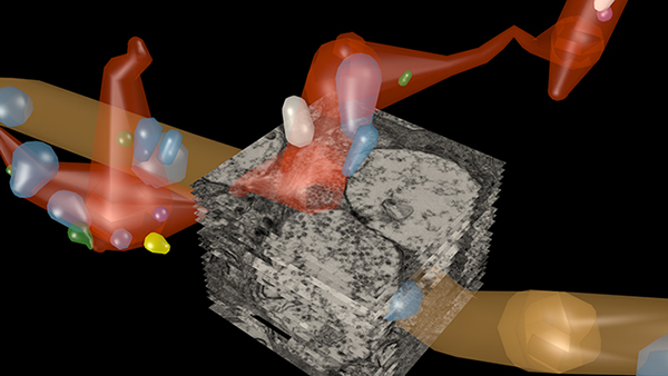
Ethan’s work has been instrumental in helping us to understanding complex gap junctional networks in our retinal connectomics initiatives. His Graffinity software package allowed us to explore multivariate graphs, and pull out complex relationships of neurons and gap junctions that would not have been easily possible with other approaches.
Ethan is now off to Google X, and we wish him the very best and look forward to many more interactions in the future.


This abstract was presented yesterday, April 29th at the 2018 Association for Research in Vision and Opthalmology (ARVO) meetings in Honolulu, Hawaii by Bryan W. Jones.
Abstract:
The retina is a complex, heterocellular tissue with most/all retinal cell classes becoming impacted or altered in retinitis pigmentosa (RP) and age-related macular degeneration (AMD) in a process called retinal remodeling. Defining disease and the stage-specific cytoarchitectural and metabolic responses in RP and AMD is critical for highlighting targets for intervention. We now know that negative plasticity and neural retinal remodeling occurs regardless of retinal insult in models of retinal degeneration as well as in human RP and in human AMD, revealing that no retinal disease fails to trigger remodeling and reprogramming.
Evidence in the literature over the past decade has improved our understanding into mechanisms of initial retinal degeneration and informed our understanding of the subsequent remodeling events in the neural retina that occur post-photoreceptor degeneration. Remodeling associated with retinal degeneration is intimately linked with insults that cause photoreceptor stress and eventually photoreceptor cell death. These phenomena result in reprogramming of cell types in retina followed by progressive neural degeneration akin to CNS neural degenerations involving both neuronal and glial classes. No cell class in the retina is spared from the effects of remodeling. The earliest cell classes involved in remodeling are horizontal, bipolar and Müller cells and the Müller glia are the last cell class left in the remodeling retina.
Our efforts are now focused on elucidating the precise wiring changes in retina, through the creation of pathological connectomes, or “patho-connectomes” to study precisely what the circuit topologies are, compared to normal topologies derived from Retinal Connectome 1 (RC1). Also, because temporal windows are critical to understanding when interventions may be possible, we are exploring when circuit topology revisions occur to understand their impact on information flow in the retina and their impact on rescues of vision loss. Precise circuit topologies in early retinal degenerative events is our first area of exploration with ultrastructural reconstructions of outer retinal neurons, bipolar cells and horizontal cells. Müller glia are also of intense interest as we are tracking the earliest metabolic and morphological changes in glia in response to retinal degenerations.
Our manuscript, The AII amacrine cell connectome: A dense network hub is now a chapter in Neural Circuits Revealed, a Frontiers Research e-book compiled by topic editors Mariano Soiza-Reilly, Peter Saggau, and Benjamin R. Arenkiel.

We have a new publication out (direct link), The rod-cone crossover connectome of mammalian bipolar cells authored by Scott Lauritzen, Crystal Sigulinsky, James Anderson, Michael Kalloniatis, Noah Nelson, Danny Emrich, Chris Rapp, Nicolas McCarthy, Ethan Kerzner, Mariah Meyer, Bryan W. Jones, and Robert Marc.
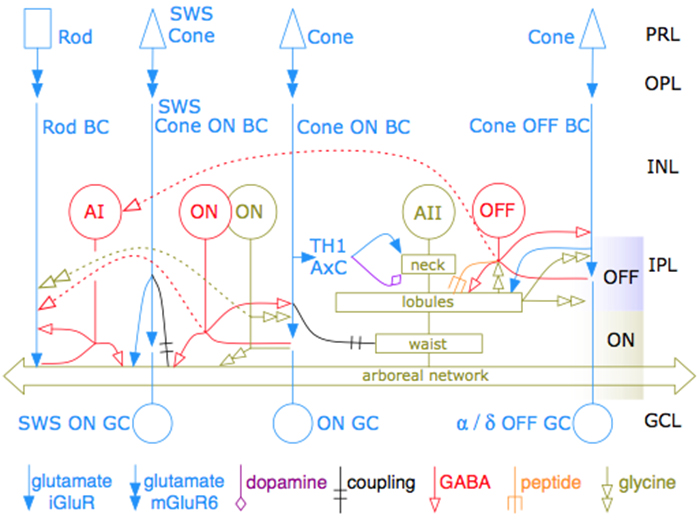
Abstract: The basis of cross-suppression between rod and cone channels has long been an enigma. Using rabbit retinal connectome RC1, we show that all cone bipolar cell (BC) classes inhibit rod BCs via amacrine cell (AC) motifs (C1-6); that all cone BC classes are themselves inhibited by AC motifs (R1-5, R25) driven by rod BCs. A sparse symmetric AC motif (CR) is presynaptic and postsynaptic to both rod and cone BCs. ON cone BCs of all classes drive inhibition of rod BCs via motif C1 wide-field GABAergic ACs (γACs) and motif C2 narrow field glycinergic ON ACs (GACs). Each rod BC receives ≈ 10 crossover AC synapses and each ON cone BC can target ≈ 10 or more rod BCs via separate AC processes. OFF cone BCs mediate monosynaptic inhibition of rod BCs via motif C3 driven by OFF γACs and GACs and disynaptic inhibition via motifs C4 and C5 driven by OFF wide-field γACs and narrow-field GACs, respectively. Motifs C4 and C5 form halos of 60-100 inhibitory synapses on proximal dendrites of AI γACs. Rod BCs inhibit surrounding arrays of cone BCs through AII GAC networks that access ON and OFF cone BC patches via motifs R1, R2, R4 R5 and a unique ON AC motif R3 that collects rod BC inputs and targets ON cone BCs. Crossover synapses for motifs C1, C4, C5 and R3 are 3-4x larger than typical feedback synapses, which may be a signature for synaptic winner-take-all switches.
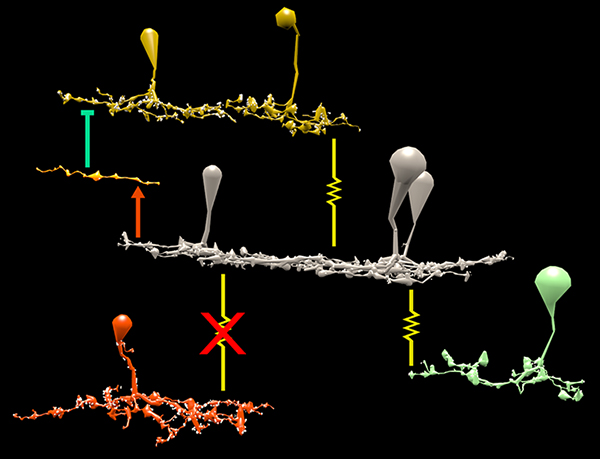
We have a new publication in Frontiers in Neuroscience, The AII Amacrine Cell Connectome: A Dense Network Hub. Authors are Robert E. Marc, James R. Anderson, Bryan W. Jones, Crystal Sigulinsky and J. Scott Lauritzen.
Abstract: The mammalian AII retinal amacrine cell is a narrow-field, multistratified glycinergic neuron best known for its role in collecting scotopic signals from rod bipolar cells and distributing them to ON and OFF cone pathways in a crossover network via a combination of inhibitory synapses and heterocellular AII::ON cone bipolar cell gap junctions. Long considered a simple cell, a full connectomics analysis shows that AII cells possess the most complex interaction repertoire of any known vertebrate neuron, contacting at least 28 different cell classes, including every class of retinal bipolar cell. Beyond its basic role in distributing rod signals to cone pathways, the AII cell may also mediate narrow-field feedback and feedforward inhibition for the photopic OFF channel, photopic ON-OFF inhibitory crossover signaling, and serves as a nexus for a collection of inhibitory networks arising from cone pathways that likely negotiate fast switching between cone and rod vision. Further analysis of the complete synaptic counts for five AII cells shows that (1) synaptic sampling is normalized for anatomic target encounter rates; (2) qualitative targeting is specific and apparently errorless; and (3) that AII cells strongly differentiate partner cohorts by synaptic and/or coupling weights. The AII network is a dense hub connecting all primary retinal excitatory channels via precisely weighted drive and specific polarities. Homologs of AII amacrine cells have yet to be identified in non-mammalians, but we propose that such homologs should be narrow-field glycinergic amacrine cells driving photopic ON-OFF crossover via heterocellular coupling with ON cone bipolar cells and glycinergic synapses on OFF cone bipolar cells. The specific evolutionary event creating the mammalian AII scotopic-photopic hub would then simply be the emergence of large numbers of pure rod bipolar cells.
This abstract was presented at the 2014 FASEB Summer Research Conference in Saxtons River, Vermont by J. Scott Lauritzen, Crystal L. Sigulinsky, Noah T. Nelson, Nathan R. Sherbotie, Danny P. Emrich, Rebecca L. Pfeiffer, Jefferson R. Brown, John V. Hoang, Joshua M. Dudleston, Carl B. Watt, Kevin Rapp, Marguerite V. Shaw, Jia-Hui Yang, James R. Anderson, Bryan W. Jones and Robert E. Marc.
Purpose: Functional mapping in tiger salamander shows that bipolar cell (BC) projective fields far exceed their axonal fields, and directly implicates wide-field GABAergic amacrine cells (wf γACs) and gap junctions (Asari & Meister, 2014). Strikingly, single BCs exert differential effects on functionally distinct ganglion cells (GCs), likely achieved by privatized amacrine cell (AC) presynaptic inhibition to specific BC-GC synaptic pairs (Asari & Meister, 2012). To address whether BC projective fields in the mammal are equally broad, wf γAC- and gap junction-dependent, and GC type unselective, we reconstructed all electrical and chemical synaptic partners of a single ON cone BC in the inner plexiform layer of the rabbit retina, and searched BC-GC synaptic pairs for differential synaptic inhibition.
Methods: Cells in retinal connectome 1 (RC1) were annotated with Viking viewer, and explored via connectivity visualizations and 3D rendering (Anderson et al., 2011). Small molecule signals embedded in RC1, e.g. GABA, glycine, and L-glutamate, combined with morphological reconstruction and connectivity analysis allow robust cell classification. We used the MacNeil et al. (2004) rabbit BC classification scheme.
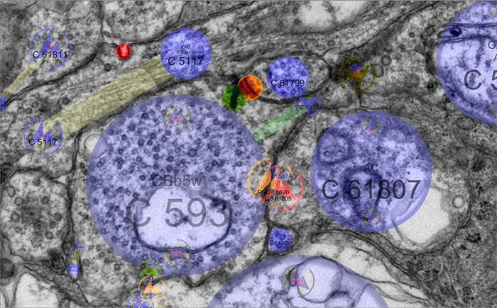
Results: CBb5w 593 is one of 20 ON cone BCs of this class in RC1. This CBb5w is presynaptic to 17 distinct GCs and 262 AC processes, and postsynaptic to 228 AC processes. The majority of these ACs are wf γACs. We estimate this BC forms synapses with 50 unique ACs. Asari & Meister (2014) found that single bipolar cell projective fields range up to 1 mm, far beyond a BC axonal field, and differentially drive multiple classes of GC. We discovered BC-BC within- and cross-class coupling and lateral inhibition that construct sign-conserving and sign-inverting projective fields to many distinct ganglion cell classes across the entire 0.25 mm diameter of RC1, much greater than a 60 µm BC axonal field. Cross-class projections access a broader set of GCs than expected from in-class projections alone. The BC-BC coupling is independent of BC-AII AC coupling. 94% of the CBb5w 593 BC-GC synaptic pairs receive feedback inhibition within the varicosity of the ribbon, but the number of feedback synapses is highly variable (coefficient of variation = 0.81). 35% of the BC-GC pairs receive feedforward inhibition within 2 microns of the postsynaptic density.
Conclusions: Mammalian BCs use novel cross-class topologies to distribute signals to a wide range of GCs and establish projective fields similar to those discovered in non-mammalian species. BC-BC within- and cross-class coupling and lateral inhibition via wf γACs establish sign-conserving and sign-inverting projective fields, respectively, up to 1 mm diameters. BC-GC synaptic pairs overwhelmingly employ feedback vs. feedforward inhibition to modulate signaling, and the numbers of feedback synapses are highly variable across these pairs, accounting for privatized and differential GC responses to the same BC drive.
This abstract was presented today at the 2014 Association for Research in Vision and Opthalmology (ARVO) meetings in Orlando, Florida by J Scott Lauritzen, Noah T. Nelson, Crystal L. Sigulinsky, Nathan Sherbotie, John Hoang, Rebecca L. Pfeiffer, James R. Anderson, Carl B. Watt, Bryan W. Jones and Robert E. Marc.
Purpose: Converging evidence suggests that large- and intermediate-scale neural networks throughout the nervous system exhibit small world’ design characterized by high local clustering of connections yet short path length between neuronal modules (Watts & Strogatz 1998 Nature; Sporns et al.2004 Trends in Cog Sci). It is suspected that this organizing principle scales to local networks (Ganmor et al. 2011 J Neurosci; Sporns 2006 BioSystems) but direct observation of synapses and local network topologies mediating small world design has not been achieved in any neuronal tissue. We sought direct evidence for synaptic and topological substrates that instantiate small world network architectures in the ON inner plexiform layer (IPL) of the rabbit retina. To test this we mined ≈ 200 ON cone bipolar cells (BCs) and ≈ 500 inhibitory amacrine cell (AC) processes in the ultrastructural rabbit retinal connectome (RC1).
Methods: BC networks in RC1 were annotated with the Viking viewer and explored via graph visualization of connectivity and 3D rendering (Anderson et al. 2011 J Microscopy). Small molecule signals embedded in RC1 e.g. GABA glycine and L-glutamate combined with morphological reconstruction and connectivity analysis allow for robust cell classification. MacNeil et al. (2004 J Comp Neurol) BC classification scheme used for clarity.
Results: Homocellular BC coupling (CBb3::CBb3 CBb4::CBb4 CBb5::CBb5) and within-class BC inhibitory networks (CBb3 → AC –| CBb3 CBb4 → AC –| CBb4 CBb5 → AC –| CBb5) in each ON IPL strata form laminar-specific functional sheets with high clustering coefficients. Heterocellular BC coupling (CBb3::CBb4 CBb4::CBb5 CBb3::CBb5) and cross-class BC inhibitory networks (CBb3 → AC –| CBb4 CBb4 → AC –| CBb3 CBb4 → AC –| CBb5 CBb5 → AC –| CBb4 CBb3 → AC –| CBb5 CBb5 → AC –| CBb3) establish short synaptic path lengths across all ON IPL laminae.
Conclusions: The retina contains a greater than expected number of synaptic hubs that multiplex parallel channels presynaptic to ganglion cells. The results validate a synaptic basis (ie. direct synaptic connectivity) and local network topology for the small world architecture indicated at larger scales providing neuroanatomical plausibility of this organization for local networks and are consistent with small world design as a fundamental organizing principle of neural networks on multiple spatial scales.
Support: NIH EY02576 (RM), NIH EY015128 (RM), NSF 0941717 (RM), NIH EY014800 Vision Core (RM), RPB CDA (BWJ), Thome AMD Grant (BWJ).
We have a new publication, this one a chapter titled: Retinal connectomics: A New Era For Connectivity Analysis in The New Visual Neurosciences (A little cheaper on Amazon here) textbook. Authors are Robert E. Marc, Bryan W. Jones, James S. Lauritzen, Carl B. Watt and James R. Anderson.