Synapse Classification And Localization In Electron Micrographs

Synapse-classification_

We have a new publication, Synapse Classification And Localization In Electron Micrographs in Pattern Recognition Letters.  Authors are: Vignesh JagadeeshJames Anderson, Bryan W. JonesRobert MarcSteven Fisher and B.S. Manjunath.

Abstract:  Classification and detection of biological structures in Electron Micrographs (EM) is a relatively new large scale image analysis problem. The primary challenges are in modeling diverse visual characteristics and development of scalable techniques. In this paper we propose novel methods for synapse detection and localization, an important problem in connectomics. We first propose an attribute based descriptor for characterizing synaptic junctions. These descriptors are task specific, low dimensional and can be scaled across large image sizes. Subsequently, techniques for fast localization of these junctions are proposed. Experimental results on images acquired from a mammalian retinal tissue compare favorably with state of the art descriptors used for object detection.

Retinal connectomics: A New Era For Connectivity Analysis in The New Visual Neurosciences

New-Visual-Neurosciences

We have a new publication, this one a chapter titled: Retinal connectomics: A New Era For Connectivity Analysis in The New Visual Neurosciences (A little cheaper on Amazon here) textbook.  Authors are Robert E. Marc, Bryan W. Jones, James S. Lauritzen, Carl B. Watt and James R. Anderson.

Robust Segmentation based Tracking using an Adaptive Wrapper for Inducing Priors

Robust-Segmentation-based-Tracing-using-an-Adaptive-Wrapper-for-Inducing-Priors

We have published another manuscript, Robust Segmentation based Tracking using an Adaptive Wrapper for Inducing Priors.  This manuscript describes the work on adaptive tracing and proposes an algorithm that adapts a generic tracing algorithm to an application of interest.  In our specific case, it is boundaries of cells in high frequency space in transmission electron microscopy images.  But the approach in this paper is applicable to biological, medical, remote sensing and surveillance data as well utilizing priors specific to the application.  The co-authors on the paper are: Vignesh JagadeeshJames Anderson, Bryan W. JonesRobert E. MarcSteven K Fisher and B.S Manjunath.

Retinal Connectomics: Toward Complete, Accurate Networks

Retinal Connectomics_600

We have a new publication, Retinal connectomics: Toward complete, accurate networks in Progress in Retinal and Eye Research.  Authors are:  Robert E. Marc, Bryan W. JonesCarl B. Watt, Crystal Sigulinsky, James R. Anderson and J. Scott Lauritzen.

Abstract:
Connectomics is a strategy for mapping complex neural networks based on high-speed automated electron optical imaging, computational assembly of neural data volumes, web-based navigational tools to explore 1012-1015 byte (terabyte to petabyte) image volumes, and annotation and markup tools to convert images into rich networks with cellular metadata. These collections of network data and associated metadata, analyzed using tools from graph theory and classification theory, can be merged with classical systems theory, giving a more completely parameterized view of how biologic information processing systems are implemented in retina and brain. Networks have two separable features: topology and connection attributes. The first findings from connectomics strongly validate the idea that the topologies complete retinal networks are far more complex than the simple schematics that emerged from classical anatomy. In particular, connectomics has permitted an aggressive refactoring of the retinal inner plexiform layer, demonstrating that network function cannot be simply inferred from stratification; exposing the complex geometric rules for inserting different cells into a shared network; revealing unexpected bidirectional signaling pathways between mammalian rod and cone systems; documenting selective feedforward systems, novel candidate signaling architectures, new coupling motifs, and the highly complex architecture of the mammalian AII amacrine cell. This is but the beginning, as the underlying principles of connectomics are readily transferrable to non-neural cell complexes and provide new contexts for assessing intercellular communication.

FASEB Bio-Art Competition Winner 2013

1254_40X_GBY_72dpi-1

Bryan W. Jones and Robert E. Marc and were selected as 2013 FASEB BioArt Winners (Press release here).  This image shows a region of an amazingly complex retina from a goldfish (Carassius auratus auratus) analyzed using tools called Computational Molecular Phenotyping (CMP) that reveal the metabolic state of the all cell types in tissues.  These cells were labeled with antibodies for the presence of two fundamental amino acid metabolites (anti-glycine in red, anti-GABA in blue) and an amino acid tracer of physiologic activity (anti-AGB in green).   These labels allow us to visualize the metabolic state and therefore, classes of bipolaramacrine and horizontal cells.  More details on the image here.


The Alternative Complement Pathway Deficiency Amerliorates Chronic Smoked-Induced Functional And Morphological Ocular Injury

Alex Woodell, Beth Coughlin, Kannan Kunchithapautham, Sarah Casey, Tucker Williamson, W. Drew Ferrell,  Carl Atkinson, Bryan Jones and Baerbel Rohrer have a new manuscript out, The Alternative Complement Pathway Deficiency Amerliorates Chronic Smoked-Induced Functional And Morphological Ocular Injury in PLOS One.

The short story is: Don’t smoke.  But then you knew that.  Where this paper contributes is that it provides clear findings that show ocular pathologies generated by cigarette smoke are dependent upon activation of the immune system, in particular complement and the alternative pathway which are critical findings in the treatment of AMD.

Constructive Retinal Plasticity After Selective Ablation of the Photoreceptors

This abstract was presented today at the Association for Research in Vision and Opthalmology (ARVO) meetings in Seattle, Washington by Corinne N. Beier, Bryan W. Jones, Philip Huie, Yannis M. Paulus, Daniel Lavinsky, Loh-Shan B. Leung, Hiroyuki Nomoto,  Robert E. Marc, Daniel V. Palanker, and Alexander Sher.

Purpose: In the rabbit retina there is evidence of constructive retinal plasticity in response to focal ablation of a small patch of the photoreceptor layer by laser photocoagulation. After a two-month healing period, healthy photoreceptors migrate inwards filling the damaged area and restoring visual sensitivity to the lesion site. We investigated the integrity and function of the neural populations above the lesion, whether the migrating photoreceptors formed new connections with deafferented bipolar cells, and to what degree the new function resembled normal retinal function.

Methods: 
Retinal photocoagulation lesions of Moderate and Barely Visible clinical grades were produced in rabbits with a 532-nm laser, using beam diameter of 200 and 400 μm. Retinal ganglion cell (RGC) responses to spatio-temporal white noise stimulus were recorded on a 512-electrode array. Inner retinal neuron cell types were identified using Computational Molecular Phenotyping (CMP). Light evoked activity of the inner retinal neurons was measured through 1-amino-4-guanidobutane (AGB) labeling. Synaptic structure between photoreceptors and bipolar cells was characterized through transmission electron microscopy (TEM) imaging.

Results: 
The lesioned areas of the retina, after a two-month healing period, regained visual sensitivity. There was no significant difference between the response kinetics of RGCs with receptive fields covering the lesioned area and RGCs with receptive fields unaffected by the lesion. Furthermore, the average receptive field sizes of RGCs covering the lesion were consistent with the average receptive field sizes of RGCs unaffected by the lesion. CMP showed that all major inner retinal neuron cell types are present above both acute and healed lesions. Light evoked activity in the retina, as measured by AGB concentration levels, was diminished in the acute lesion but returned to within 10% of normal after two months. TEM images showed normal photoreceptor synaptic structure inside the healed lesion area.

Conclusions: 
Migrating photoreceptors establish new functional connectivity to deafferented bipolar cells and have normal synaptic structure. The new circuitry results in spatial and temporal properties of the RGC responses that resemble those of the healthy retina. In summary, the rewiring restores normal visual response in the lesioned area, indicating constructive retinal plasticity.

Support: Burroughs Wellcome Fund Career Award at the Scientific Interface; the Pew Charitable Trusts Scholarship in the Biomedical Sciences (A.S.), RPB CDA, Thome Foundation (BWJ), NIH EY02576, NIH EY015128, NSF 0941717, NIH EY014800 Vision Core (R.M.); NIH 5R01EY18608, AFOSR FA9550- 10-1-0503, DoD W81XWH-12-10575, Stanford University Bio-X (D.P.), Edward N. and Della L. Thome Memorial Foundation grant for Age-Related Macular Degeneration Research (BWJ) RPB unrestricted award (Moran Eye Center)

Retinal Metabolic Response to Cigarette Smoke

This abstract was presented today at the Association for Research in Vision and Opthalmology (ARVO) meetings in Seattle, Washington by Alexandra D. Butler, William D. Ferrell, Alex Woodell, Carl Atkinson, Baerbel Rohrer, Robert E. Marc and Bryan W. Jones.

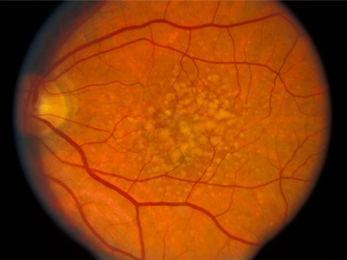
Purpose:  Smoking is the single largest risk factor for age-related macular degeneration, aside from age. Several of the main genetic risk factors for AMD are polymorphisms occurring in complement genes involved in the alternative, classical and common terminal pathways. To better understand the metabolic impact of smoking on the retina, we used computational molecular phenotyping (CMP) and examined the effects of cigarette smoke on wild type (wt) retinas and mice in which either the alternative pathway (complement factor B, CfB) or the common terminal pathway (complement component 3, C3) was removed.

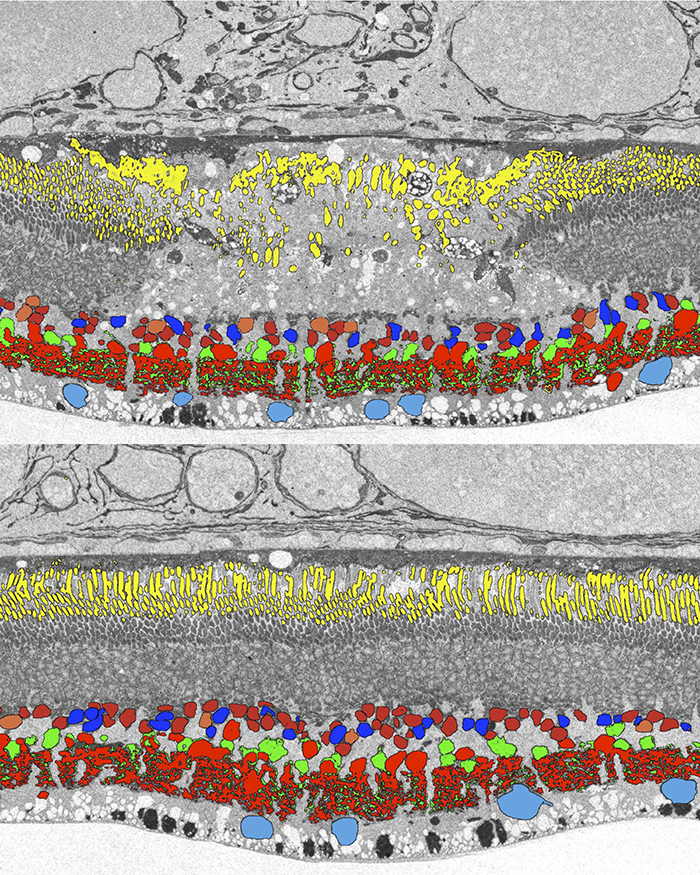
Methods:  Mice were exposed to either cigarette smoke or filtered air. Cigarette smoke (CS) was generated using an automated cigarette-smoking machine (Model TE-10, Teague Enterprises, Davis, CA) by burning 3R4F reference cigarettes (2.45 mg nicotine per cigarette; purchased from the Tobacco Health Research Institute, University of Kentucky, Lexington, KY). Mice were exposed to CS for 6 hours/day, 5 days/week for 6 months. Age matched room filtered air exposed mice were used as controls. Eyes were enucleated immediately post-mortem, fixed in 1% paraformaldehyde, 2.5% glutaraldehyde, dehydrated in graded methanols, embedded in eponates and histologically analyzed with CMP.

Results:  Alterations in retinal small molecule signatures from mice exposed to cigarette smoke were observed compared to retinas from non-smoked mice in wt, CfB and C3 knockout mice. Signal changes with arginine, glutamine and glutathione progressively increased in the retinas of smoked exposed wt, CfB and C3 knockout mice, indicating increased response profiles to cell stress. Both Müller cells and photoreceptors of wt smoked retinas demonstrated changes relative to non- smoked retinas.

Conclusions:  Arginine, glutamine and glutathione, amino acids known to be involved in cellular stress responses, were increased in retinal neurons and glial cells upon smoke exposure. Eliminating essential components of the complement system, a cascade required for the maintenance of the immune privilege of the eye, appears to exacerbate responses to cigarette smoke in oxidative damage response related pathways. Understanding complement-dependent alterations in the eye will aid in our understanding of AMD pathology and may open new avenues for novel treatment strategies.

Support:  RPB CDA (BWJ), Thome AMD Grant (BWJ), NIH EY02576 (RM), NIH EY015128 (RM), NSF 0941717 (RM), NIH EY014800 Vision Core (RM), NIH EY019320 (BR), VA merit award RX000444 (BR), grant to MUSC from RPB