Tag Archives: Bryan W. Jones

Proteomic changes in the lens of a congenital cataract mouse model lead to reduced levels of glutathione and taurine

This abstract was presented today, May 4th at the 2022  Association for Research in Vision and Opthalmology (ARVO) meetings in Denver, Colorado by Sheldon Rowan @SheldonRowan, Eloy Bejarano, Elizabeth Whitcomb, Rebecca Pfeiffer @BeccaPfeiffer19, Kristie Rose, Kevin Schey, Bryan Jones @BWJones, Allen Taylor.

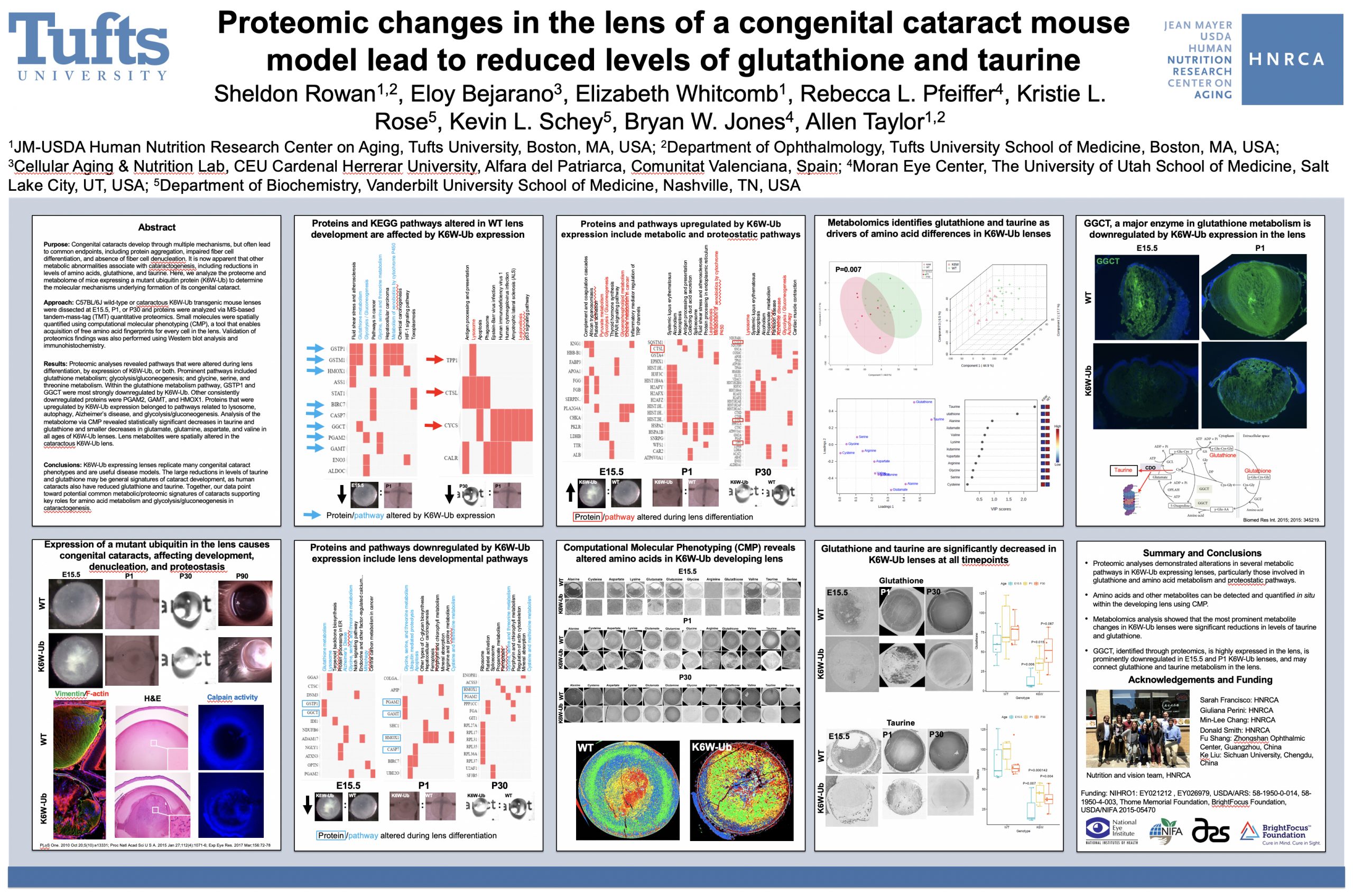
Purpose: Congenital cataracts develop through multiple mechanisms, but often lead to common endpoints, including protein aggregation, impaired fiber cell differentiation, and absence of fiber cell denucleation. It is now apparent that other metabolic abnormalities associate with cataractogenesis, including reductions in levels of amino acids, glutathione, and taurine. Here, we analyze the proteome and metabolome of mice expressing a mutant ubiquitin protein (K6W-Ub) to determine the molecular mechanisms underlying formation of its congenital cataract.

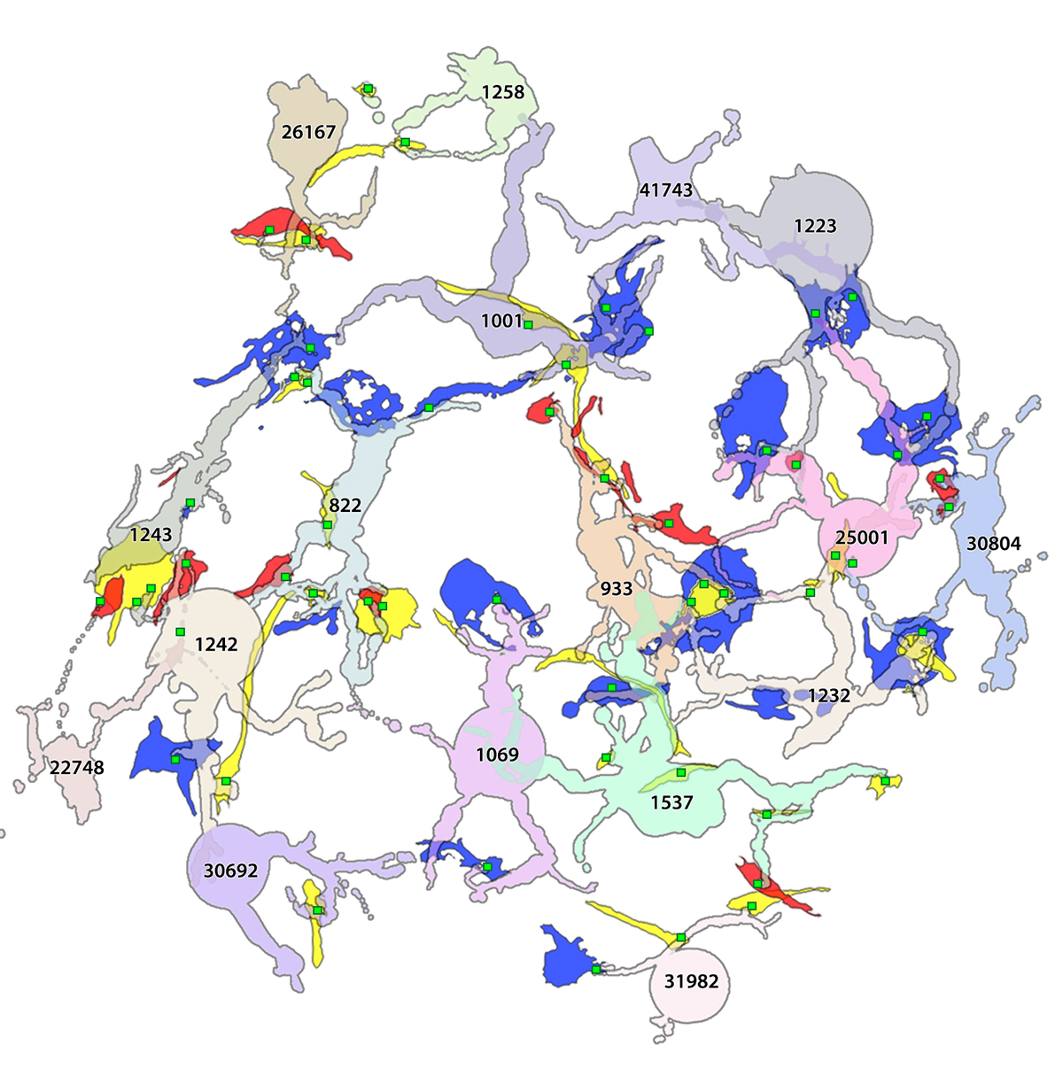
Methods: C57BL/6J wild-type or cataractous K6W-Ub transgenic mouse lenses were dissected at E15.5, P1, or P30 and proteins were analyzed via MS-based tandem-mass-tag (TMT) quantitative proteomics. Small molecules were spatially quantified using computational molecular phenotyping (CMP), a tool that enables acquisition of free amino acid fingerprints for every cell in the lens. Validation of proteomics findings was also performed using Western blot analysis and immunohistochemistry.

Results: Proteomic analyses revealed pathways that were altered during lens differentiation, by expression of K6W-Ub, or both. Prominent pathways included glutathione metabolism; glycolysis/gluconeogenesis; and glycine, serine, and threonine metabolism. Within the glutathione metabolism pathway, GSTP1 and GGCT were most strongly downregulated by K6W-Ub. Other consistently downregulated proteins were PGAM2, GAMT, and HMOX1. Proteins that were upregulated by K6W-Ub expression belonged to pathways related to lysosome, autophagy, Alzheimer’s disease, and glycolysis/gluconeogenesis. Analysis of the metabolome via CMP revealed statistically significant decreases in taurine and glutathione and smaller decreases in glutamate, glutamine, aspartate, and valine in all ages of K6W-Ub lenses. Lens metabolites were spatially altered in the cataractous K6W-Ub lens.

Conclusions: K6W-Ub expressing lenses replicate many congenital cataract phenotypes and are useful disease models. The large reductions in levels of taurine and glutathione may be general signatures of cataract development, as human cataracts also have reduced glutathione and taurine. Key roles for amino acid metabolism and glycolysis/gluconeogenesis in cataractogenesis are emerging. Together our data point toward potential common metabolic/proteomic signatures of cataracts.

ARVO Mini-Symposium: Pathoconnectomics in Retinal Degeneration

Lab PI, Bryan Jones delivered a talk at the ARVO 2022 mini-symposium on Pathoconnectomics in Retinal Degeneration.

Abstract: Connectomics has demonstrated that synaptic networks and their topologies are precise and directly correlate with physiology and behavior. The next extension of connectomics is pathoconnectomics: to map neural network synaptology and circuit topologies corrupted by neurological disease in order to identify robust targets for therapeutics. The retina is ideal for pathoconnectomics approaches, and reveals common rules of how neural systems are wired, and how they break in neurodegenerative disease.

Primary Cilia in Amacrine Cells in Retinal Development

We have a new collaborative manuscript out in iOVS, Primary Cilia in Amacrine Cells in Retinal Development. (pdf here)

Authors: Ke Ning; Brent E. Sendayen; Tia J. Kowal; Biao WangBryan W. Jones @BWJones; Yang Hu; and Yang Sun.

Abstract:

Purpose: Primary cilia are conserved organelles found in polarized cells within the eye that regulate cell growth, migration, and differentiation. Although the role of cilia in photoreceptors is well-studied, the formation of cilia in other retinal cell types has received little attention. In this study, we examined the ciliary profile focused on the inner nuclear layer of retinas in mice and rhesus macaque primates.

Methods: Retinal sections or flatmounts from Arl13b-Cetn2 tg transgenic mice were immunostained for cell markers (Pax6, Sox9, Chx10, Calbindin, Calretinin, ChaT, GAD67, Prox1, TH, and vGluT3) and analyzed by confocal microscopy. Primate retinal sections were immunostained for ciliary and cell markers (Pax6 and Arl13b). Optical coherence tomography (OCT) and ERGs were used to assess visual function of Vift88 mice.

Results: During different stages of mouse postnatal eye development, we found that cilia are present in Pax6-positive amacrine cells, which were also observed in primate retinas. The cilia of subtypes of amacrine cells in mice were shown by immunostaining and electron microscopy. We also removed primary cilia from vGluT3 amacrine cells in mouse and found no significant vision defects. In addition, cilia were present in the outer limiting membrane, suggesting that a population of Müller glial cells forms cilia.

Conclusions: We report that several subpopulations of amacrine cells in inner nuclear layers of the retina form cilia during early retinal development in mice and primates.


Subretinal Rather Than Intravitreal Adeno-Associated Virus–Mediated Delivery of a Complement Alternative Pathway Inhibitor Is Effective in a Mouse Model of RPE Damage

We have a new manuscript out in iOVS, Subretinal Rather Than Intravitreal Adeno-Associated Virus–Mediated Delivery of a Complement Alternative Pathway Inhibitor Is Effective in a Mouse Model of RPE Damage. (pdf here)

Authors: Balasubramaniam Annamalai; Nathaniel Parsons; Crystal Nicholson; Elisabeth Obert; Bryan W. Jones @BWJones; and Bärbel Rohrer.

Abstract:

Purpose: The risk for age-related macular degeneration has been tied to an overactive complement system. Despite combined attempts by academia and industry to develop therapeutics that modulate the complement response, particularly in the late geographic atrophy form of advanced AMD, to date, there is no effective treatment. We have previously demonstrated that pathology in the smoke-induced ocular pathology (SIOP) model, a model with similarities to dry AMD, is dependent on activation of the alternative complement pathway and that a novel complement activation site targeted inhibitor of the alternative pathway can be delivered to ocular tissues via an adeno-associated virus (AAV).

Methods: Two different viral vectors for specific tissue targeting were compared: AAV5-VMD2-CR2-fH for delivery to the retinal pigment epithelium (RPE) and AAV2YF-smCBA-CR2-fH for delivery to retinal ganglion cells (RGCs). Efficacy was tested in SIOP (6 months of passive smoke inhalation), assessing visual function (optokinetic responses), retinal structure (optical coherence tomography), and integrity of the RPE and Bruch’s membrane (electron microscopy). Protein chemistry was used to assess complement activation, CR2-fH tissue distribution, and CR2-fH transport across the RPE.

Results: RPE- but not RGC-mediated secretion of CR2-fH was found to reduce SIOP and complement activation in RPE/choroid. Bioavailability of CR2-fH in RPE/choroid could be confirmed only after AAV5-VMD2-CR2-fH treatment, and inefficient, adenosine triphosphate–dependent transport of CR2-fH across the RPE was identified.

Conclusions: Our results suggest that complement inhibition for AMD-like pathology is required basal to the RPE and argues in favor of AAV vector delivery to the RPE or outside the blood-retina barrier.

A pathoconnectome of early neurodegeneration: Network changes in retinal degeneration

We have a new manuscript out in Experimental Eye Research, A pathoconnectome of early neurodegeneration: Network changes in retinal degeneration. (pdf here)

Authors: Rebecca L. Pfeiffer @BeccaPfeiffer19, James R. Anderson, Jeebika Dahal, Jessica C. Garcia, Jia-Hui Yang, Crystal L. Sigulinsky @CLSigulinsky, Kevin Rapp, Daniel P. Emrich, Carl B. Watt, Hope AB Johnstun, Alexis R. Houser, Robert E. Marc @robertmarc60, and Bryan W. Jones @BWJones.

Abstract: Connectomics has demonstrated that synaptic networks and their topologies are precise and directly correlate with physiology and behavior. The next extension of connectomics is pathoconnectomics: to map neural network synaptology and circuit topologies corrupted by neurological disease in order to identify robust targets for therapeutics. In this report, we characterize a pathoconnectome of early retinal degeneration. This pathoconnectome was generated using serial section transmission electron microscopy to achieve an ultrastructural connectome with 2.18nm/px resolution for accurate identification of all chemical and gap junctional synapses. We observe aberrant connectivity in the rod-network pathway and novel synaptic connections deriving from neurite sprouting. These observations reveal principles of neuron responses to the loss of network components and can be extended to other neurodegenerative diseases.


Müller Cell Metabolic Signatures: Evolutionary Conservation and Disruption in Disease

We have a new manuscript out in Trends in Endocrinology & Metabolism, Müller Cell Metabolic Signatures: Evolutionary Conservation and Disruption in Disease.

Authors: Rebecca L. Pfeiffer @BeccaPfeiffer19, Robert E. Marc @robertmarc60, and Bryan William Jones @BWJones.

This manuscript functions as both a review and presents some exciting new data demonstrating how the glutamate cycle is disrupted during retinal degenerative disease.

Abstract: Müller cells are glia that play important regulatory roles in retinal metabolism. These roles have been evolutionarily conserved across at least 300 million years. Müller cells have a tightly locked metabolic signature in the healthy retina, which rapidly degrades in response to insult and disease. This variation in metabolic signature occurs in a chaotic fashion, involving some central metabolic pathways. The cause of this divergence of Müller cells, from a single class with a unique metabolic signature to numerous separable metabolic classes, is currently unknown and illuminates potential alternative metabolic pathways that may be revealed in disease. Understanding the impacts of this heterogeneity on degenerate retinas and the implications for the metabolic support of surrounding neurons will be critical to long-term integration of retinal therapeutics for the restoration of visual perception following photoreceptor degeneration.

Optic Cup Morphogenesis Requires Neural Crest-Mediated Basement Membrane Assembly

We have a new manuscript out in Development, Optic cup morphogenesis requires neural crest-mediated basement membrane assembly.

Authors: Chase D. Bryan @CDBE30, Macaulie A. Casey, Rebecca L. Pfeiffer @BeccaPfeiffer19, Bryan W. Jones @BWJones, and Kristen M. Kwan @BlockInTheBack

This is a collaborative project out of the Kwan lab that we helped out with some of the ultrastructural work and analysis.

Abstract: Organogenesis requires precise interactions between a developing tissue and its environment. In vertebrates, the developing eye is surrounded by a complex extracellular matrix as well as multiple mesenchymal cell populations. Disruptions to either the matrix or periocular mesenchyme can cause defects in early eye development, yet in many cases, the underlying mechanism is unknown. Here, using multidimensional imaging and computational analyses in zebrafish, we establish that cell movements in the developing optic cup require neural crest. Ultrastructural analysis reveals that basement membrane formation around the developing eye is also dependent on neural crest, but only specifically around the retinal pigment epithelium. Neural crest cells produce the extracellular matrix protein nidogen: impairing nidogen function disrupts eye development, and strikingly, expression of nidogen in the absence of neural crest partially restores optic cup morphogenesis. These results demonstrate that eye formation is regulated in part by extrinsic control of extracellular matrix assembly.

An Update On Retinal Prostheses

We have a new manuscript out in Clinical Neurophysiology, An Update on Retinal Prostheses. PubMedDirect Link PDF here.

Authors: Lauren N. Ayton, Nick Barnes, Gislin Dagnelie, Takashi Fujikado, Georges Goetz, Ralf Hornig, Bryan W. Jones, Mahiul M.K. Muqit, Daniel L. Rathbun, Katarina Stingl, James D. Weiland, Matthew A. Petoe.

Abstract: Retinal prostheses are designed to restore a basic sense of sight to people with profound vision loss. They require a relatively intact posterior visual pathway (optic nerve, lateral geniculate nucleus and visual cor- tex). Retinal implants are options for people with severe stages of retinal degenerative disease such as retinitis pigmentosa and age-related macular degeneration.

There have now been three regulatory-approved retinal prostheses. Over five hundred patients have been implanted globally over the past 15 years. Devices generally provide an improved ability to localize high-contrast objects, navigate, and perform basic orientation tasks. Adverse events have included con- junctival erosion, retinal detachment, loss of light perception, and the need for revision surgery, but are rare. There are also specific device risks, including overstimulation (which could cause damage to the retina) or delamination of implanted components, but these are very unlikely.
Current challenges include how to improve visual acuity, enlarge the field-of-view, and reduce a com- plex visual scene to its most salient components through image processing. This review encompasses the work of over 40 individual research groups who have built devices, developed stimulation strategies, or investigated the basic physiology underpinning retinal prostheses. Current technologies are summarized, along with future challenges that face the field.


Contrast Sensitivity Isocontours Of The Central Visual Field

We have a new manuscript out in Nature Scientific Reports, Contrast Sensitivity Isocontours Of The Central Visual Field.

Authors: Agnes Y. J. Choi, Lisa Nivison-Smith, Jack Phu, Barbara Zangerl, Sieu K. Khuu, Bryan W. Jones, Rebecca L. Pfeiffer, Robert E. Marc & Michael Kalloniatis.

Abstract: Standard automated perimetry (SAP), the most common form of perimetry used in clinical practice, is associated with high test variability, impacting clinical decision making and efficiency. Contrast sensitivity isocontours (CSIs) may reduce test variability in SAP by identifying regions of the visual field with statistically similar patterns of change that can be analysed collectively and allow a point (disease)-to-CSI (normal) comparison in disease assessment as opposed to a point (disease)-to-point (normal) comparison. CSIs in the central visual field however have limited applicability as they have only been described using visual field test patterns with low, 6° spatial sampling. In this study, CSIs were determined within the central 20° visual field using the 10-2 test grid paradigm of the Humphrey Field Analyzer which has a high 2° sampling frequency. The number of CSIs detected in the central 20° visual field was greater than previously reported with low spatial sampling and stimulus size dependent: 6 CSIs for GI, 4 CSIs for GII and GIII, and 3 CSIs for GIV and GV. CSI number and distribution were preserved with age. Use of CSIs to assess visual function in age-related macular degeneration (AMD) found CSI guided analysis detected a significantly greater deviation in sensitivity of AMD eyes from normal compared to a standard clinical pointwise comparison (−1.40 ± 0.15 dB vs −0.96 ± 0.15 dB; p < 0.05). This work suggests detection of CSIs within the central 20° is dependent on sampling strategy and stimulus size and normative distribution limits of CSIs can indicate significant functional deficits in diseases affecting the central visual field such as AMD.

Coupling architecture of the retinal Aii/ON cone bipolar cell network and alteration in degeneration

This poster was presented today, July 28th at the 2019 International Gap Junction Conference in Victoria, Canada by Crystal L. Sigulinsky, Rebecca L. PfeifferJames R. Anderson, Christopher Rapp, Jeebika Dahal, Jessica C Garcia, Jia-Hui Yang, Daniel P. Emrich, Hope Morrison, Kevin D. Rapp, Carl B. Watt, Mineo Kondo, Hiroko Terasaki, Robert E. Marc and Bryan W. Jones.

Almost full resolution version here.

Authors:

Crystal L Sigulinsky1, Rebecca L Pfeiffer1, James R. Anderson1, Christopher N. Rapp1, Jeebika Dahal1, Jessica C Garcia1, Jia-Hui Yang1, Daniel P. Emrich1, Hope Morrison1, Kevin D. Rapp1, Carl B. Watt1, Mineo Kondo2, Hiroko Terasaki3, Robert E. Marc1, Bryan W. Jones1
1Moran Eye Center/ Ophthalmology, University of Utah, Salt Lake City, Utah, United States; 2Mie University, Tsu, Japan; 3Nagoya University, Nagoya-shi, Japan;

ABSTRACT:

Background and aim:
Gap junctions are prevalent throughout the neural retina, with expression by every major neuronal class and at every level of signal processing. Yet, the functional roles and expressing cells/participating networks for many remain unknown. Spontaneous network spontaneous hyperactivity observed during retinal degeneration contributes to visual impairment and requires gap junctional coupling in the Aii amacrine cell/ON cone bipolar cell (CBC) network.  However, it remains unclear whether this hyperactivity reflects changes in the underlying circuitry or dysfunction of the normative circuitry. Here, we used connectomics-based mapping of retinal circuitry to 1) define the coupling architecture of the Aii/ON CBC network in healthy adult rabbit retina using connectome RC1 and 2) evaluate changes in coupling motifs in RPC1, a pathoconnectome from a rabbit retinal degeneration model.

Methods:

RC1 and RPC1 are connectomes built by automated transmission electron microscopy at ultrastructural (2 nm/pixel) resolution. RC1 is a 0.25 mm diameter volume of retina from a 13-month old, light adapted female Dutch Belted rabbit. RPC1 is a 0.07 mm diameter volume of degenerate retina from a transgenic P347L model of autosomal dominant retinitis pigmentosa (10-months old, male, New Zealand White background) presenting with ~50% rod loss. ON CBCs, Aii amacrine cells, and their coupling partners were annotated using the Viking application. Coupling motifs and features were explored with 3D rendering and network graph visualization. Gap junctions were validated by 0.25 nm resolution recapture with goniometric tilt when necessary.

Results:

Complete reconstruction of 37 ON CBCs in RC1 yielded 1339 gap junctions and revealed pervasive in- and cross-class coupling motifs among ON CBCs that produce complex network topologies within the coupled Aii network. Robust rulesets underlie class-specific coupling profiles with specificity defined beyond geometric opportunity. These coupling profiles enabled classification of all 145 ON CBCs contained within RC1 into 7 distinct classes. In RPC1, two ON CBC classes appear to retain their class-specific coupling profiles, accepting and rejecting specific combinations of Aii and ON CBC class partnerships. However, aberrant partnerships exist, including both loss of motifs and acquisition of novel ones.

Conclusions:

Gap junctions formed by ON CBCs are prominent network components, with specificity rivaling that of chemical synapses. These gap junctions not only subserve canonical signal transfer for night vision, but also extensive coupling within and across the parallel processing streams. Clearly aberrant morphological and synaptic changes exist in RPC1, including changes in the coupling specificity of both Aii and ON CBCs. Thus, circuit topology is altered prior to complete loss of rods, with substantial implications for therapeutic interventions for blinding diseases that depend upon the surviving retinal network.