Tag Archives: Rebecca L. Pfeiffer

Rod Bipolar Cell Networks in Early Retinal Remodeling

Rebecca Pfeiffer, a post-doc in the laboratory presented her work on “Rod Bipolar Cell Networks in Early Retinal Remodeling” as a platform presentation at the ISER 2018 meeting in Belfast, Northern Ireland.

Authors: Rebecca Pfeiffer, James R. Anderson, Daniel P. Emrich, Jeebika Dahal, Crystal L Sigulinsky, Hope AB Morrison, Jia-Hui Yang, Carl B. Watt, Kevin D. Rapp, Jessica C Garcia, Mineo Kondo, Hiroko Terasaki, Robert E. Marc, and Bryan W. Jones.

Abstract: Retinal remodeling is a form of negative plasticity that occurs as a consequence of retinal degenerative diseases. Part of retinal remodeling involves anomalous sprouting of processes, termed neurites. The synaptic structures and partners of the neurites are not yet defined, leading to uncertainty about the consistency of network motifs between healthy and degenerate retina. Our goal is to map out the identities and network relationships of bipolar cell networks using a connectomics strategy. Retinal connectomes or ultrastructural maps of neuronal connectivity have substantially contributed to our understanding of retinal network topology, providing ground truth against which pathological network topologies can be evaluated. We have generated the first pathoconnectome (RPC1), or connectome of pathological tissues, of early retinal remodeling at 2nm/pixel, and are currently investigating the impact of remodeling on network architecture.
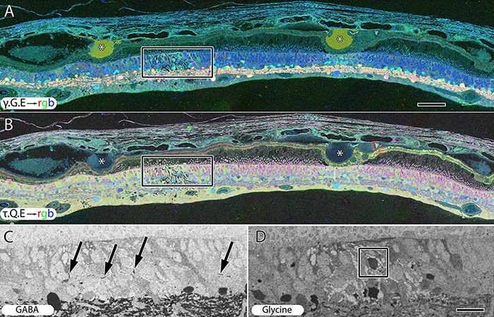
The tissue for RPC1 was obtained from a 10mo transgenic P347L rabbit model of autosomal dominant retinitis pigmentosa. Tissue was fixed in mixed aldehydes, osmicated, dehydrated, embedded in epon resin, and sectioned at 70nm. Serial sections were placed on grids, stained, and imaged using a JEOL JEM-1400 TEM using SerialEM software. Every 30th section was reserved for computational molecular phenotyping (CMP), and probed for small molecules: glutamate, glutamine, glycine, GABA, taurine, glutathione; or TEM compatible proteins GFAP and GS. The pathoconnectome volume is explored and annotated using the Viking software suite.
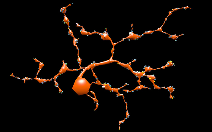
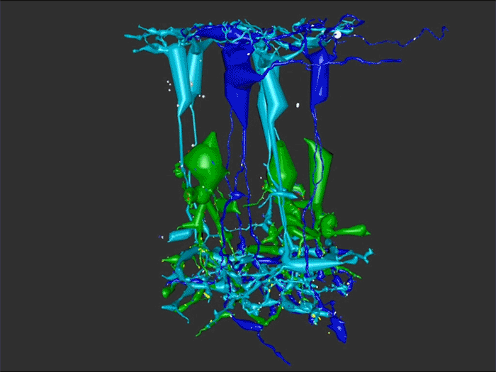
RPC1 was selected as an example of early retinal remodeling, demonstrating Muller cell hypertrophy, metabolic dysregulation, and degeneration of rod outer segments, indicating phase 1 remodeling and neuronal sprouting. We have observed the presence of both cone pedicles and rod spherules within the OPL to be synaptically active with neurites from some rod bipolar cells forming functional synapses with both rod spherules and cone pedicles. These rod bipolar cells also exhibit structurally altered ribbon synapses. We are currently evaluating network motifs and comparing them to networks established from our previous connectome, RC1, generated from a healthy rabbit.
These findings allow us to evaluate and analyze the impact of retinal remodeling on retinal networks which may have important implications for therapeutic interventions being developed which rely on inner retina network integrity.

Coupling Architecture Of The Aii/ON Cone Bipolar Cell Network In The Degenerate Retina

Crystal Sigulinsky, a post-doc in the lab, presented her work on “coupling architecture of the
Aii/ON cone bipolar cell network in the degenerate retina” at the RD2018 meeting in Killarney, Ireland today.  Authors are: Crystal L Sigulinsky, Rebecca L Pfeiffer, James R Anderson, Jeebika Dahal, Hope Morrison, Daniel P. Emrich, Jessica C Garcia, Jia-Hui Yang, Carl B. Watt, Kevin D. Rapp, Mineo Kondo, Hiroko Terasaki, Robert E. Marc, and Bryan W. Jones.

Purpose: Retinal network hyperactivity within degenerative retinal networks is a component of the disease process with implications for therapeutic interventions for blinding diseases that depend upon the surviving retinal network. Connexin36-containing gap junctions centered on the Aii amacrine cell network appear to mediate the aberrant signaling observed in mouse models of retinal degeneration. However, it remains unclear whether this hyperactivity reflects changes in the underlying circuitry or dysfunction/dysregulation of the normative circuitry. Mapping retinal circuitry in the ultrastructural rabbit Retinal Connectome, RC1, has revealed Aii network topologies explicitly involving gap junctions. In addition to canonical Aii-to-Aii and Aii-to-ON cone bipolar cell (CBC) coupling, we describe pervasive in- and cross-class coupling motifs among ON CBCs that extend and dramatically expand the coupled Aii network topologies. Since virtually every gap junction in the inner plexiform layer contains Connexin36, these circuits likely participate in the aberrant signaling of degenerate retinas. This study examines these Aii and ON CBC coupling motifs in Retinal PathoConnectome 1 (RPC1), an ultrastructural pathoconnectome of a rabbit model of retinitis pigmentosa.

Approach: RPC1 is a 2nm/pixel resolution volume of retina from a 10 month old, transgenic P347L rabbit model of autosomal dominant retinitis pigmentosa in early phase 1 retinal remodeling, a time point where cone and rod photoreceptors are still present, albeit going through cell stress. RPC1 spans the vitreous to basal outer nuclear layer and was built by automated transmission electron microscopy and computational assembly. ON CBCs, Aii amacrine cells, and their coupling partners were annotated using the Viking application and explored with 3D rendering and graph visualization of connectivity. Gap junctions were validated by 0.25 nm resolution recapture with goniometric tilt when necessary. Motifs were compared to those discovered in RC1. RC1 is a 2 nm resolution, 0.25 mm diameter volume of a light-adapted adult female Dutch Belted rabbit retina spanning the ganglion cell through inner nuclear layers.

Conclusions: RPC1 shows degeneration of rod outer segments, Müller cell hypertrophy and neuronal sprouting, characteristic of early stage retinal degeneration and phase 1 remodeling, when retinal hyperactivity and its reliance on gap junctional coupling has likely already initiated and human patients would still have some vision. All major coupling motifs (Aii-to-Aii, Aii-to-ON CBC, and ON CBC-to-ON CBC) were observed. Preliminary examinations indicate that several ON CBC classes retained their class-specific coupling profiles, accepting and rejecting specific combinations of Aii and ON CBC class partnerships. However, recent findings reveal aberrant partnerships in the coupled network, including both loss of prominent motifs and acquisition of novel ones. Thus, clear aberrant morphological and synaptic changes have been identified in RPC1, including changes in the coupling specificity and gap junction distributions of both Aii amacrine cells and ON CBCs (Figure 6). This suggests that the Aii/ON CBC circuit topology is already altered during early phase 1 remodeling, with substantial implications for therapeutic interventions in human subjects. The full coupling network is actively being examined and progress has begun on RPC2, a second pathoconnectome for examining later, phase 2 remodeling in this same model.

An almost full size poster available here in pdf format.

Retinal Remodeling And Metabolic Alterations in Human AMD

We have a new publication out (direct link, open access), Müller Cell Metabolic Chaos During Retinal Degeneration authored by Bryan W. JonesRebecca Pfeiffer, William Ferrell, Carl Watt, James Tucker, and Robert Marc.

Abstract:

Age-related macular degeneration (AMD) is a progressive retinal degeneration resulting in central visual field loss, ultimately causing debilitating blindness. AMD affects 18% of Americans from 65 to 74, 30% older than 74 years of age and is the leading cause of severe vision loss and blindness in Western populations. While many genetic and environmental risk factors are known for AMD, we currently know less about the mechanisms mediating disease progression. The pathways and mechanisms through which genetic and non-genetic risk factors modulate development of AMD pathogenesis remain largely unexplored. Moreover, current treatment for AMD is palliative and limited to wet/exudative forms. Retina is a complex, heterocellular tissue and most retinal cell classes are impacted or altered in AMD. Defining disease and stage-specific cytoarchitectural and metabolic responses in AMD is critical for highlighting targets for intervention. The goal of this article is to illustrate cell types impacted in AMD and demonstrate the implications of those changes, likely beginning in the retinal pigment epithelium (RPE), for remodeling of the the neural retina. Tracking heterocellular responses in disease progression is best achieved with computational molecular phenotyping (CMP), a tool that enables acquisition of a small molecule fingerprint for every cell in the retina. CMP uncovered critical cellular and molecular pathologies (remodeling and reprogramming) in progressive retinal degenerations such as retinitis pigmentosa (RP). We now applied these approaches to normal human and AMD tissues mapping progression of cellular and molecular changes in AMD retinas, including late-stage forms of the disease.

Pattern Recognition Reveals Different Visual Field Signature Patterns When Using Spatially Equated Test Sizes Compared To Standard Goldmann III Alone

This abstract was presented today, May 2th at the 2016 Association for Research in Vision and Opthalmology (ARVO) meetings in Seattle, Washington by Michael Kalloniatis, Robert E. Marc, Sieu K. Khuu, Jack Phu, Barbara Zangerl, Lisa Nivison-Smith, Bryan W. Jones, and Rebecca L. Pfeiffer. 

Abstract Number: 4745

Author Block: Michael Kalloniatis, Robert E. Marc, Sieu K. Khuu, Jack Phu, Barbara Zangerl, Lisa Nivison-Smith, Bryan W. Jones, Rebecca L. Pfeiffer
1 Centre for Eye Health, SOVS, University of New South Wales, Kensington, New South Wales, Australia; 2 SOVS, UNSW, Sydney, New South Wales, Australia; 3 Univ of Utah/Moran Eye Center, Salt Lake City, Utah, United States

Disclosure Block:Michael Kalloniatis, 2014/094035 A1 (USA) and 13865419.9 (EU) (Code P (Patent) ); Robert E. Marc, None; Sieu K. Khuu, 2014/094035 A1 (USA) and 13865419.9 (EU) (Code P (Patent) ); Jack Phu, None; Barbara Zangerl, None; Lisa Nivison-Smith, None; Bryan W. Jones, None; Rebecca L. Pfeiffer, None

Purpose:To identify areas within the visual field with matching contrast sensitivity (CS) signature patterns as a function of age using pattern recognition and determine the discrimination of CS data when using spatially equated test stimuli compared to the single size Goldmann (G)III alone.

Methods:52 subjects (classified in decade age groups from 20-60+ years) were tested using the Humphrey Visual Field Analyser 30-2 paradigm in full threshold mode for GI to GV. At least two thresholds were obtained per size. Two visual field maps were analyzed: a spatially equated visual field where GI was used centrally, GII mid-peripherally and GIII in the outer rings to place the test size at or close to complete spatial summation and a second where a single GIII was used at all locations. Thresholds were expressed as dB* (Khuu & Kalloniatis, IOVS 2015), converted to pixel values and analyzed using an unsupervised classification using isodata clustering (PCI, Geomatica, Canada). Class separation was extracted across the ages to develop dot plots of decade measures of CS.

Results:The 77 data points across the central 60° visual field can be distilled into 6 functional classes using the spatially equated visual field (Class separation 1). The 6 classes reflect areas in visual space that change in a similar manner across the ages. The use of the single GIII target resulted in only 4 classes displaying a poorer discrimination over the central visual field (Class separation 2). Extracted dot plots from class separation illustrated average CS within each class could be assessed across the decades.

Conclusions:When using spatially equated visual field testing, concentric areas were separated into distinct CS signatures consistent with known visual field sensitivity. We confirmed these areas change systematically with age. GIII failed to discriminate central areas of the 30-2 that likely reflects the fact that this size operates outside complete spatial summation and thus may not be the optimal test size for assessing visual function in the central visual field. More importantly, we showed pattern recognition can be applied to complex visual field data sets to identify common features and age-related visual function changes. This analysis allows regions to be averaged as they are statistically identical: this approach will likely assist structure-function studies.

Progressive Retinal Remodeling In A Transgenic Rabbit Model Of Retinitis Pigmentosa

This poster was presented today, May 2th at the 2016 Association for Research in Vision and Opthalmology (ARVO) meetings in Seattle, Washington by Rebecca L. Pfeiffer, Bryan W. Jones, and Robert E. Marc.

Posterboard #: D0246

Abstract Number: 2256 – D0246

Author Block: Rebecca L. Pfeiffer1,2 , Bryan W. Jones1,2 , Robert E. Marc1,2 
1 Ophthalmology, University of Utah, Salt Lake City, Utah, United States; 2 Interdepartmental Program in Neuroscience, University of Utah, Salt Lake City, Utah, United States

Purpose:Retinal degenerations are a collection of neural disorders, usually precipitated by photoreceptor degeneration. All display progressive metabolic alterations and neural loss following the death of the photoreceptors. Although it has been demonstrated that the metabolism of Müller cells (MCs) is drastically altered in degeneration, the full impact of these changes on surrounding neurons and long-term characterization of remodeling was previously unavailable, due to short lifespans of model organisms.

Methods:Retinal samples were collected from WT and Tg P347L rabbits at ages ranging from 3 months to 6 years. Following enucleation, retinas were divided into fragments and incubated for 10 minutes at 35 degrees C in D-isomers of Glutamate (dE), Glutamine (dQ), or Aspartate (dD) and Ames-bicarbonate medium to explore retinal transport capabilities at each stage of degeneration. Retinas were then fixed in mixed aldehyde buffer and processed for transmission electron microscope connectomics, immunocytochemistry for a range of macromolecules, and computational molecular phenotyping for small molecules (CMP) (J Comp Neurol. 464:1, 2003).

Results:CMP reveals that single metabolic phenotype of MCs splits and diverges into many phenotypes continuously throughout degeneration. Further, all neuronal classes continue to die throughout degeneration. By 6 years, over 90% of neurons are lost, and the remaining glutamatergic neurons have altered metabolic signatures with a large increase in aspartate levels, at times exceeding glutamate. Transport of the D-isomers indicates that glutamate transport capabilities remain intact until the latest stages of degeneration. This may not be true of their GABA transporters.

Conclusions:These results suggest three main conclusions. First, retinal remodeling in degeneration is relentlessly progressive long after all photoreceptors have disappeared. Second, cell types previously thought to remain after degeneration onset, such as ganglion cells, will also ultimately die and the cells not lost often will change their metabolism. The consequence of this metabolic change in neurons is not yet fully explored. Third, the persistent robust glutamate transport capabilities of Müller cells indicate Müller cells can metabolize glutamate despite the massive loss of glutamine synthetase activity, likely unmasking alternate metabolic pathways.

2-nm Resolution Anatomy of Retinal Neuro-Glial-Vascular Architecture

This abstract was presented today, May 2th at the 2016 Association for Research in Vision and Opthalmology (ARVO) meetings in Seattle, Washington by Jefferson R. Brown, Rebecca L. Pfeiffer, Crystal Sigulinsky, Felix Vazquez-Chona, Daniel Emrich, Bryan W. Jones, Robert E. Marc.

Abstract Number: 995

Author Block: Jefferson R. Brown, Rebecca L. Pfeiffer, Crystal Sigulinsky, Felix Vazquez-Chona, Daniel Emrich, Bryan W. Jones, Robert E. Marc
1 Dept of Ophthalmology, University of Utah, Salt Lake City, Utah, United States

Disclosure Block:Jefferson R. Brown, None; Rebecca L. Pfeiffer, None; Crystal Sigulinsky, None; Felix Vazquez-Chona, None; Daniel Emrich, None; Bryan W. Jones, None; Robert E. Marc, Signature Immunologics (Code I (Personal Financial Interest) )

Purpose:Retinal vasculature is strongly affected by degenerative pathologies and in turn, may also contribute to their progression. However, much of what we understand about the normal, healthy interaction between neurons, glia, and blood vessels at the ultrastructural level is limited to single section electron microscopy. The technology of serial section transmission electron microscopy (ssTEM) extends the high definition of TEM imaging into three dimensions to create volumes, allowing for more thorough visualization and analysis of the vascular-glial-neuronal complex.

Methods:RC2 is a 40TB ssTEM volume of over 1,400 horizontal sections of retinal tissue derived from an adult female C57BL/6J mouse. The tissue sample is 250 um in diameter and spans the outer nuclear layer to the vitreal surface. Baseline resolution is 2.18nm per pixel. Visualization, navigation and metadata annotations of the database are made via the Viking software suite.

Results:Much of the retinal vascular basement membrane directly contacts Muller cells. In the ganglion cell layer, direct basement membrane contact with astrocytes is frequent. Microglia commonly contact the basement membrane, and occasionally direct contact of neurons onto basement membrane was observed. Full 3D reconstruction of all vascular pathways with associated endothelia and pericytes within the volume was completed, demonstrating that all the retinal capillary layers are continuous with one another [Figure].

Conclusions:The presence of occasional direct neuronal contact onto vascular basement membrane supports earlier work by Ochs and colleagues (2000) and suggests the blood-retina barrier does not universally involve retinal glia. However, since such contacts are extremely sparse, it remains to be seen whether this finding has biologic significance, though their existence suggests significance. The RC2 volume is a valuable resource to aid in discovery of defining characteristics of wild type neurovascular architecture.


The intro figure is a side view of reconstruction of all vasculature within the RC2 volume. Vessels at the top of the figure correspond to the outer plexiform layer, while those at the bottom correspond to the ganglion cell layer. This capillary plexus is one continuous structure. Visualization by VikingView software.

Webvision Chapter: Retinal Degeneration, Remodeling and Plasticity

We have published a new chapter in Webvision, Retinal Degeneration, Remodeling and Plasticity that covers the history of the study of retinal degenerations and some of the implications for vision rescue.  Authors are Bryan W. Jones, Rebecca L. Pfeiffer and Robert E. Marc.  It will, like other Webvision chapters evolve over time, which is the whole point of Webvision, but we hope it will generate some discussion.

Ultrastructural Connectomics Reveals The Entire Chemical And Electrical Synaptic Cohort Of An ON Cone Bipolar Cell In The Inner Plexiform Layer Of The Rabbit Retina

593-Horizontal

This abstract was presented at the 2014 Society for Neuroscience meeting in Washington D.C. by J. Scott Lauritzen, Crystal L. Sigulinsky, Danny P. Emrich, Joshua M. Dudleston, Noah T. Nelson, Rebecca L. Pfeiffer, Nathan R. Sherbotie, John V. Hoang, Jefferson R. Brown, Carl B. WattJames R. Anderson, Bryan W. Jones and Robert E. Marc.

Purpose: Despite large-scale efforts aimed at mapping the mammalian nervous system, the entire synaptic cohort of a single mammalian neuron of any class has never been mapped. To this end we reconstructed all chemical and electrical synaptic partners of a single ON cone bipolar cell (ON CBC) in the inner plexiform layer (IPL) of the rabbit retina. We then searched all members of the same cell class for repeating network motifs and explored postsynaptic cell sampling topologies from this bipolar cell (BC).

Methods: Cells in retinal connectome 1 (RC1) were annotated with Viking viewer, and explored via graph visualization of connectivity and 3D rendering (Anderson et al., 2011 J Microscopy). Small molecule signals in RC1, e.g. GABA, glycine, and L-glutamate, combined with morphological reconstruction and connectivity analysis allow robust cell classification. The default resolution of RC1 is 2.18nm/pixel, however goniometric recapture at 0.273 nm/pixel was performed as needed for synapse verification.

Results: ON CBC 593 is one of 20 BCs of this class in RC1, the axonal arbors of which tile with gap junctions between nearest neighbors at their distal axonal tips. ON CBC 593 contains 194 ribbons, 274 postsynaptic densities, 20 gap junctions, and 66 conventional synapses, for a total of 554 synaptic connections. Twenty ganglion cells sample the glutamatergic output. ON CBC 593 is presynaptic to 262 amacrine cell (AC) processes, and is postsynaptic to 228 AC processes. Of these, 33% form reciprocal connections. We approximate that ON CBC 593 forms synapses with 50 distinct ACs. ON CBC 593 is routinely pre- and postsynaptic to within-class, cross-class, feedback, and feedforward inhibition motifs, including 1 instance of OFF-ON crossover inhibition. ON CBC 593 forms 12 gap junctions with at least 2 AII ACs, 7 with 5 ON CBCs, and 1 with itself. We searched for repeating network motifs across all ON CBCs of this class in RC1. Thus far, 80% of these form in-class inhibitory motifs, and 75% form cross-class inhibitory motifs. All ACs and GCs discovered to contact multiple branches of ON CBC 593 form synapses on every branch.

Conclusions: An individual bipolar cell is inherently multi-kinetic, receiving inhibition driven by all ON CBC classes, sharing these signals via gap junctions with ON CBCs of the same class, and driving inhibition of all ON CBC classes. This constitutes a substrate for multi-channel coordination throughout the IPL, and predicts multi-kinetic BC responses. The results establish a normative framework against which members of the same and different classes may be compared, and foster interpretation of BC physiological behavior under different stimulus regimes.

Retinal Prosthetics, Optogenetics and Photoswitches

Retinal-Prosthetics-Optogenetics-and-Photoswitches

We have a new publication, Retinal Prosthetics, Optogenetics and Photoswitches in ACS Chemical Neuroscience.  Authors are:  Robert E. MarcRebecca L. Pfeiffer, and Bryan W. Jones.

Abstract:

Three technologies have emerged as therapies to restore light sensing to profoundly blind patients suffering from late-stage retinal degenerations: (1) retinal prosthetics, (2) optogenetics, and (3) chemical photoswitches. Prosthetics are the most mature and the only approach in clinical practice. Prosthetic implants require complex surgical intervention and provide only limited visual resolution but can potentially restore navigational ability to many blind patients. Optogenetics uses viral delivery of type 1 opsin genes from prokaryotes or eukaryote algae to restore light responses in survivor neurons. Targeting and expression remain major problems, but are potentially soluble. Importantly, optogenetics could provide the ultimate in high-resolution vision due to the long persistence of gene expression achieved in animal models. Nevertheless, optogenetics remains challenging to implement in human eyes with large volumes, complex disease progression, and physical barriers to viral penetration. Now, a new generation of photochromic ligands or chemical photoswitches (azobenzene-quaternary ammonium derivatives) can be injected into a degenerated mouse eye and, in minutes to hours, activate light responses in neurons. These photoswitches offer the potential for rapidly and reversibly screening the vision restoration expected in an individual patient. Chemical photoswitch variants that persist in the cell membrane could make them a simple therapy of choice, with resolution and sensitivity equivalent to optogenetics approaches. A major complexity in treating retinal degenerations is retinal remodeling: pathologic network rewiring, molecular reprogramming, and cell death that compromise signaling in the surviving retina. Remodeling forces a choice between upstream and downstream targeting, each engaging different benefits and defects. Prosthetics and optogenetics can be implemented in either mode, but the use of chemical photoswitches is currently limited to downstream implementations. Even so, given the high density of human foveal ganglion cells, the ultimate chemical photoswitch treatment could deliver cost-effective, high-resolution vision for the blind.

Ultrastructural Reconstruction of ON Cone Bipolar Cell Projective Fields In The Innter Plexiform Layer of The Rabbit Retina

retina reconstruction

This abstract was presented at the 2014 FASEB Summer Research Conference in Saxtons River, Vermont by J. Scott Lauritzen, Crystal L. Sigulinsky, Noah T. Nelson, Nathan R. Sherbotie, Danny P. Emrich, Rebecca L. Pfeiffer, Jefferson R. Brown, John V. Hoang, Joshua M. Dudleston, Carl B. Watt, Kevin Rapp, Marguerite V. Shaw, Jia-Hui Yang, James R. Anderson, Bryan W. Jones and Robert E. Marc.

Purpose: Functional mapping in tiger salamander shows that bipolar cell (BC) projective fields far exceed their axonal fields, and directly implicates wide-field GABAergic amacrine cells (wf γACs) and gap junctions (Asari & Meister, 2014). Strikingly, single BCs exert differential effects on functionally distinct ganglion cells (GCs), likely achieved by privatized amacrine cell (AC) presynaptic inhibition to specific BC-GC synaptic pairs (Asari & Meister, 2012). To address whether BC projective fields in the mammal are equally broad, wf γAC- and gap junction-dependent, and GC type unselective, we reconstructed all electrical and chemical synaptic partners of a single ON cone BC in the inner plexiform layer of the rabbit retina, and searched BC-GC synaptic pairs for differential synaptic inhibition.

Methods: Cells in retinal connectome 1 (RC1) were annotated with Viking viewer, and explored via connectivity visualizations and 3D rendering (Anderson et al., 2011). Small molecule signals embedded in RC1, e.g. GABA, glycine, and L-glutamate, combined with morphological reconstruction and connectivity analysis allow robust cell classification. We used the MacNeil et al. (2004) rabbit BC classification scheme.

Results: CBb5w 593 is one of 20 ON cone BCs of this class in RC1. This CBb5w is presynaptic to 17 distinct GCs and 262 AC processes, and postsynaptic to 228 AC processes. The majority of these ACs are wf γACs. We estimate this BC forms synapses with 50 unique ACs. Asari & Meister (2014) found that single bipolar cell projective fields range up to 1 mm, far beyond a BC axonal field, and differentially drive multiple classes of GC. We discovered BC-BC within- and cross-class coupling and lateral inhibition that construct sign-conserving and sign-inverting projective fields to many distinct ganglion cell classes across the entire 0.25 mm diameter of RC1, much greater than a 60 µm BC axonal field. Cross-class projections access a broader set of GCs than expected from in-class projections alone. The BC-BC coupling is independent of BC-AII AC coupling. 94% of the CBb5w 593 BC-GC synaptic pairs receive feedback inhibition within the varicosity of the ribbon, but the number of feedback synapses is highly variable (coefficient of variation = 0.81). 35% of the BC-GC pairs receive feedforward inhibition within 2 microns of the postsynaptic density.

Conclusions: Mammalian BCs use novel cross-class topologies to distribute signals to a wide range of GCs and establish projective fields similar to those discovered in non-mammalian species. BC-BC within- and cross-class coupling and lateral inhibition via wf γACs establish sign-conserving and sign-inverting projective fields, respectively, up to 1 mm diameters. BC-GC synaptic pairs overwhelmingly employ feedback vs. feedforward inhibition to modulate signaling, and the numbers of feedback synapses are highly variable across these pairs, accounting for privatized and differential GC responses to the same BC drive.